. 2024 Nov 15;16(11):1777.
doi: 10.3390/v16111777.

Porcine Airway Organoid-Derived Well-Differentiated Epithelial Cultures as a Tool for the Characterization of Swine Influenza a Virus Strains

Affiliations

Porcine Airway Organoid-Derived Well-Differentiated Epithelial Cultures as a Tool for the Characterization of Swine Influenza a Virus Strains

Nora M Gerhards et al. Viruses. .

Abstract

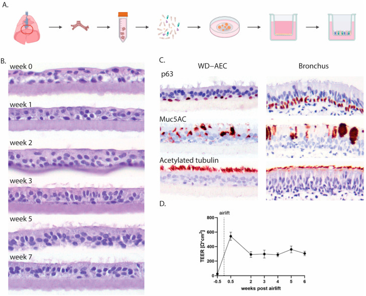
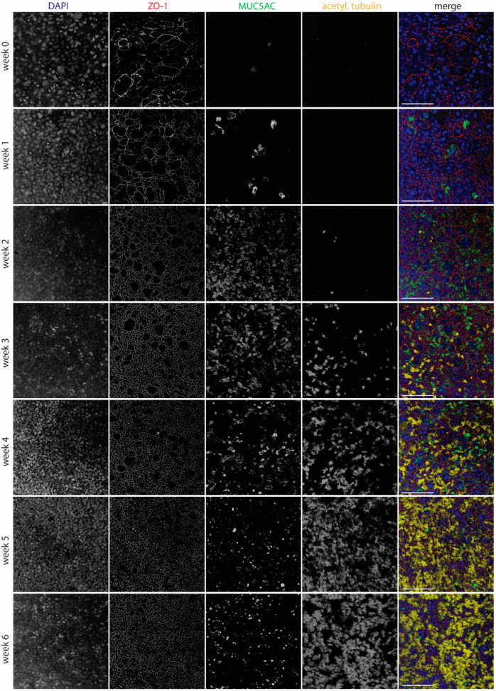
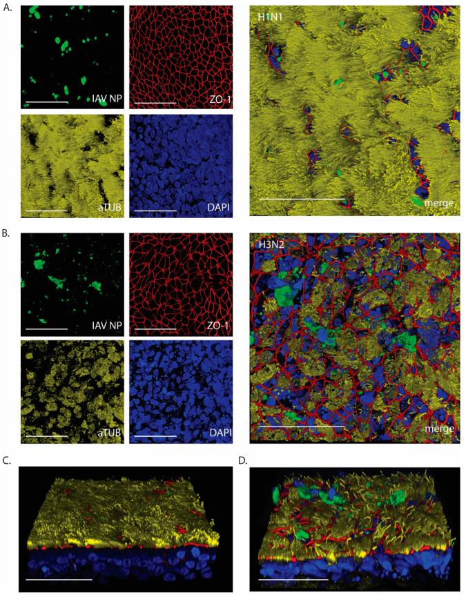
Swine influenza A viruses (IAVsw) are important causes of disease in pigs but also constitute a public health risk. IAVsw strains show remarkable differences in pathogenicity. We aimed to generate airway organoids from the porcine lower respiratory tract and use these to establish well-differentiated airway epithelial cell (WD-AEC) cultures grown at an air-liquid interface (ALI) for in vitro screening of IAVsw strain virulence. Epithelial cells were isolated from bronchus tissue of juvenile pigs, and airway organoids were cultured in an extracellular matrix in a culture medium containing human growth factors. Single-cell suspensions of these 3D organoids were seeded on Transwell filters and differentiated at ALI to form a pseudostratified epithelium containing ciliated cells, mucus-producing cells and tight junctions. Inoculation with a low dose of IAVsw in a low volume inoculum resulted in virus replication without requiring the addition of trypsin, and was quantified by the detection of viral genome loads in apical washes. Interestingly, inoculation of an H3N2 strain known to cause severe disease in pigs induced a greater reduction in trans-epithelial resistance and more damage to tight junctions than H1N2 or H1N1 strains associated with mild disease in pigs. We conclude that the porcine WD-AEC model is useful in assessing the virulence of IAVsw strains.

Keywords: Transwell cultures; airway epithelial cells; air–liquid interface; organoids; pig; swine influenza virus.


Conflict of interest statement

The authors declare no conflicts of interest. The funders had no role in the design of the study; in the collection, analyses, or interpretation of data; in the writing of the manuscript; or in the decision to publish the results.

Figures

Figure 1
Figure 1
Establishment of AO-derived WD-AECs. (A) Schematic outline of the isolation of porcine bronchial epithelial cells from the primary bronchi which were subsequently grown as 3D organoids in an extracellular matrix before seeding in 2D on Transwell filters and culturing at air–liquid interface upon confluency. (B) Transverse histology sections over the course of 7 weeks showing the development of a pseudostratified ciliated respiratory epithelium. Hematoxylin and eosin stain, 40× objective. (C) Immunohistochemistry (IHC) staining of WD-AECs 7 weeks post-airlift and porcine bronchus epithelial cells. P63 staining to visualize basal cells, Muc5AC staining to visualize mucus (goblet cells) and acetylated tubulin staining to visualize cilia. WD-AEC resembles an in vivo bronchial epithelium in cellular composition and morphology, despite a reduced thickness. (D) Development of transepithelial electrical resistance (TEER) over the course of differentiation of WD-AECs. After an initial increase post-airlift, TEER values remained consistent. For B, C and D: Differentiation was followed for three separate donors. Representative data from one experiment are shown.
Figure 2
Figure 2
Development of WD-AECs over time. Confocal laser scanning microscopy images showing the development of tight junctions (ZO-1), goblet cells (Muc5ac) and cilia (acetylated tubulin) over the course of 6 weeks after airlifting Transwell filters. Goblet cells were most abundant at 2–4 weeks, while cilia appeared after 3 weeks. Tight junctions were detected from week 0 onwards, but only started to show a regular pattern from week 2 onwards. Merged images show nuclei in blue, tight junctions in red, goblet cells in green and cilia in yellow. Scale bar represents 100 µm. 40× objective. Representative data from one experiment are shown.
Figure 3
Figure 3
Infection of porcine WD-AECs with IAVsw. (A) Experimental design of IAVsw inoculation of porcine WD-AEC cultures. Three different IAVsw strains were used (H1N1, H1N2, H3N2) and applied apically at a dose of 102 or 103 TCID50 in a volume of 10 µL. After 1 h incubation, Transwell filters were washed 4 times. An apical wash was collected at 16 h, 24 h and 48 h post-inoculation, and transepithelial electrical resistance (TEER) was measured at 0 hpi, 24 hpi and 48 hpi. At 48 hpi, filters were fixed and stained by hematoxylin and eosin (HE) and against IAV nucleoprotein (NP). (B) Viral RNA loads in apical washes. All viruses replicated in porcine WD-AECs. For H1N2, a dose–response curve was observed over all evaluated time points, which was only observed at the earlier time points for the other two viruses. (C) TEER values as a measure of epithelial integrity. TEER values declined for H3N2- and H1N1-inoculated WD-AECs over 48 h but remained constant in H1N2-inoculated WD-AECs. (D) Transverse histology sections stained by HE at 48 h post-infection. The epithelial cell layer remained intact for H1N2-inoculated WD-AECs, while there was substantial thinning of the cell layer in H3N2-inoculated WD-AECs. For H1N1, the higher dose seemed to cause more cell loss compared to the lower dose. 40× objective. B, C and D show representative results from one out of three experiments (pig04).
Figure 4
Figure 4
IAV NP expression after 24 h post-infection with 103 TCID50. Confocal microscopy images to visualize expression of IAV NP and epithelial composition. (A) H1N1-inoculated WD-AECs showed NP expression (green), continuous tight junctions (red), a thick layer of cilia (yellow) and predominantly intact nuclei (blue). (B) H3N2-inoculated filters showed NP expression while at the same time tight junctions were compromised, cilia expression was reduced and more fragmented nuclei were visible. (C) Angled side-view of WD-AEC shown in (A). (D) Angled side-view of WD-AEC shown in (B). Note that IAV NP staining (green) is visible within or above the apical side of WD-AEC cultures. All images: Scale bar represents 50 µm. 100× objective. Representative data from one experiment are shown.

Similar articles

References

    1. Castrucci M.R., Donatelli I., Sidoli L., Barigazzi G., Kawaoka Y., Webster R.G. Genetic reassortment between avian and human influenza A viruses in Italian pigs. Virology. 1993;193:503–506. doi: 10.1006/viro.1993.1155. - DOI - PubMed
    1. Ma W. Swine influenza virus: Current status and challenge. Virus Res. 2020;288:198118. doi: 10.1016/j.virusres.2020.198118. - DOI - PMC - PubMed
    1. Anderson T.K., Chang J., Arendsee Z.W., Venkatesh D., Souza C.K., Kimble J.B., Lewis N.S., Davis C.T., Vincent A.L. Swine Influenza A Viruses and the Tangled Relationship with Humans. Cold Spring Harb. Perspect. Med. 2021;11:a038737. doi: 10.1101/cshperspect.a038737. - DOI - PMC - PubMed
    1. Cogdale J., Kele B., Myers R., Harvey R., Lofts A., Mikaiel T., Hoschler K., Banyard A.C., James J., Mollett B.C., et al. A case of swine influenza A(H1N2)v in England, November 2023. Euro Surveill. 2024;29:2400002. doi: 10.2807/1560-7917.ES.2024.29.3.2400002. - DOI - PMC - PubMed
    1. Hennig C., Graaf A., Petric P.P., Graf L., Schwemmle M., Beer M., Harder T. Are pigs overestimated as a source of zoonotic influenza viruses? Porc. Health Manag. 2022;8:30. doi: 10.1186/s40813-022-00274-x. - DOI - PMC - PubMed

Publication types

MeSH terms

LinkOut - more resources