Review
. 2024 Nov 12:11:1440670.
doi: 10.3389/fmolb.2024.1440670. eCollection 2024.

Esophageal adenocarcinoma models: a closer look

Affiliations
Review

Esophageal adenocarcinoma models: a closer look

Nadeem Bhat et al. Front Mol Biosci. .

Abstract

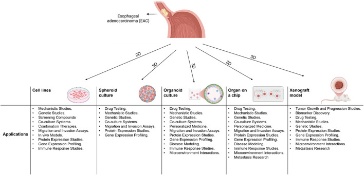
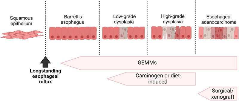
Esophageal adenocarcinoma (EAC) is a subtype of esophageal cancer with significant morbidity and mortality rates worldwide. Despite advancements in tumor models, the underlying cellular and molecular mechanisms driving EAC pathogenesis are still poorly understood. Therefore, gaining insights into these mechanisms is crucial for improving patient outcomes. Researchers have developed various models to better understand EAC and evaluate clinical management strategies. However, no single model fully recapitulates the complexity of EAC. Emerging technologies, such as patient-derived organoids and immune-competent mouse models, hold promise for personalized EAC research and drug development. In this review, we shed light on the various models for studying EAC and discuss their advantages and limitations.

Keywords: 3D culture models; GEMMs; animal models; esophageal adenocarcinoma; patient-derived organoids.


Conflict of interest statement

The authors declare that the research was conducted in the absence of any commercial or financial relationships that could be construed as a potential conflict of interest.

Figures

FIGURE 1
FIGURE 1
A schematic diagram showing 2D and 3D models that are used in Esophageal Adenocarcinoma (EAC) and their applications.
FIGURE 2
FIGURE 2
A schematic diagram illustrating the progression from Barrett’s Esophagus (BE) to Esophageal Adenocarcinoma (EAC) and correlating it with the various in vivo models employed to investigate each stage.

References

    1. Abdulnour-Nakhoul S., Nakhoul N. L., Wheeler S. A., Haque S., Wang P., Brown K., et al. (2007). Characterization of esophageal submucosal glands in pig tissue and cultures. Dig. Dis. Sci. 52, 3054–3065. 10.1007/s10620-006-9739-3 - DOI - PubMed
    1. Abuwatfa W. H., Pitt W. G., Husseini G. A. (2024). Scaffold-based 3D cell culture models in cancer research. J. Biomed. Sci. 31, 7. 10.1186/s12929-024-00994-y - DOI - PMC - PubMed
    1. Aikou S., Aida J., Takubo K., Yamagata Y., Seto Y., Kaminishi M., et al. (2013). Columnar metaplasia in a surgical mouse model of gastro-esophageal reflux disease is not derived from bone marrow-derived cell. Cancer Sci. 104, 1154–1161. 10.1111/cas.12213 - DOI - PMC - PubMed
    1. Attwood S. E., Harrison L. A., Preston S. L., Jankowski J. A. (2008). Esophageal adenocarcinoma in mice and men: back to basics. Am. J. Gastroenterol. 103, 2367–2372. 10.1111/j.1572-0241.2008.02004.x - DOI - PubMed
    1. Ballout F., Lu H., Bhat N., Chen L., Peng D., Chen Z., et al. (2024). Targeting SMAD3 improves response to oxaliplatin in esophageal adenocarcinoma models by impeding DNA repair. Clin. Cancer Res. 30, 2193–2205. 10.1158/1078-0432.CCR-24-0027 - DOI - PMC - PubMed

LinkOut - more resources