. 2024 Nov 12:15:1477976.
doi: 10.3389/fphys.2024.1477976. eCollection 2024.

Lipin1 as a therapeutic target for respiratory insufficiency of duchenne muscular dystrophy

Affiliations

Lipin1 as a therapeutic target for respiratory insufficiency of duchenne muscular dystrophy

Alexandra Brown et al. Front Physiol. .

Abstract

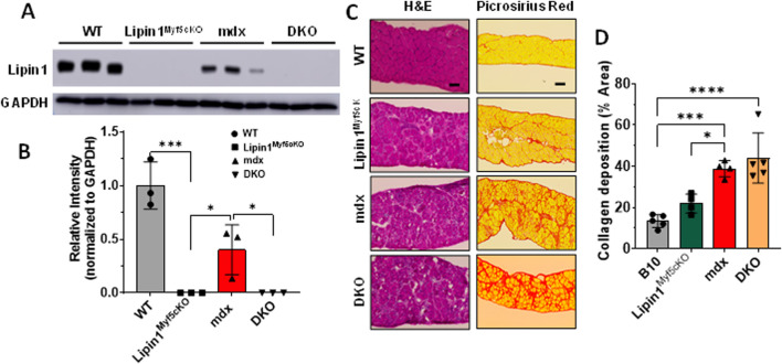
In Duchenne muscular dystrophy (DMD), diaphragm muscle dysfunction results in respiratory insufficiency which is a leading cause of death in patients. Mutations to the dystrophin gene result in myocyte membrane instability, contributing to the structural deterioration of the diaphragm muscle tissues. With previous works suggesting the importance of lipin1 for maintaining skeletal muscle membrane integrity, we explored the roles of lipin1 in the dystrophic diaphragm. We found that the protein expression levels of lipin1 were reduced by 60% in the dystrophic diaphragm. While further knockdown of lipin1 in the dystrophic diaphragm leads to increased necroptosis, restoration of lipin1 in the dystrophic diaphragm results in reduced inflammation and fibrosis, decreased myofiber death, and improved respiratory function. Our results demonstrated that lipin1 restoration improved respiratory function by enhancing membrane integrity and suggested that lipin1 could be a potential therapeutic target for preventing respiratory insufficiency and respiratory failure in DMD. Continued investigation is required to better understand the mechanisms behind these findings, and to determine the role of lipin1 in maintaining muscle membrane stability.

Keywords: DMD; diaphragm; dystrophin; lipin1; membrane integrity; muscular dystrophy; skeletal muscle; therapeutic target.


Conflict of interest statement

The authors declare that the research was conducted in the absence of any commercial or financial relationships that could be construed as a potential conflict of interest.

Figures

FIGURE 1
FIGURE 1
Histology analysis of the diaphragm muscles of WT, lipin1 Myf5cKO , mdx, and dystrophin/lipin1-DKO mice. (A) Representative Western blot and (B) densitometric analysis of lipin1 in the diaphragm muscles of WT, lipin1 Myf5cKO , mdx, and DKO mice (n = 3 mice/group). (C) representative H&E and Sirius red staining images, and (D) collagen deposition quantification analysis of diaphragm transverse sections (n = 5-6 mice/group). Scale bar = 200 μm. *p < 0.0001.
FIGURE 2
FIGURE 2
Protein expression levels of cell death, inflammation, and fibrosis markers in the diaphragm muscles of WT, lipin1 Myf5cKO , mdx, and dystrophin/lipin1-DKO mice. (A) Western blot and (B) densitometric analysis of necroptotic, apoptotic, inflammation, and fibrosis markers in the diaphragm of WT, lipin1 Myf5cKO , mdx, and DKO mice (n = 3 mice/group). *p < 0.0001.
FIGURE 3
FIGURE 3
Muscle damage in the diaphragms of WT, lipin1 Myf5cKO , mdx, and dystrophin/lipin1-DKO mice. (A) Representative images of EBD uptake (red) in diaphragm muscle sections of WT, lipin1 Myf5cKO , mdx, and DKO mice. WGA (green) was used to visualize the borders of muscle fibers and DAPI (blue) stained nuclei. (B) Quantification analysis of EBD-positive muscle fiber expressed as the percentage of the total number of muscle fibers in each mouse (n = 4−8 mice/group). Scale bar = 200 μm. **p < 0.01; ***p < 0.001; ****p < 0.0001.
FIGURE 4
FIGURE 4
Histology analysis of the diaphragm muscles of WT, mdx, and mdx:lipin1 Tg/0 mice. (A) Representative Western blot and (B) densitometric analysis of lipin1 in the diaphragm muscles of WT, mdx, and mdx:lipin1 Tg/0 mice (n = 3 mice/group). (C) Representative H&E and Sirius red staining images, and (D) collagen deposition quantification analysis of diaphragm transverse sections (n = 4–5 mice/group). Scale bar = 200 μm. **p < 0.01; ***p < 0.001; ****p < 0.0001.
FIGURE 5
FIGURE 5
Protein expression levels of cell death, inflammation, and fibrosis markers in the diaphragm muscles of WT, mdx, and mdx:lipin1 Tg/0 mice. (A) Western blot and (B) quantification analysis of necroptotic, apoptotic, inflammatory, and fibrosis markers in the diaphragm of WT, mdx, and mdx:lipin1 Tg/0 mice. Representative immunostaining of (C) CD86+ (red) and (D) CD206+ (red) macrophages in diaphragms of indicated mice. WGA (green) was used to visualize the borders of muscle fibers and DAPI (blue) stained nuclei. Scale bar = 100 μm. n = 3 mice/group. *p < 0.05 **p < 0.01; ***p < 0.001; ****p < 0.0001.
FIGURE 6
FIGURE 6
Muscle damage in the diaphragms of WT, mdx, and mdx:lipin1 Tg/0 mice. (A) Representative images of EBD uptake (red) in diaphragm muscle sections of WT, mdx, and mdx:lipin1 Tg/0 mice. WGA (green) was used to visualize the borders of muscle fibers and DAPI (blue) stained nuclei. Scale bar = 200 μm. (B) Quantification analysis of EBD-positive muscle fiber expressed as the percentage of the total number of muscle fibers in each mouse (n = 4−9 mice per group). *p < 0.05 **p < 0.01.
FIGURE 7
FIGURE 7
Respiratory function measurement of WT, lipin1 Myf5cKO , mdx, DKO, and mdx:lipin1 Tg/0 mice. (A) Respiratory rate, (B) tidal volume, and (C) minute volume in WT, lipin1 Myf5cKO , mdx, DKO, and mdx:lipin1 Tg/0 mice. All were measured by whole-body plethysmography. Respiratory measurements were taken over a 10-minute interval and averaged to obtain the data points for each mouse (n = 6–7 mice per group). *p < 0.05 **p < 0.01; ***p < 0.001; ****p < 0.0001.

Similar articles

References

    1. Acharyya S., Villalta S. A., Bakkar N., Bupha-Intr T., Janssen P. M., Carathers M., et al. (2007). Interplay of IKK/NF-kappaB signaling in macrophages and myofibers promotes muscle degeneration in Duchenne muscular dystrophy. J. Clin. Investig. 117 (4), 889–901. 10.1172/JCI30556 - DOI - PMC - PubMed
    1. Alshudukhi A. A., Zhu J., Huang D. T., Jama A., Smith J. D., Wang Q. J., et al. (2018). Lipin-1 regulates Bnip3-mediated mitophagy in glycolytic muscle. Faseb J. 32 (12), 6796–6807. 10.1096/fj.201800374 - DOI - PMC - PubMed
    1. Barbe F., Quera-Salva M. A., McCann C., Gajdos P., Raphael J. C., de Lattre J., et al. (1994). Sleep-related respiratory disturbances in patients with Duchenne muscular dystrophy. Eur. Respir. J. 7 (8), 1403–1408. 10.1183/09031936.94.07081403 - DOI - PubMed
    1. Bez Batti Angulski A., Hosny N., Cohen H., Martin A. A., Hahn D., Bauer J., et al. (2023). Duchenne muscular dystrophy: disease mechanism and therapeutic strategies. Front. Physiol. 14, 1183101. PMCID: 10330733. 10.3389/fphys.2023.1183101 - DOI - PMC - PubMed
    1. Brown A., Morris B., Kaumau J. K., Alshudukhi A. A., Jam A., Ren H. (2023). Automated image analysis pipeline development to monitor disease progression in muscular dystrophy using cell profiler. Arch. Microbio Immunol. 7 (1), 178–187. 10.26502/ami.936500115 - DOI - PMC - PubMed

LinkOut - more resources