. 2025 Jan 21;46(4):380-393.
doi: 10.1093/eurheartj/ehae782.

Enhanced Parkin-mediated mitophagy mitigates adverse left ventricular remodelling after myocardial infarction: role of PR-364

Affiliations

Enhanced Parkin-mediated mitophagy mitigates adverse left ventricular remodelling after myocardial infarction: role of PR-364

Lizhuo Ai et al. Eur Heart J. .

Abstract

Background and aims: Almost 30% of survivors of myocardial infarction (MI) develop heart failure (HF), in part due to damage caused by the accumulation of dysfunctional mitochondria. Organelle quality control through Parkin-mediated mitochondrial autophagy (mitophagy) is known to play a role in mediating protection against HF damage post-ischaemic injury and remodelling of the subsequent deteriorated myocardium.

Methods: This study has shown that a single i.p. dose (2 h post-MI) of the selective small molecule Parkin activator PR-364 reduced mortality, preserved cardiac ejection fraction, and mitigated the progression of HF. To reveal the mechanism of PR-364, a multi-omic strategy was deployed in combination with classical functional assays using in vivo MI and in vitro cardiomyocyte models.

Results: In vitro cell data indicated that Parkin activation by PR-364 increased mitophagy and mitochondrial biogenesis, enhanced adenosine triphosphate production via improved citric acid cycle, altered accumulation of calcium localization to the mitochondria, and initiated translational reprogramming with increased expression of mitochondrial translational proteins. In mice, PR-364 administered post-MI resulted in widespread proteome changes, indicating an up-regulation of mitochondrial metabolism and mitochondrial translation in the surviving myocardium.

Conclusions: This study demonstrates the therapeutic potential of targeting Parkin-mediated mitophagy using PR-364 to protect surviving cardiac tissue post-MI from progression to HF.

Keywords: Heart failure; Mitochondrial function; Multi-omics; Myocardial infarction; Parkin-dependent mitophagy; Proteomics; Translational reprogramming.


Figures

Structured Graphical Abstract
Parkin activator proposed therapeutic mechanisms. Inactive Parkin (red) is activated by PR-364; activated Parkin (green) promotes enhanced mitophagy and mito-biogenesis, improves mitochondrial function via increased mitochondrial calcium level and biogenesis, and ultimately leads to limited post-myocardial infarction cardiac remodelling and heart failure. MI, myocardial infarction.
Figure 1
Figure 1
PR-364 selectively activates Parkin and promotes Parkin-dependent mitophagy in vitro. (A) Recombinant His6-SUMO-Parkin (50 nM) was incubated with DMSO or PR-364 at indicated concentrations in the presence of MBP-TcPINK1 (2 nM) and ubiquitination reaction was carried out for 90 min at room temperature. EC50 value is reported as mean ± standard deviation (n = 3). (B) Per cent activation for Parkin and per cent inhibition for other E3 ligases was plotted and EC50 was determined (n = 3). (C) Western blot probed with anti-Ubiquitin antibody (VU-1) after PR-364. (D) PhosphoSer65ubiquitin (50 nM) was used as a Parkin activation control in the western blot. The signal intensities of PR-364 doses relative to pUb signal were shown on the graph, by one-way analysis of variance. (E) Western blot analyses of Parkin and p62 to measure mitophagy activation (n = 3/group) in H9c2 cells. (F and G) Western blot quantifications of Parkin and p62. Two-way analysis of variance with Šidák correction for multiple analyses was used to demonstrate Parkin levels after silencing. p62 up-regulation was significant by PR-364 in siControl cells but not in siParkin cells. (H) Western blot analysis of vehicle- vs. PR-364-treated cells with the addition of bafilomycin at 3 h before harvesting (n = 3/group). (I) Quantification of p62 in of mitochondrial p62 vs. total p62. Error bars represent mean ± standard deviation. Two-way analysis of variance with interaction was used to demonstrate the effects of PR-364 on p62 protein levels with or without bafilomycin (50 nmoles), which inhibits mitophagy flux. (J) 60× images of live H9c2 cells with PR-364 treatment and stained with mitophagy dye, to demonstrate mitophagy. Scale bars represent 50 µm. (K) Cells were treated with either vehicle (DMSO) or PR-364 for 6 h, with or without addition of bafilomycin (50 nmoles) 3 h before harvesting and being analysed by western blot for protein expression. (L) Western blot quantification of Pgc-1α protein level (n = 3/group), by t-test. (M–O) Western blot and quantification of Tfam and Tom70 (n = 3/group), by multiple comparisons post two-way analysis of variance
Figure 2
Figure 2
PR-364 up-regulates metabolite and protein expression, improves mitochondrial respiration and calcium ion uptake in AC16 cardiomyocytes. (A) Experimental design for in vitro AC16 studies (graph created using biorender.com). (B) Numbers of proteins were identified and metabolites that were measured using mass spectrometry–based untargeted proteomics or targeted metabolomics. (C) Heat map of 39 metabolites and 188 proteins that were significantly different between PR-364 and vehicle AC16 cells. The scale bar indicates z-scores of protein intensity values, with highly abundant proteins depicted in dark red and lower abundance proteins depicted in dark blue. n = 6, P < .05 by unpaired t-test for each metabolite measured or protein identified. (D) Pathway analysis of the 39 metabolites revealed enrichment of the TCA cycle by PR-364. Metabolites showing mean values at each dot; n = 6, by t-test for each metabolite. Specifically, mean mass spectrometry peak intensity ATPvehicle = 34 660, mean mass spectrometry peak intensity ATPPR-364 = 218 512; n = 6, P < .0001 by t-test, statistics not shown in the figure (TCA cycle illustration was created using Biorender.com). (E) Seahorse mito-stress assay reveals increased ATP production and maximal respiration after PR-364 administration; mean ATPvehicle = 0.148 pmol/min/μg, mean ATPPR-364 = 0.312 pmol/min/μg; n = 3; P < .05 by t-test. (F) Proteomics pathway analysis showed PR-364 increased expression of some proteins involved in mitochondrial calcium ion transport. Proteins showing mean values at each dot; n = 6; P < .05 by t-test for each protein, statistics not shown on graph. (G) Proteomics pathway analysis showed PR-364 increased expression of proteins involved in mitochondrial translation. Proteins showing mean values at each dot; n = 6; P < .05 by t-test for each protein, statistics not shown on graph
Figure 3
Figure 3
PR-364 increases mitochondrial translation and mitophagy in 6 h. (A) Experimental design of the 6 h mouse cohort with myocardial infarction at 2 h to study the proteomic effect of PR-364 post-myocardial infarction in vivo (graph created using biorender.com). (B) Western blot image of LC3-I and LC3-II detection in total left ventricle lysate (top) and mitochondrial enriched fraction lysate (bottom). (C and D) Quantifications of mito LC3-II to mito ponceau stain and LC3-II to LC3-I ratio in the total left ventricle lysate (n = 3, mean ± standard deviation, by t-test). (E) Experimental design of the 6 h control mouse cohort to study the proteomic effect of PR-364 to healthy heart in vivo (graph created using biorender.com). (F) Heat map of the top 25 proteins that are significantly altered by PR-364 (the scale bar indicates z-scores of protein intensity values; n = 4, P < .05 each protein by t-test). (G) Principal component analysis plot of the proteomic profiles between PR-364 mice and vehicle mice. (H) Gene set enrichment analysis of the proteomic data showed enriched mitochondrial translation by PR-364. (I) MitoPLEX measurement of the heart tissue samples (the scale bar indicates z-scores of protein intensity values; n = 5, proteins changed with P < .005 are boxed in black, by unpaired t-test)
Figure 4
Figure 4
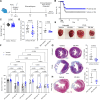
PR-364 protects myocardium by maintaining cardiac function, preventing cardiac remodelling, and reducing mortality post-myocardial infarction. (A) Twenty-eight-day in vivo paradigm (graph created using biorender.com). (B) Survival curve of the PR-364- or vehicle-treated wild-type mice post-permanent coronary artery ligation. (C) Measurement of body weight of each wild-type mouse in both vehicle and PR-364 groups, not significant by t-test. (D) Heart weight to tibia length ratios in vehicle vs. PR-364-treated wild-type and Parkin knockout mice, respectively, mean ± standard deviation, by unpaired t-test. (E) Representative pictures of hearts from untreated (no permanent coronary artery ligation) and permanent coronary artery ligation wild-type mice in both vehicle- and PR-364-treated groups. (F) Echocardiogram recordings of the wild-type and Parkin knockout mice on baseline (before permanent coronary artery ligation), Day 14, and Day 28. Ejection fractions were calculated based on the M-mode echo at each time point, by two-way analysis of variance with Šídák’s multiple comparisons test. (G) 60× representative heart sections after Masson Trichrome staining to examine infarct size in hearts of untreated and permanent coronary artery ligation wild-type mice after vehicle vs. PR-364 treatment, mean ± standard deviation, by unpaired t-test. (H) Representative Parkin knockout heart sections after Masson Trichrome staining to examine cardiac infarct size with vehicle vs. PR-364 treatment post-permanent coronary artery ligation. Mean ± standard deviation, by unpaired t-test
Figure 5
Figure 5
PR-364 reduces mortality and leads to translational reprogramming of the heart in mice post-permanent coronary artery ligation. (A) Experimental schematic of the 3-day (A) and 7-day (B) mouse cohorts (graph created using biorender.com). (B) Seven-day cohort survival curve. (C) The numbers of total proteins detected by mass spectrometry in the two cohorts. (D) Sankey diagrams showing the proteomic changes in two cohorts. Left: From the 3826 identified proteins in the Day 3 mice, 904 proteins (∼24% of total detected proteome) had significant changes either between vehicle sham control group vs. vehicle permanent coronary artery ligation group or between vehicle permanent coronary artery ligation group vs. PR-364 permanent coronary artery ligation group (cut-off P < .05 by t-test). Seventy proteins (646 up + 80 down—656 no change) showed significant changes in both comparisons. Right: from the 3994 identified proteins in the Day 7 mice, 1115 proteins (∼28% of total detected proteome) showed significant changes either between vehicle sham control group vs. vehicle permanent coronary artery ligation group or between vehicle permanent coronary artery ligation group vs. PR-364 permanent coronary artery ligation group (cut-off P < .05 by t-test). Two hundred and eighty-seven proteins (617 up + 191 down—521 no change) showed significant changes in both comparisons. (E) Principal component analysis plots of the 70 proteins from 3 days (left) and 287 proteins from 7 days (right), indicating PR-364 translationally reprogrammed a subgroup of proteome in the permanent coronary artery ligation hearts back to the normal healthy state (vehicle sham). (F) Bubble plots of GO network analysis of the 3-day vehicle permanent coronary artery ligation and PR-364 permanent coronary artery ligation group (114 up + 134 down proteins). (G and H) Bubble plots of GO network analysis of the 7-day vehicle permanent coronary artery ligation and PR-364 permanent coronary artery ligation group (355 up + 239 down proteins)

References

    1. Jenča D, Melenovský V, Stehlik J, Staněk V, Kettner J, Kautzner J, et al. . Heart failure after myocardial infarction: incidence and predictors. ESC Heart Fail 2021;8:222–37. 10.1002/ehf2.13144 - DOI - PMC - PubMed
    1. Neubauer S. The failing heart—an engine out of fuel. N Engl J Med 2007;356:1140–51. 10.1056/NEJMra063052 - DOI - PubMed
    1. Knowlton AA, Chen L, Malik ZA. Heart failure and mitochondrial dysfunction: the role of mitochondrial fission/fusion abnormalities and new therapeutic strategies. J Cardiovasc Pharmacol 2014;63:196–206. 10.1097/01.fjc.0000432861.55968.a6 - DOI - PMC - PubMed
    1. Poznyak AV, Ivanova EA, Sobenin IA, Yet S-F, Orekhov AN. The role of mitochondria in cardiovascular diseases. Biology (Basel) 2020;9:137. 10.3390/biology9060137 - DOI - PMC - PubMed
    1. Zhang R, Krigman J, Luo H, Ozgen S, Yang M, Sun N. Mitophagy in cardiovascular homeostasis. Mech Ageing Dev 2020;188:111245. 10.1016/j.mad.2020.111245 - DOI - PMC - PubMed

MeSH terms

Substances