. 2025 Jan 31;99(1):e0185324.
doi: 10.1128/jvi.01853-24. Epub 2024 Nov 27.

The respective roles of TMPRSS2 and cathepsins for SARS-CoV-2 infection in human respiratory organoids

Affiliations

The respective roles of TMPRSS2 and cathepsins for SARS-CoV-2 infection in human respiratory organoids

Masatoshi Kakizaki et al. J Virol. .

Abstract

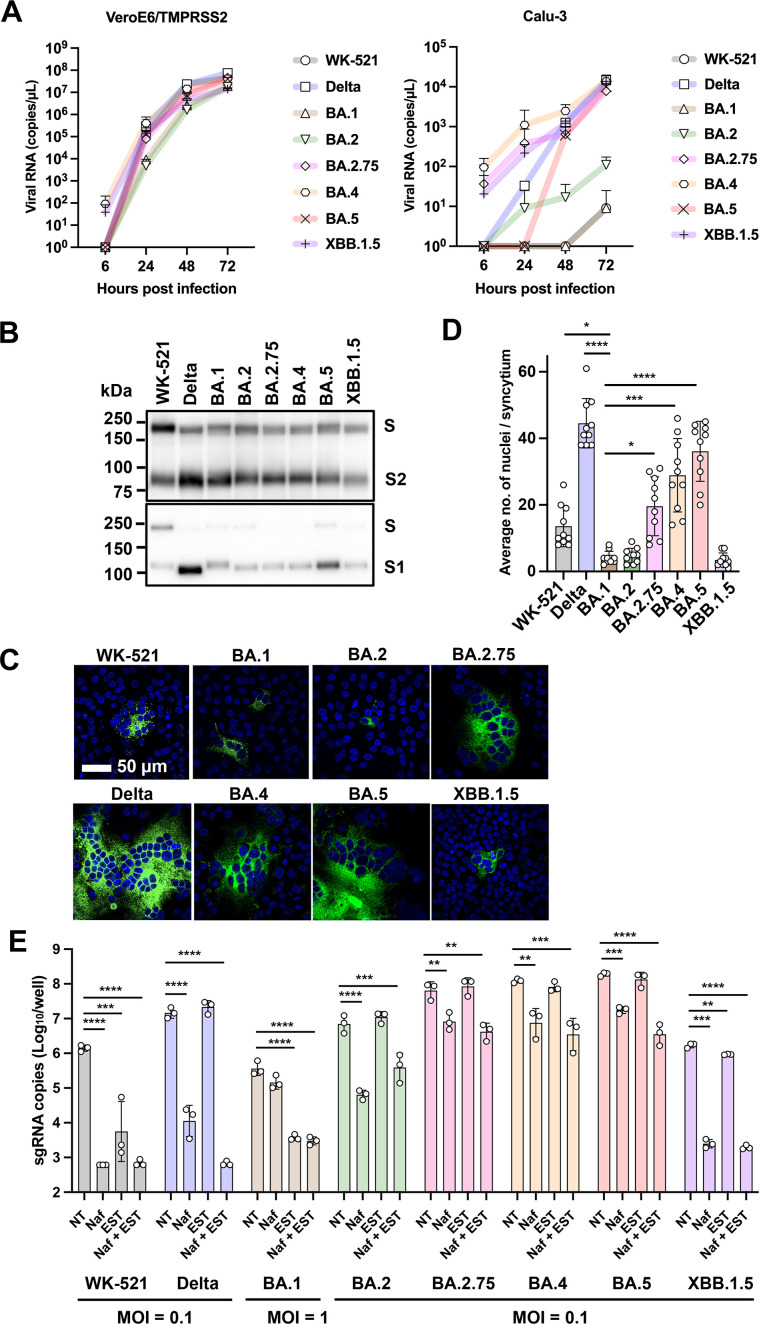
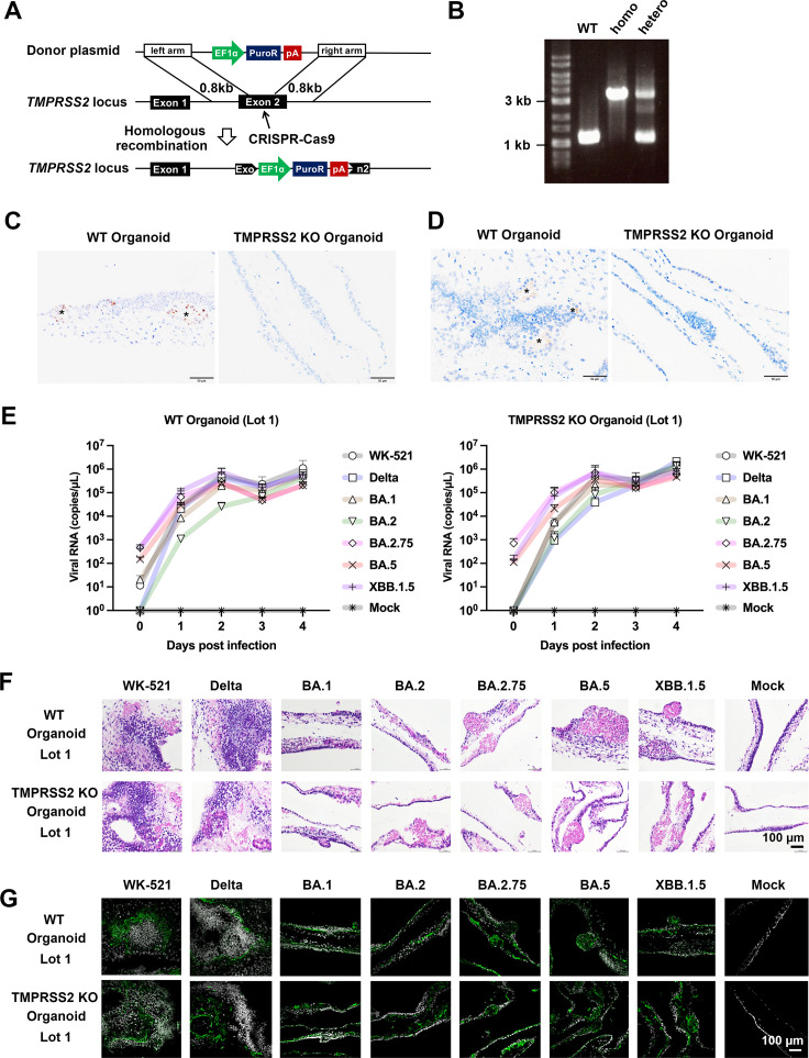
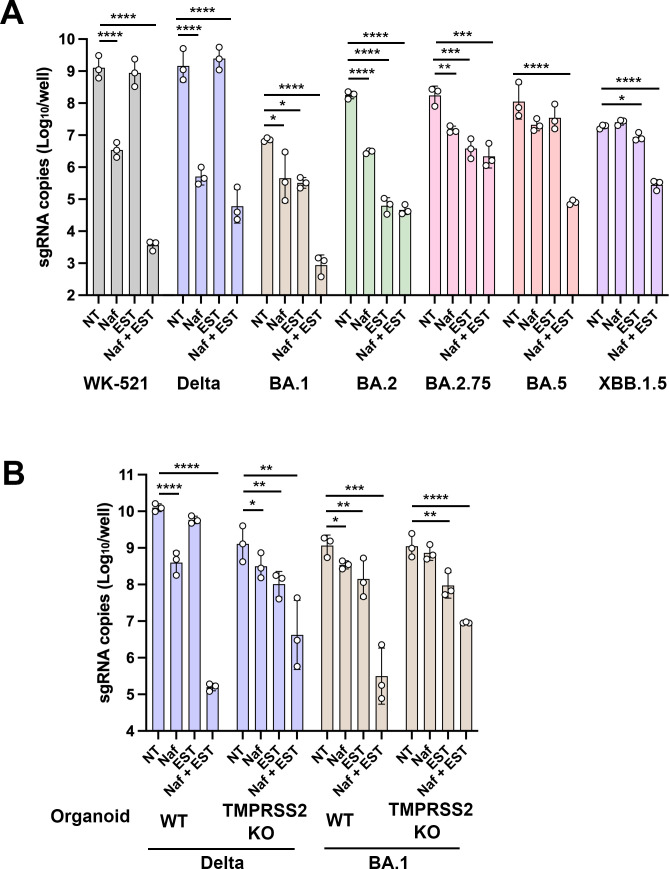
A critical aspect of the mechanism of severe acute respiratory syndrome coronavirus 2 (SARS-CoV-2) infection is the protease-mediated activation of the viral spike (S) protein. The type II transmembrane serine protease TMPRSS2 is crucial for SARS-CoV-2 infection in lung epithelial Calu-3 cells and murine airways. However, the importance of TMPRSS2 needs to be re-examined because the ability to utilize TMPRSS2 is significantly reduced in the Omicron variants that spread globally. For this purpose, replication profiles of SARS-CoV-2 were analyzed in human respiratory organoids. All tested viruses, including Omicron variants, replicated efficiently in these organoids. Notably, all SARS-CoV-2 strains retained replication ability in TMPRSS2-gene knockout (KO) respiratory organoids, suggesting that TMPRSS2 is not essential for SARS-CoV-2 infection in human respiratory tissues. However, TMPRSS2-gene knockout significantly reduces the inhibitory effect of nafamostat, indicating the advantage of TMPRSS2-utilizing ability for the SARS-CoV-2 infection in these organoids. Interestingly, Omicron variants regained the TMPRSS2-utilizing ability in recent subvariants. The basal infectivity would be supported mainly by cathepsins because the cathepsin inhibitor, EST, showed a significant inhibitory effect on infection with any SARS-CoV-2 strains, mainly when used with nafamostat. A supplementary contribution of other serine proteases was also suggested because the infection of the Delta variant was still inhibited partially by nafamostat in TMPRSS2 KO organoids. Thus, various proteases, including TMPRSS2, other serine proteases, and cathepsins, co-operatively contribute to SARS-CoV-2 infection significantly in the respiratory organoids. Thus, SARS-CoV-2 infection in the human respiratory tissues would be more complex than observed in cell lines or mice.

Importance: We explored how the severe acute respiratory syndrome coronavirus 2 (SARS-CoV-2) virus infects human respiratory organoids, which are a cultured cell model made to mimic the physiological conditions of the human airways. We focused on understanding the role of different proteases of host cells in activating the virus spike proteins. Specifically, we looked at TMPRSS2, a transmembrane serine protease, and cathepsin L, a lysosomal enzyme, which helps the virus enter cells by cutting the viral spike protein. We discovered that while TMPRSS2 is crucial for the virus in certain cells and animal models, other proteases, including cathepsins and various serine proteases, also play significant roles in the SARS-CoV-2 infection of human respiratory organoids. We suggest that SARS-CoV-2 uses a more complex mechanism involving multiple proteases to infect human airways, differing from what we see in conventional cell lines or animal models. This complexity might help explain how different variants can spread and infect people effectively.

Keywords: SARS-CoV-2; TMPRSS2; cathepsin; cleavage.


Conflict of interest statement

The authors declare no conflict of interest.

Figures

Fig 1
Fig 1
Infection in VeroE6/TMPRSS2 and Calu-3 cells. (A) Replication kinetics in cells infected with viruses at a multiplicity of infection (MOI) of 0.1. Viral RNA copy numbers in the culture supernatants were quantified at 6, 24, 48, and 72 h post-infection (p.i.). Error bars indicate the standard deviations of three biological replicates. Three confirmatory experiments were conducted. Mean values with positive standard deviations are shown. (B) S proteins detected in the culture supernatant of infected VeroE6/TMPRSS2 cells. SDS-PAGE and western blot analysis. Three confirmatory experiments were conducted. (A, B) (C) Syncytium formation in cells transfected with the S protein expression plasmid. S protein was detected by an indirect immunofluorescent assay (green) and nuclei were detected by DAPI (blue) staining. Three confirmatory experiments were conducted. (D) The number of nuclei in 10 individual, randomly selected syncytia shown in C was counted. Mean values ± standard deviations are shown, and significant differences were determined with one-way ANOVA. Multiple comparisons between the BA.1 spike and other variants spike were adjusted with Dunn’s multiple comparison test, *P < 0.05, **P < 0.01, ***P < 0.001, ****P < 0.0001. (E) Cells were infected with viruses at an MOI of 0.1 or 1.0 in the absence (NT) or the presence of either nafamostat (Naf) or EST or both (Naf +EST). At 24 h p.i., viral RNAs in cells were quantified by real-time RT-PCR. Error bars indicate the standard deviations of three biological replicates. Three confirmatory experiments were conducted. Statistical significance was determined with two-way ANOVA. Multiple comparisons between NT and different groups were adjusted with Dunnett’s multiple comparison tests, *P < 0.05, **P < 0.01, ***P < 0.001, ****P < 0.0001.
Fig 2
Fig 2
Replication ability in human respiratory organoids. (A) A schematic diagram of the targeting strategy for TMPRSS2 locus. The following donor plasmids were used to target the TMPRSS2 locus. Donor plasmids: EF1α; elongation factor 1 alpha promoter, PuroR; puromycin resistant protein, pA; polyadenylation sequence. The CRISPR-Cas9 system was utilized to produce TMPRSS2 sequence-specific double-strand breaks. (B) Genotyping was performed to examine whether the human iPS cells were correctly targeted. (C) RNA in situ hybridization. Representative images showing TMPRSS2 mRNA. Target signals were visualized by 3′,3′ diaminobenzidine (DAB), brown; counterstaining, hematoxylin. Asterisks, glandular structure; bars, 50 µm. (D) Immunohistochemistry. Representative images showing TMPRSS2 antigens. Target signals were visualized by DAB, brown; counter-staining, hematoxylin. Asterisks, glandular structure; bars, 50 µm. (E) Replication kinetics in WT and TMPRSS2-KO human respiratory organoids (Lot 1) infected with viruses at an MOI of 0.1. Viral RNA copy numbers in the culture supernatants were quantified at 1, 2, 3, and 4 days post-infection (p.i.). Error bars indicate the standard deviations of three biological replicates. Mean values with positive standard deviations are shown. (F) H&E staining of uninfected or infected human respiratory organoids. (G) Immunofluorescence images of SARS-CoV-2 N protein (green) in uninfected or infected human respiratory organoids. Nuclei were counterstained with DAPI (blue).
Fig 3
Fig 3
Entry phenotype in human respiratory organoids. (A) Wild-type human respiratory organoids were infected with viruses at an MOI of 1.0 in the absence (NT) or presence of either nafamostat (Naf) or EST or both (Naf +EST). At 24 h p.i., viral RNAs in cells were quantified by real-time RT-PCR. Error bars indicate the standard deviations of three biological replicates. Two confirmatory experiments were conducted. Statistical significance was determined using two-way ANOVA. Multiple comparisons between NT and different groups were adjusted with Dunnett’s multiple comparison tests, *P < 0.05, **P < 0.01, ***P < 0.001, ****P < 0.0001. (B) Wild-type or TMPRSS2-gene knockout human respiratory organoids were infected with viruses at an MOI of 0.1 in the absence (NT) or presence of either nafamostat (Naf) or EST or both (Naf +EST). At 24 h p.i., viral RNAs in cells were quantified by real-time RT-PCR. Error bars indicate the standard deviations of three biological replicates. Two confirmatory experiments were conducted. Statistical significance was determined using two-way ANOVA. Multiple comparisons between NT and different groups were adjusted with Dunnett’s multiple comparison tests, *P < 0.05, **P < 0.01, ***P < 0.001, ****P < 0.0001.

References

    1. Zhou P, Yang XL, Wang XG, Hu B, Zhang L, Zhang W, Si HR, Zhu Y, Li B, Huang CL, et al. . 2020. A pneumonia outbreak associated with a new coronavirus of probable bat origin. Nature 579:270–273. doi:10.1038/s41586-020-2012-7 - DOI - PMC - PubMed
    1. Wu F, Zhao S, Yu B, Chen YM, Wang W, Song ZG, Hu Y, Tao ZW, Tian JH, Pei YY, Yuan ML, Zhang YL, Dai FH, Liu Y, Wang QM, Zheng JJ, Xu L, Holmes EC, Zhang YZ. 2020. A new coronavirus associated with human respiratory disease in China. Nature 579:265–269. doi:10.1038/s41586-020-2008-3 - DOI - PMC - PubMed
    1. Takeda M. 2022. Proteolytic activation of SARS-CoV-2 spike protein. Microbiol Immunol 66:15–23. doi:10.1111/1348-0421.12945 - DOI - PMC - PubMed
    1. Jackson CB, Farzan M, Chen B, Choe H. 2022. Mechanisms of SARS-CoV-2 entry into cells. Nat Rev Mol Cell Biol 23:3–20. doi:10.1038/s41580-021-00418-x - DOI - PMC - PubMed
    1. Benton DJ, Wrobel AG, Xu P, Roustan C, Martin SR, Rosenthal PB, Skehel JJ, Gamblin SJ. 2020. Receptor binding and priming of the spike protein of SARS-CoV-2 for membrane fusion. Nature 588:327–330. doi:10.1038/s41586-020-2772-0 - DOI - PMC - PubMed