Multicenter Study
. 2024 Nov 4;65(13):60.
doi: 10.1167/iovs.65.13.60.

Novel Uveal Melanoma Patient-Derived Organoid Models Recapitulate Human Disease to Support Translational Research

Affiliations
Multicenter Study

Novel Uveal Melanoma Patient-Derived Organoid Models Recapitulate Human Disease to Support Translational Research

Lauren A Dalvin et al. Invest Ophthalmol Vis Sci. .

Abstract

Purpose: A lack of representative human disease models has limited the translation of new and more effective treatments in uveal melanoma (UM), the most common primary adult intraocular malignancy. To fill this critical need, we developed and characterized a multicenter biobank of UM patient-derived organoids (PDOs).

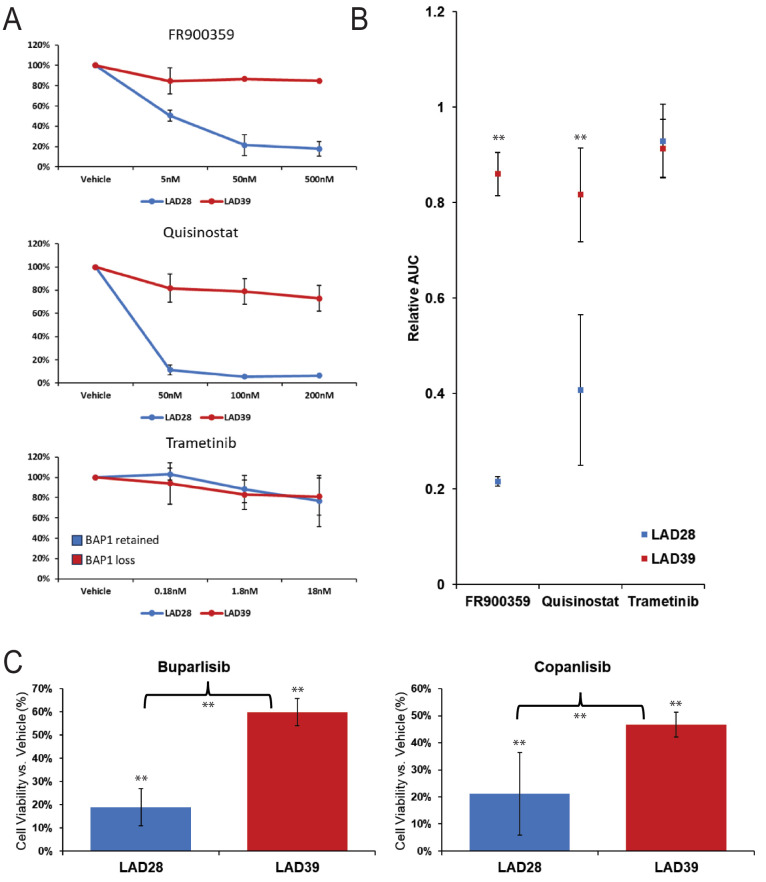
Methods: UM patients requiring enucleation from 2019 to 2024 donated tumor tissue for PDO generation. PDOs were cultured in Cultrex and compared to donor primary tumor using exome sequencing, RNA sequencing, and immunohistochemistry. The ability of PDOs to maintain the transformed phenotype was evaluated in an orthotopic xenograft model and monitored with fundus imaging. ATAC sequencing and drug response assays were done in a subset of PDOs to explore the feasibility of their use for mechanistic and translational studies.

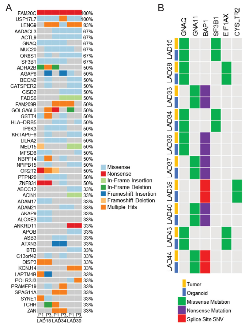
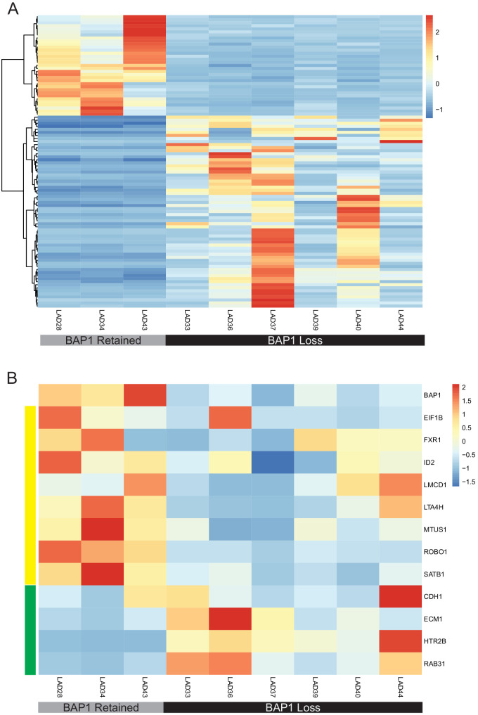
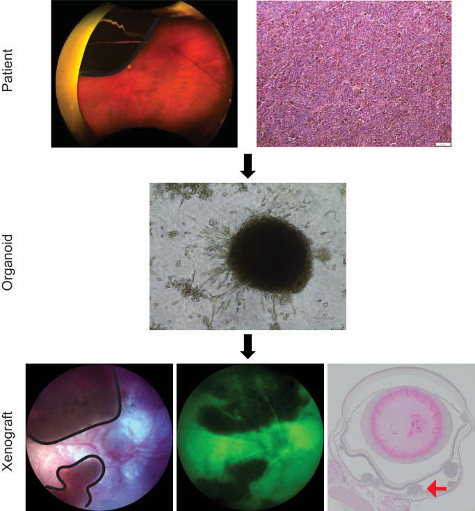
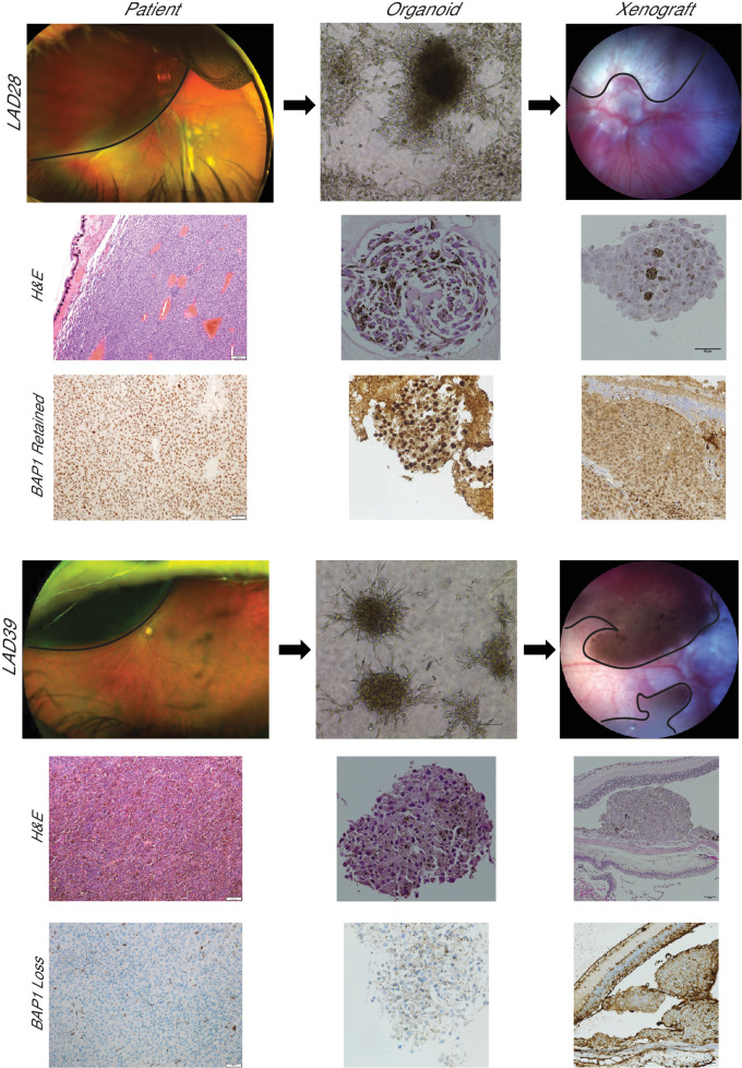
Results: PDOs were successfully established in 40 of 44 cases (91%), retained clinically relevant mutations and molecular markers from the primary tumor, and displayed similar gene expression profiles and well-validated clinical prognostic markers of the disease. PDOs retained tumorigenic capacity in an in vivo model resembling human disease progression. Finally, we demonstrated that PDOs were a feasible platform to identify and evaluate novel therapeutic targets and investigate differential, personalized drug response.

Conclusions: PDO models offer a new platform with improved representation of human UM to aid in translational research for this dismal condition.


Conflict of interest statement

Disclosure: L.A. Dalvin, IDEAYA Biosciences (C); C.M. Andrews-Pfannkoch, None; D.R. Miley, None; T.L. Hogenson, None; S.A. Erickson, None; S. Malpotra, None; K.J. Anderson, None; M.E. Omer, None; L.L. Almada, None; C. Zhang, None; H. Li, None; D.R. Salomao, None; C.L. Shields, None; S.E. Lally, None; R.M. Malsch, None; J.A. Armitage, None; H.L. Holmes, None; M.F. Romero, None; M.P. Fautsch, None; S.N. Markovic, None; M.E. Fernandez-Zapico, None

Figures

Figure 1.
Figure 1.
Patient-derived organoids can be established from uveal melanoma tumors. (A) UM PDO pipeline. FNAB specimens are taken from the primary tumor and dissociated, and PDOs are propagated. PDOs are characterized with immunofluorescence (IF) and immunohistochemistry (IHC) to confirm melanocytic origin and similarities to the clinical sample. PDOs can be further characterized with omics analyses and are used for drug studies. The inset shows transillumination to mark the tumor in an enucleated globe followed by FNAB through the marked area to harvest tumor tissue for PDO generation. (B) H&E histopathology images of primary UM tumors from which PDOs were derived demonstrate UM of spindle (LAD15, LAD55, LAD57) or mixed (LAD39) cell type. Scale bars: 1 mm for LAD15, 100 µm for all others. Corresponding brightfield images of organoids are shown. Scale bars: 100 µm. (C) Uveal melanoma PDOs characterized by immunofluorescence with 4′,6-diamidino-2-phenylindole (DAPI) nuclear stain. PDOs have cytoplasmic staining for Melan-A (green) and nuclear staining for SOX10 (blue), confirming the melanocytic origin of the cells. (D) Uveal melanoma PDOs demonstrate histopathological features consistent with the corresponding clinical sample. The hallmark feature of nuclear BAP1 retention or loss is consistent between the clinical sample and the PDO.
Figure 2.
Figure 2.
Uveal melanoma patient-derived organoids retain genomic features of the corresponding clinical tumor. (A) Oncoplot of PDOs at P1 and P3 shows relative stability and retention of key uveal melanoma hallmark mutations in GNAQ and SF3B1 through passaging. (B) Genetic comparison of clinically relevant markers between the PDO and the corresponding donor tumor (n = 10) shows retention of key driver and prognostic mutations in the PDOs. Splice site single nucleotide variants were not detected in PDOs by whole exome sequencing but were confirmed by PCR resequencing for the variant identified on clinical testing.
Figure 3.
Figure 3.
UM PDOs have differential gene expression based on BAP1 status. (A) Heatmap of z-scores for differential gene expression between BAP1-retained and BAP1-loss PDOs using DeSeq2. Differential expression determined using ±1 log fold change and P ≤ 0.1. (B) Heatmap of z-scores for gene expression of a subset of genes used on a well-validated clinical prognostic gene expression profiling test and further analyzed in TCGA samples shows that uveal melanoma PDOs simulated expression patterns seen in the clinical samples. BAP1 loss is a known poor prognostic indicator. Genes with low expression in high-risk primary UM by gene expression profiling and TCGA are denoted by the yellow panel, and those with high expression in high-risk tumors are denoted by the green panel. High-risk PDOs, defined by those with BAP1 loss, followed the expected pattern of prognostic gene expression.
Figure 4.
Figure 4.
UM orthotopic xenografts can be generated from patient-derived material. Patients requiring enucleation presented with a unilateral, large, pigmented choroidal mass (black outline) shown on Optos pseudocolor fundus photographs. Tumor was confirmed by histopathology evaluation on H&E-stained sections. PDOs were generated from a FNAB of the primary clinical tumor. Patient-derived material was injected suprachoroidally in nude mice (n = 4) and produced viable tumor (black outline). The tumor was confirmed to be subretinal by fluorescein angiography (green image), which shows dark tumor and overlying hyperfluorescent retinal vasculature. Subretinal tumor was confirmed by H&E staining (red arrow). Representative images from LAD28 are shown.
Figure 5.
Figure 5.
UM orthotopic xenografts generated from PDOs resemble the corresponding clinical tumor. Patients with a unilateral, large, pigmented choroidal mass (black outline) consistent with primary UM donated tumor tissue for LAD28 (BAP1 retained) and LAD39 (BAP1 loss). H&E-stained slides are shown of the primary clinical tumor (100×). BAP1 immunohistochemistry of the primary tumors of origin for LAD28 and LAD39 revealed retention or loss of nuclear BAP1 expression, respectively (200×). PDOs were generated from a FNAB of each clinical sample, and light microscopy (100×) and H&E (400×) images of LAD28 and LAD39 are shown. BAP1 immunohistochemistry of PDOs (400×) showed recapitulation of BAP1 retention or loss, matching the primary tumor. Murine orthotopic xenografts were generated from LAD28 (n = 2) and LAD39 (n = 2) via suprachoroidal injection of approximately 150,000 cells. The injected eye developed pigmented choroidal masses (black outline), simulating in vivo human pathology. H&E and nuclear BAP1 immunohistochemistry of the tumors from enucleated mouse eyes matched the primary human tumor.
Figure 6.
Figure 6.
UM PDOs have differential drug responses based on BAP1 status. (A) Line graph showing response for UM PDOs with retained (LAD28) or lost (LAD39) BAP1 expression indicate differential responses to small molecule inhibitor therapy including FR900359, quisinostat, and trametinib. Assays were performed in triplicate with standard deviation. (B) Areas under the curve corresponding to the line graphs show significant differential drug responses to FR900359 (P = 0.003) and quisinostat (P = 0.01) with similarly poor treatment response of LAD28 and LAD39 to trametinib (P = 0.85). (C) Treatment with two different PI3K inhibitors (buparlisib and copanlisib, 5000 nm at 72 hours) showed selective treatment resistance of UM PDOs with BAP1 loss (buparlisib BAP1 retained vs. loss, P < 0.001; copanlisib BAP1 retained vs. loss, P = 0.03; all drug vs. vehicle control, P < 0.001). Data were analyzed using a two-tailed paired t-test. **Statistically significant.

References

    1. Singh AD, Turell ME, Topham AK.. Uveal melanoma: trends in incidence, treatment, and survival. Ophthalmology. 2011; 118(9): 1881–1885. - PubMed
    1. Singh AD, Bergman L, Seregard S.. Uveal melanoma: epidemiologic aspects. Ophthalmol Clin North Am. 2005; 18(1): 75–84, viii. - PubMed
    1. Xu TT, Moser JC, Dalvin LA.. Uveal melanoma: laboratory advances and new frontiers in patient care. Curr Opin Ophthalmol. 2021; 32(3): 301–308. - PubMed
    1. Eskelin S, Pyrhönen S, Summanen P, Hahka-Kemppinen M, Kivelä T. Tumor doubling times in metastatic malignant melanoma of the uvea: tumor progression before and after treatment. Ophthalmology. 2000; 107(8): 1443–1449. - PubMed
    1. Rantala ES, Hernberg M, Kivelä TT.. Overall survival after treatment for metastatic uveal melanoma: a systematic review and meta-analysis. Melanoma Res. 2019; 29(6): 561–568. - PMC - PubMed

Publication types

Substances