

Review on Symptomatic pedunculated leiomyomas in pregnancy with special consideration of an example case
- PMID: 39601812
- PMCID: PMC12055881
- DOI: 10.1007/s00404-024-07840-4
Review on Symptomatic pedunculated leiomyomas in pregnancy with special consideration of an example case
Abstract
Objectives/hypothesis: Symptomatic pedunculated leiomyomas in pregnancy; review of the literature with special consideration of an example case.
Study design: Retrospective narrative review with an example case.
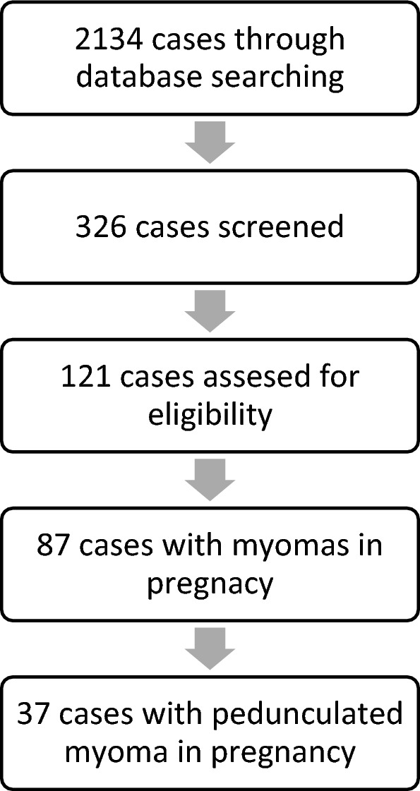
Methods: Systematic evaluation of 37 reports.
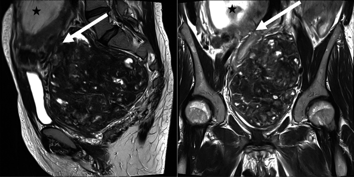
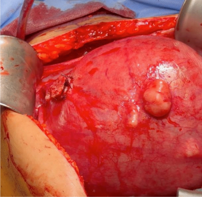
Example case: A 36-year-old Caucasian primigravida was referred symptomatic at 16 + 0 weeks due to a 13,5 cm myoma causing pain, constipation, urine retention and dysesthesias. Our patient underwent myomectomy at 17 + 0 weeks. One pedunculated leiomyoma was successfully removed.
Conclusion: Myomectomy can be performed and is safe for pedunculated fibroids in pregnancy. Depending on the clinical scenario, surgical removal may be indicated. Based on the size of the fibroids and expected adhesions, a laparotomy is a safe option and is not a contraindication for vaginal birth in the case of pedunculated fibroids. Myomas larger than 10 cm should be removed by laparotomy.
Keywords: Fibroids; Laparoscopy; Laparotomy; Myomectomy; Pedunculated myoma; Pregnancy.
© 2024. The Author(s).
Conflict of interest statement
Declarations. Conflict of interest: U.J. received travel money from pfm. C.D. is funded by Roche, AstraZeneca, TEVA, Mentor, and MCI Healthcare. All other authors declare no conflict of interest. Informed consent and Ethical approval: Informed consent for research and publication purposes was obtained from the patient mentioned in the study before collecting data.
Figures

References
-
- Schatteman Makar, Vergote (1989) Myomectomy during pregnancy: uncommon case report. Acta Chirurgica Belgica 89:212–214 - PubMed
Publication types
MeSH terms
LinkOut - more resources
Full Text Sources
Medical
