. 2024 Dec 3;121(49):e2408304121.
doi: 10.1073/pnas.2408304121. Epub 2024 Nov 27.

A widespread family of ribosomal peptide metallophores involved in bacterial adaptation to metal stress

Affiliations

A widespread family of ribosomal peptide metallophores involved in bacterial adaptation to metal stress

Laura Leprevost et al. Proc Natl Acad Sci U S A. .

Erratum in

Abstract

Ribosomally synthesized and posttranslationally modified peptides (RiPPs) are a structurally diverse group of natural products that bacteria employ in their survival strategies. Herein, we characterized the structure, the biosynthetic pathway, and the mode of action of a RiPP family called bufferins. With thousands of homologous biosynthetic gene clusters throughout the bacterial phylogenetic tree, bufferins form by far the largest family of RiPPs modified by multinuclear nonheme iron-dependent oxidases (MNIO, DUF692 family). Using Caulobacter vibrioides bufferins as a model, we showed that the conserved Cys residues of their precursors are transformed into 5-thiooxazoles, further expanding the reaction range of MNIO enzymes. This rare modification is installed in conjunction with a partner protein of the DUF2063 family. Bufferin precursors are rare examples of bacterial RiPPs found to feature an N-terminal Sec signal peptide allowing them to be exported by the ubiquitous Sec pathway. We reveal that bufferins are involved in copper homeostasis, and their metal-binding propensity requires the thiooxazole heterocycles. Bufferins enhance bacterial growth under copper stress by complexing excess metal ions. Our study thus describes a large family of RiPP metallophores and unveils a widespread but overlooked metal homeostasis mechanism in bacteria.

Keywords: metallophore; multinuclear non-heme iron-dependent oxidase (MNIO); ribosomally synthesized and post-translationally modified peptide (RiPP).


Conflict of interest statement

Competing interests statement:The authors declare no competing interest.

Figures
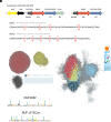
Fig. 1.
Fig. 1.
In silico analyses of the bufferin family. (A) Gene composition of the buf1 and buf2 BGCs and sequences of BufA1 and BufA2 with the Cys residues labeled (numbering according to the core peptides). The signal peptide and core peptide sequences are in pale and dark gray, respectively. In the vicinity of buf1ABCD are genes coding for SigF and the anti-sigma factor NrsF that regulate buf1 and buf2 expression. (B) SSN analysis of bufferin-like precursors genetically associated with MNIOs. DUF2282 and BUF_6/12Cys proteins, including BufA1 and BufA2 of C. vibrioides, belong to the largest two clusters (red and yellow dots), respectively. The oxazolins are shown as black dots. Small clusters in orange contain chryseobasins (7) that are predicted to harbor signal peptides. (C) Weblogos for the DUF2282 and BUF_6/12Cys bufferins. (D) SSN analysis of MNIOs (at 80% identity). MNIOs genetically associated with DUF2282 and BUF_6/12Cys proteins, including BufB1 and BufB2 of C. vibrioides, are in red and yellow, respectively. Those associated with oxazolins are colored black. MNIOs associated with chryseobasins, methanobactins, TglA-type pearlins, and aminopyruvatides are found in small clusters numbered 1 to 4.
Fig. 2.
Fig. 2.
Regulation and function of the bufferins. (A) Reporter assays with bufA1-lacZ and bufA2-lacZ transcriptional fusions. Bacteria were grown for 16 h with the indicated concentrations of CuSO4, FeSO4, ZnSO4, or MnCl2. Nonparametric, one-way ANOVA Kruskal–Wallis (two-sided) test followed by a Dunn’s multiple-comparison tests were used to analyze the differences between treated cultures and the control (n = 4; * in the Upper Left graph indicates P = 0.047). (B) Effect of copper on bacterial growth. The CvDKO, Cvbuf1ABCD (buf1), and Cvbuf2ABC (buf2) strains were grown without or with 225 μM CuSO4. In (C), expression of buf1 in E. coli BL21(pCA24-buf1AstrBCD) grown in minimal medium was induced by IPTG, and the bacteria were challenged with 0.3 mM CuSO4 for 3 h before serial dilutions. BL21(pCA24-psmCA) expressing the unrelated pseudomycoidin operon psmCA expressed from the same plasmid backbone was used as a control. In (D), expression of buf1 in E. coli BL21(pCA24-buf1AstrBCD) grown in rich medium was induced by IPTG, followed by 2 h in microaerobic conditions with CuSO4 and Cu2+-reducing ascorbate as indicated, before serial dilutions. The control was as in (C). (E) Assay of bacterial lysis by D. discoidum. Bacterial densities in the zones of contact were determined using ImageJ analyses. A nonparametric Mann–Whitney test (two-sided) was used to analyze the differences between the CvWT strain (n = 5) and the CvDKO strain (n = 4; * indicates P = 0.0159). The medians are shown.
Fig. 3.
Fig. 3.
Posttranslational modifications of bufferin 1. (A) Top–down LC-MS analyses of cell extracts of Cvbuf1(pSigF) compared with CvDKO(pSigF). Left panel: total ion chromatograms, Right panels: deconvoluted mass spectra of the compounds detected for Cvbuf1(pSigF). Their Mw corresponds to the core peptide (i.e., without signal peptide; calculated monoisotopic Mw = 6,978.35 Da), with mass shifts of −10 Da ([M+H]+ at m/z 6,969.32) and −12 Da ([M+H]+ at m/z 6,967.30). (B) Bottom–up analysis of Buf1. MS/MS spectrum of the central tryptic peptide ([M+2H]2+ at m/z 917.36). The two Cys residues carry carbamidomethyl groups resulting from alkylation with iodoacetamide (+57.02 Da), together with −4.03 Da mass shifts. (C) UV/vis spectra of affinity-purified Buf1 and the CysIISer+CysIIISer mutant. (D) NMR ROESY/TOCSY experiments to assign the resonances of the 19-mer peptide. (E) HNCACB experiment on the uniformly 13C, 15N-labeled peptide: sequential walk for the sequence following CysII. (F) The HNCO planes at the 15N frequency of the indicated residues show classical ~175 ppm values for all carbonyl resonances, except for that downstream of a modified Cys, whose carbonyl carbon resonates at 184 ppm. (G) HNCO planes through the resonance of Lys24 that follows CysII while modifying the offset of the 13C decoupling pulse during the C=O evolution period. Only when centered at 120 ppm is the 90-Hz carbon–carbon coupling refocused. (H) Measured NMR parameters and proposed structure for the Buf1 posttranslational modifications (shown for CysII).
Fig. 4.
Fig. 4.
Analyses of the bufferin 1-copper complex formed in vivo. (A) Native MS analysis of the purified complex: isotopic patterns of the major charge state (6+) species. The spectra in black and red show Buf1 from a nonsupplemented culture and from cultures supplemented with CuSO4, respectively. The calculated isotopic patterns for [M+6H]6+ and [M+4H+CuII]6+ species are shown underneath. (B) Evidence for Cu2+ binding to Buf1 by EPR spectroscopy. Spectra of Echo-Detected Field-Swept of CuSO4 (red) and of the Buf1–Cu2+ complex (black) are shown with the pseudo-modulation of the spectra underneath. The Right panels show the Easyspin fits (red) and the experimental spectra (black) for the Buf1–Cu2+ complex (Top) and for CuSO4 (Bottom). (C) Experimental and fitted EXAFS spectra of the Buf1–Cu2+ complex (Upper) and corresponding Fourier transforms (Lower). (D) Superposition of the 1H, 15N HSQC spectra of apo Buf1 (black) and the Buf1–Cu2+ complex (red). sc = side chain.
Fig. 5.
Fig. 5.
Biosynthesis of bufferin 1. (A) Effect of Cu on the growth of C. vibrioides strains expressing the indicated genes. (B) In vitro reconstitution of Buf1 biosynthesis. The deconvoluted mass spectra for the products of the in vitro reactions are shown, with the added proteins indicated in the corresponding panels. Heat-denatured BufB1 and BufC1 were used for the control reaction. (C) LC-MS analysis of the 19-residue peptide of in vitro modified SUMO-Buf1. The extracted ion chromatogram (EIC) of [M+3H]3+ (at m/z 573.8945) and the deconvoluted mass are shown. (D) Role of the signal peptide for the PTMs. A schematic representation of BufA1 variants with signal peptide truncations is shown on Top. LC-MS analyses of modified BufA1(-8 aa) (Left) and modified BufA1(CP) (Right): the MS spectra of the most abundant charge states [M+9H]9+ and [M+8H]8+, corresponding to deconvoluted monoisotopic masses [M+H]+ of 8391.1281 and 8277.9398 (blue), respectively, are compared to the theoretical spectra of the unmodified peptides (black).

References

    1. Arnison P. G., et al. , Ribosomally synthesized and post-translationally modified peptide natural products: Overview and recommendations for a universal nomenclature. Nat. Prod. Rep. 30, 108–160 (2013). - PMC - PubMed
    1. Li Y., Rebuffat S., The manifold roles of microbial ribosomal peptide-based natural products in physiology and ecology. J. Biol. Chem. 295, 34–54 (2020). - PMC - PubMed
    1. Montalban-Lopez M., et al. , New developments in RiPP discovery, enzymology and engineering. Nat. Prod. Rep. 38, 130–239 (2021). - PMC - PubMed
    1. Burkhart B. J., Hudson G. A., Dunbar K. L., Mitchell D. A., A prevalent peptide-binding domain guides ribosomal natural product biosynthesis. Nat. Chem. Biol. 11, 564–570 (2015). - PMC - PubMed
    1. Chekan J. R., Ongpipattanakul C., Nair S. K., Steric complementarity directs sequence promiscuous leader binding in RiPP biosynthesis. Proc. Natl. Acad. Sci. U.S.A. 116, 24049–24055 (2019). - PMC - PubMed

LinkOut - more resources