. 2024 Nov 27;16(775):eadp5730.
doi: 10.1126/scitranslmed.adp5730. Epub 2024 Nov 27.

Seeding-competent TDP-43 persists in human patient and mouse muscle

Affiliations

Seeding-competent TDP-43 persists in human patient and mouse muscle

Eileen M Lynch et al. Sci Transl Med. .

Abstract

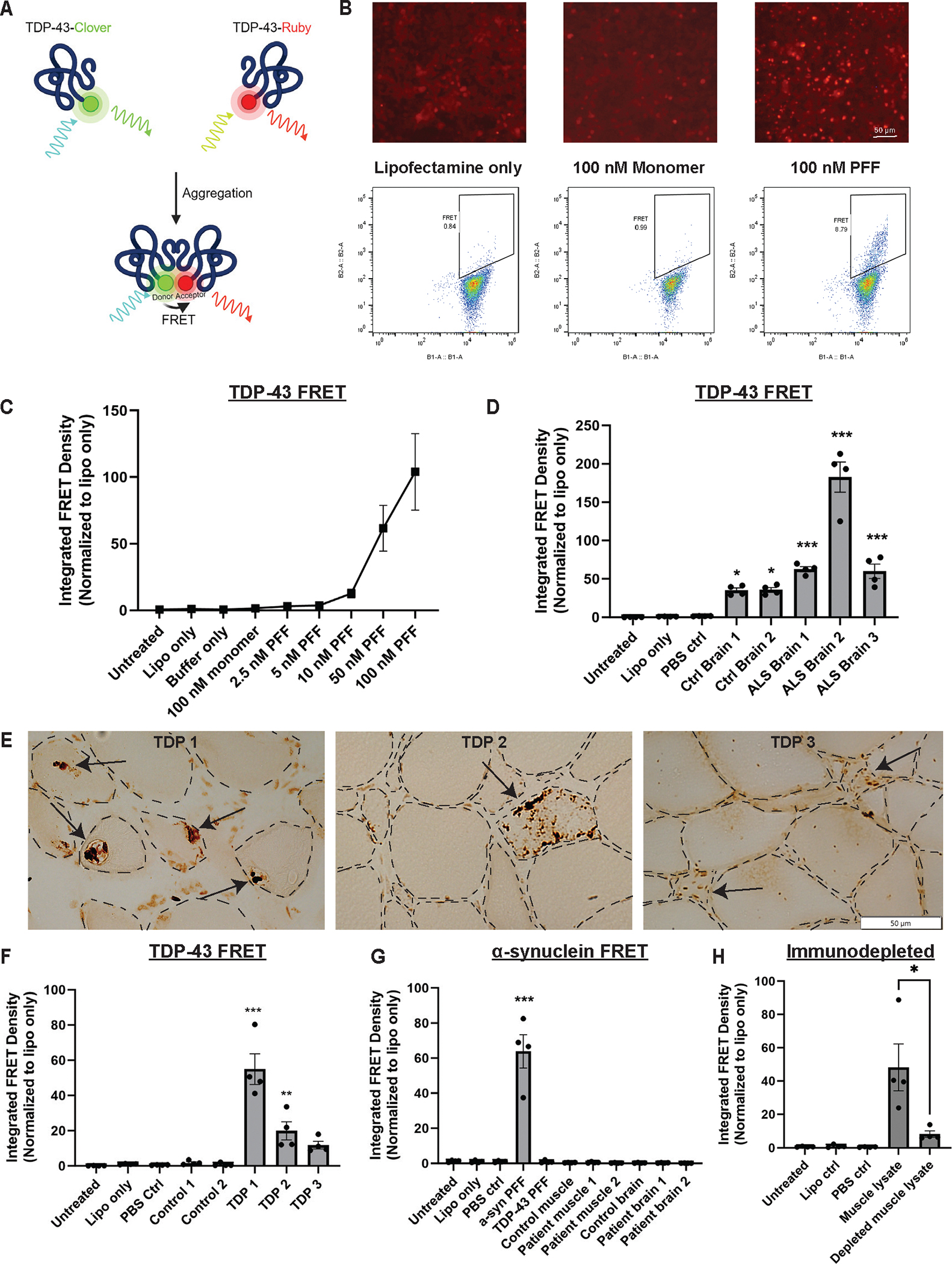
TAR DNA binding protein 43 (TDP-43) is an RNA binding protein that accumulates as aggregates in the central nervous systems of some patients with neurodegenerative diseases. However, TDP-43 aggregation is also a sensitive and specific pathologic feature found in a family of degenerative muscle diseases termed inclusion body myopathy. TDP-43 aggregates from amyotrophic lateral sclerosis (ALS) and frontotemporal dementia brain lysates may serve as self-templating aggregate seeds in vitro and in vivo, supporting a prion-like spread from cell to cell. Whether a similar process occurs in patient muscle is not clear. We developed a mouse model of inducible, muscle-specific cytoplasmic localized TDP-43. These mice develop muscle weakness with robust accumulation of insoluble and phosphorylated sarcoplasmic TDP-43, leading to eosinophilic inclusions, altered proteostasis, and changes in TDP-43-related RNA processing that resolve with the removal of doxycycline. Skeletal muscle lysates from these mice also have seeding-competent TDP-43, as determined by a FRET-based biosensor, that persists for weeks upon resolution of TDP-43 aggregate pathology. Human muscle biopsies with TDP-43 pathology also contain TDP-43 aggregate seeds. Using lysates from muscle biopsies of patients with sporadic inclusion body myositis (IBM), immune-mediated necrotizing myopathy (IMNM), and ALS, we found that TDP-43 seeding capacity was specific to IBM. TDP-43 seeding capacity anticorrelated with TDP-43 aggregate and vacuole abundance. These data support that TDP-43 aggregate seeds are present in IBM skeletal muscle and represent a unique TDP-43 pathogenic species not previously appreciated in human muscle disease.


Conflict of interest statement

Competing interests:

None to disclose.

Figures

Fig. 1.
Fig. 1.. Confirmation of TDP-43 seeding in patient tissue using a FRET sensor line.
(A) TDP-43 FRET biosensors express two independently tagged c-terminal fragments (aa262–414) of human TDP-43 fused to mruby and mclover. Upon aggregation, an energy transfer occurs between the two fluorophores. (B) Representative images of the FRET biosensor and flow cytometry gating strategy used. (C) A dose response curve of the TDP-43 biosensors treated with increasing concentrations of recombinant TDP-43 PFF. (D) The RIPA-insoluble fraction of human ALS patient autopsy brain tissue induces TDP-43 aggregation in the FRET biosensors. (E) Representative images of TDP-43 staining of human muscle biopsies with TDP-43 inclusions (arrows). (F) The RIPA-insoluble fraction from control muscle or muscle biopsies confirmed to have positive TDP-43 aggregate staining were added to the FRET biosensors to confirm seeding. (G) Samples from muscle and brain lysates with established TDP-43 seeding ability were added to an α-synuclein FRET biosensors to test for TDP-43 seeding specificity. (H) A human muscle sample shows decreased FRET activity following immunodepletion of TDP-43. *P<0.05, **P<0.01, ***P<0.001 by one-way ANOVA followed by Dunnett’s multiple comparisons test to lipofectamine control. In (H) an unpaired two-tailed Student’s t-test was used to compare immunodepleted to control lysate.
Fig. 2.
Fig. 2.. Mice expressing cytoplasmic hTDP-43 develop a myopathy with premature death.
(A) HSA-rtTA mice were crossed with tetO-hTDP-43ΔNLS mice to create doxycycline-inducible muscle-specific expression of hTDP-43ΔNLS. Created with BioRender.com. (B) A cohort of mice were studied over the course of a 4 week dox treatment (shaded in green on the survival curve), tracking total body weight, strength, and survival time. N = 13 for survival curve, N = 11 for weight and grip strength, N = 7 for hanging wire as 4 mice were too frail to perform the test. (C) After 4 weeks of dox treatment, H&E staining of tibialis anterior muscles showed mild myopathic changes with varied fiber sizes. Non-nuclear eosinophilic staining in H&E and pale regions on NADH staining (arrows) highlight sarcoplasmic inclusion. (D) Mice treated with dox chow for 4 weeks compared to age-matched no dox controls showed decreased hind limb muscle weights as well as decreased total body weight compared to untreated controls. (E) Example x-ray images of spinal kyphosis in HSA-hTDP-43ΔNLS mice after 3 weeks on dox chow and the calculated kyphosis index. *P<0.05, **P<0.01, ***P<0.001; unpaired two-tailed Student’s t-test.
Fig. 3.
Fig. 3.. HSA-hTDP-43ΔNLS mice develop abundant sarcoplasmic insoluble phosphorylated TDP-43 aggregates which disrupt proteostasis and splicing.
(A) Representative immunofluorescent images of hindlimb cross sections of HSA-hTDP-43ΔNLS mice after 4 weeks of transgene activation, showing abundant TDP-43 and pTDP-43 aggregates. (B) Representative western blots of gastrocnemius muscle lysates from HSA-hTDP-43ΔNLS mice show increased amounts of total, soluble, and insoluble TDP-43 and pTDP-43 over time. The total GAPDH can be found in part D. Similar blots were repeated in 4 independent experiments. (C) Mice treated with dox chow for 4 weeks were evaluated for TDP-43 transgene expression in non-skeletal muscle tissue. Blots were repeated twice. (D) Representative blots of gastrocnemius muscle lysates from HSA-hTDP-43ΔNLS mice processed at the indicated time points and evaluated for markers of disrupted proteostasis. Similar blots were repeated in 3 independent experiments. (E) Representative dual fluorescent imaging of pTDP-43 (red) and p62 (green upper panel) or ubiquitin (green lower panel) at 2 weeks of dox treatment. (F) Agarose gel electrophoresis of products from the RT-PCR amplification of TDP-43 splicing targets Sh3bgr and Tns1 from TA muscles of HSA-hTDP-43ΔNLS mice with and without dox treatment. A more slowly migrating band is consistent with exon inclusion. Schematic diagram created with BioRender.com.
Fig. 4.
Fig. 4.. TDP-43 aggregates are cleared and proteostasis resolved following removal of doxycycline.
(A) Representative immunofluorescent images of quadriceps femoris muscles stained for pTDP-43 in HSA-hTDP43ΔNLS mice at the indicated timepoints when treated with dox chow for 2 weeks followed by a return to a regular chow diet. (B) A representative western blot of HSA-hTDP43ΔNLS mouse gastrocnemius muscles shows accumulation and then resolution of insoluble TDP-43 and pTDP-43 over the time course of two weeks doxycycline followed by 3 weeks of recovery. Similar blots were repeated in 5 independent experiments, as quantified at the 2 week on dox and 2 week recovery timepoints in (C). ***P<0.001; unpaired two-tailed Student’s t-test. (D) SDD-AGE was performed on recombinant monomeric TDP-43 or TDP-43 PFF as well as the insoluble fractions of HSA-hTDP43ΔNLS mouse muscle at the same timepoints used in A and B. During recovery there is a shift towards higher molecular weight (arrow head) and an initial increase in low molecular weight species (arrow) during the recovery phase. Only higher molecular weight species are positive for pTDP-43. (E) Representative blots of gastrocnemius muscle lysates from HSA-hTDP43ΔNLS mice processed at the indicated time points and evaluated for changes in markers of proteostasis. Similar blots were repeated in 3 independent experiments. (F) Representative H&E and NADH images of quadriceps femoris muscle after 3 weeks of recovery as compared to no dox controls. Note the presence of smaller fibers with centralized nuclei (arrows).
Fig. 5.
Fig. 5.. Ultrastructural analysis of HSA-hTDP-43ΔNLS mouse muscle.
(A) Control TA muscle fiber with myonuclei. (B-D) HSA-hTDP-43ΔNLS mice treated for two weeks with doxycycline. Note large granular amorphous inclusions that are subsarcolemmal and myonuclei-adjacent (arrows). (D) Higher magnification of the granular structure of an aggregate from a mouse on dox treatment for 2 weeks. (E-F) HSA-hTDP-43ΔNLS mice treated for two weeks with doxycycline and then changed to normal chow for three weeks. Note autophagic debris and vacuolation in and around the inclusion (arrowheads).
Fig. 6.
Fig. 6.. TDP-43 seeding in HSA-hTDP-43ΔNLS mouse muscle is detected by FRET assay.
(A) The insoluble fractions from muscle of HSA-hTDP43ΔNLS mice either treated with no dox or 3 weeks of dox were applied to the TDP-43 biosensor and α-synuclein biosensor to confirm seeding specificity. (B) Immunodepletion of TDP-43 from the insoluble fraction of 4 week on dox HSA-hTDP43ΔNLS mouse muscle significantly reduced FRET signal, further indicating TDP-43 specificity (P = 0.001). (C) HEK293 cells expressing a tetracycline-inducible mcherry-tagged TDP-43ΔNLS were activated for 16 hours then treated with the insoluble fraction from 4 week on dox HSA-hTDP43ΔNLS mouse muscle for 72 hours. Insoluble fractionation western blot of the cell lysates shows a smear of insoluble protein in the lanes treated with dox-treated muscle lysates, indicating successful aggregate seeding. (D) HSA-hTDP43ΔNLS mice were treated with dox for two weeks and allowed to recover for three weeks, as in Figure 4. Seeding peaked at 1 week of recovery and persisted through three weeks. (E) A follow-up study demonstrated seeding activity out to 8 weeks of recovery. *P>0.05, **P<0.01, ***P<0.001; One-way ANOVA with Dunnett’s multiple comparison tests to lipofectamine only control. In (B) an unpaired two-tailed Student’s t-test was used to compare immunodepleted to untreated lysate.
Fig. 7.
Fig. 7.. TDP-43 seeding is present in IBM patient muscle but does not correlate with histological pathology.
(A) The insoluble fraction from muscle biopsies of 24 IBM patients, along with 5 healthy controls, 10 with IMNM, and 10 with ALS were added to the TDP-43 FRET sensor line. Of the IBM samples, 14 were statistically significant (P<0.05). *P<0.05, **P<0.01, ***P<0.001 One-way ANOVA with Dunnett’s multiple comparisons test to lipofectamine only control. Dots on each bar represent technical replicates. (B) Representation of each patient’s average integrated FRET density (IFD). P*<0.05 with one-way ANOVA and Dunnett’s multiple comparisons test to healthy controls. The amount of seeding activity had a negative correlation with histological hallmarks of IBM such as TDP-43 aggregates (C) and rimmed vacuoles (D). (E) Representative images of a disease control, low seeding sample, and high seeding sample with their corresponding FRET values (samples indicated by arrows in (A). Scalebars represent 50 μm.
Fig. 8.
Fig. 8.. TDP-43 seed uptake and conversion occurs in skeletal muscle in vitro and in vivo.
(A) Immunostaining for TDP-43 in mouse myoblasts treated with buffer or 50 nM of recombinant TDP-43 PFF for five days at time of differentiation. The bottom row shows the same staining without the DAPI channel to better visualize the loss of endogenous nuclear TDP-43 in the seeded cells compared to untreated. (B) Soluble and insoluble lysates from TDP-43 PFF treated and untreated mouse myoblasts were subjected to immunoblot using anti-TDP-43 and pTDP-43 antibodies. Note the presence of insoluble TDP-43 species in PFF treated cells. Similar blots were repeated in 2 independent experiments. (C) The tibialis anterior muscles of C57 wild type mice were injected with 10 μg of recombinant TDP-43 PFF or a buffer control in the contralateral leg. Individual muscle fibers are outlined with dotted white lines for better visualization. Immunofluorescence for pTDP-43 on sections obtained one month post injection demonstrates positive inclusions in fibers from PFF treated muscle (enlarged yellow box). (D) Representative western blots of soluble and insoluble lysates from TDP-43 PFF treated and untreated TAsshowing an increase in insoluble TDP-43 fragments in the injected TA muscle as compared to the buffer injected contralateral muscle. Similar blots were repeated in 3 independent experiments.

Update of

References

    1. Jo M et al. , The role of TDP-43 propagation in neurodegenerative diseases: integrating insights from clinical and experimental studies. Exp Mol Med 52, 1652–1662 (2020). - PMC - PubMed
    1. Gao J, Wang L, Huntley ML, Perry G, Wang X, Pathomechanisms of TDP-43 in neurodegeneration. J Neurochem, (2018). - PMC - PubMed
    1. de Boer EMJ et al. , TDP-43 proteinopathies: a new wave of neurodegenerative diseases. J Neurol Neurosurg Psychiatry 92, 86–95 (2020). - PMC - PubMed
    1. Soto C, Pritzkow S, Protein misfolding, aggregation, and conformational strains in neurodegenerative diseases. Nat Neurosci 21, 1332–1340 (2018). - PMC - PubMed
    1. Porta S et al. , Patient-derived frontotemporal lobar degeneration brain extracts induce formation and spreading of TDP-43 pathology in vivo. Nat Commun 9, 4220 (2018). - PMC - PubMed

Publication types

MeSH terms

LinkOut - more resources