. 2024 Nov 29;10(48):eadr2261.
doi: 10.1126/sciadv.adr2261. Epub 2024 Nov 27.

The Hox protein Antennapedia orchestrates Drosophila adult flight muscle development

Affiliations

The Hox protein Antennapedia orchestrates Drosophila adult flight muscle development

Gabriela Poliacikova et al. Sci Adv. .

Abstract

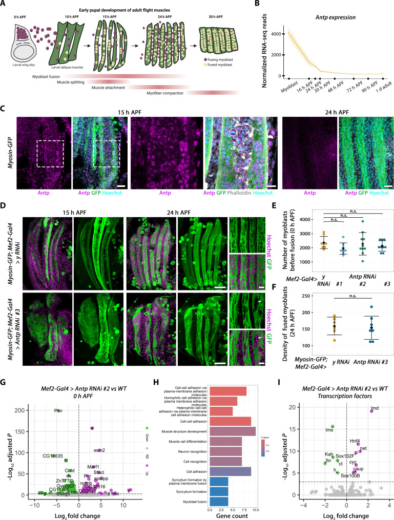
Muscle development and diversity require a large number of spatially and temporally regulated events controlled by transcription factors (TFs). Drosophila has long stood as a model to study myogenesis due to the highly conserved key TFs involved at all stages of muscle development. While many studies focused on the diversification of Drosophila larval musculature, how distinct adult muscle types are generated is much less characterized. Here, we identify an essential regulator of Drosophila thoracic flight muscle development, the Hox TF Antennapedia (Antp). Correcting a long-standing belief that flight muscle development occurs without the input of Hox TFs, we show that Antp intervenes at several stages of flight muscle development, from the establishment of the progenitor pool in the embryo to myoblast differentiation in the early pupa. Furthermore, the precisely regulated clearance of Hox in the developing flight muscle fibers is required to allow for fibrillar muscle fate diversification, setting these muscles apart from all other adult tubular muscle types.


Figures

Fig. 1.
Fig. 1.. Antp is expressed in flight muscle myoblasts from the P2.
(A) Confocal sections of 0-hour APF wing discs expressing UAS-y RNAi (left) or UAS-Antp RNAi #1 (right) driven by Mef2-Gal4, stained with anti-Antp (8c11) and anti-Twi. (B) Ratio of Antp intensity in the myoblast area versus anterior epithelium. The mean and SD are shown. Each dot represents a value from one disc (n = 14 discs from at least seven animals). (C) Normalized Antp intensity under Antp RNAi conditions (each RNAi line is depicted by with a “#”). Each dot represents a value from one disc (nWT = 14, n#1 = 10, n#2 = 12, and n#3 = 11 wing discs from at least five animals). (D) Antp gene and transcripts (coding exons are dark yellow, and noncoding exons are light yellow smaller boxes). P1 and P2 represent two distinct promoters, and A1 and A2 represent two alternative polyadenylation sites. (E and F) Confocal sections of 0-hour APF wing discs expressing UAS-GFP nls driven by Antp P1-Gal4 (E) or Antp P2-Gal4 (F), stained with anti-GFP and anti-Twi. Scale bars, 50 μm (top view) and 20 μm (bottom view).
Fig. 2.
Fig. 2.. Antp mutation leads to the absence of adult flight muscles.
(A) Schematic representation of adult flight myogenesis. (B and C) Confocal sections of stage 15 embryos labeled with antibodies against Antp and Twi. Genotypes are (B) WT and (C) AntpP2-null mutant: Antpnsrv-c3 (null mutation) over Antps1 (P2 mutation). T1, T2, and T3 depict the three thoracic segment, and A1 depicts the first abdominal segment. Scale bars, 20 μm (large views) and 5 μm (zoomed views on T2 AMPs). (D and E) Confocal sections of wing discs at the L3 feeding stage labeled with antibodies against Antp and Twi. Genotypes are identical to (B) and (C). Scale bars, 50 μm (large views) and 25 μm (zoomed views). (F) Confocal projections of 24-hour APF pupal IFMs labeled with GFP to visualize myosin. Genotypes are identical to (B) and (C) with weeP26 transgene (Myosin-GFP) added. Scale bars, 50 μm. (G) Confocal sections of pupal IFM at the pharate stage, labeled with phalloidin. Genotypes are identical to (B) and (C). Scale bars, 100 μm. DLM, dorsal longitudinal muscles; DVM, dorsal-ventral muscles; h, hours.
Fig. 3.
Fig. 3.. Antp regulates flight muscle differentiation.
(A) Schematic representation of pupal flight myogenesis. d, day. (B) Antp mRNA normalized expression, analyzed from (29). (C) Confocal sections of 15- and 24-hour APF flight muscles expressing Myosin-GFP, stained with anti-Antp, anti-GFP (to visualize myosin), phalloidin, and Hoechst. Scale bars, 20 μm [large views (left)] and 10 μm [zoomed views (right)], depicted by the white dashed box. (D) Confocal sections (left) and projections (right) of 15- and 24-hour APF flight muscles expressing Myosin-GFP; Mef2-Gal4 driving UAS-y RNAi as control (top) or UAS-Antp RNAi #3 (bottom), stained with anti-GFP and Hoechst. Scale bars, 50 μm (large sections) and 15 μm (zoomed views). Arrow depicts zones of high nuclei density. (E) Quantification of the number of myoblasts in 0-hour APF wing disc upon Antp KD. Mean and SD are shown; each dot represents a value from one disc (nWT = 8, n#1 = 8, n#2 = 10, and n#3 = 10 wing discs from at least five animals). n.s., not significant. (F) Same as in (E) at 24 hours APF (nWT = 6 and n#3 = 8 animals). (G) Volcano plot of DEGs between WT and UAS-Antp RNAi #2 driven by the Mef2-Gal4 driver. n.s., not significant. (H) GO analysis using significantly up-regulated genes. (I) Volcano plot of TFs, the same conditions as (G).
Fig. 4.
Fig. 4.. Antp overexpression suppresses fibrillary muscle fate.
(A and B) Confocal sections of adult female DLMs stained with phalloidin and Hoechst (top), with zoomed views on sarcomeres (bottom). Scale bars, 100 μm (large views) and 10 μm (zoomed views). Genotypes are UAS-mCherry-nls (A) and UAS-Antp (B) driven by the Act88F-Gal4 driver. (C) Transmission electron microscopy (TEM) micrographs of adult DLMs, cut in longitudinal sections. Scale bars, 2 μm (large views) and 500 nm (zoomed views). (D) Same as (C), cut in cross sections. Scale bars, 1 μm (large views) and 500 nm (zoomed views). (E) TEM micrographs of leg muscles. Scale bars, 2 μm. For (C) to (E), genotypes are WT (top) and UAS-Antp (bottom) driven by the Act88F-Gal4 driver. (F) Volcano plot of sarcomeric DEGs between UAS-GFP and UAS-Antp driven by the Act88F-Gal4 driver. (G) Confocal sections of adult female DLMs stained with anti-GFP (to visualize Salm), phalloidin, and Hoechst. Scale bars, 5 μm. (H) Quantification of normalized Salm intensity; represented is the mean and SD where each point represents a value of a single nucleus (nmcherry-nls = 60 nuclei and nAntp = 48 nuclei from at least three animals). Genotypes in (G) and (H) are identical to (A) and (B), with the spalt::GFP transgene added.

References

    1. Poovathumkadavil P., Jagla K., Genetic control of muscle diversification and homeostasis: Insights from Drosophila. Cells 9, 1543 (2020). - PMC - PubMed
    1. Michelson A. M., Muscle pattern diversification in Drosophila is determined by the autonomous function of homeotic genes in the embryonic mesoderm. Development 120, 755–768 (1994). - PubMed
    1. Lawrence P. A., Johnston P., The genetic specification of pattern in a drosophila muscle. Cell 36, 775–782 (1984). - PubMed
    1. Hooper J. E., Homeotic gene function in the muscles of Drosophila larvae. EMBO J. 5, 2321–2329 (1986). - PMC - PubMed
    1. Capovilla M., Kambris Z., Botas J., Direct regulation of the muscle-identity gene apterous by a Hox protein in the somatic mesoderm. Development 128, 1221–1230 (2001). - PubMed

MeSH terms