. 2024 Nov;63(11):e70013.
doi: 10.1002/gcc.70013.

Epigenetic Modeling of Jumping Translocations of 1q Heterochromatin in Acute Myeloid Leukemia After 5'-Azacytidine Treatment

Affiliations

Epigenetic Modeling of Jumping Translocations of 1q Heterochromatin in Acute Myeloid Leukemia After 5'-Azacytidine Treatment

Anair Graciela Lema Fernandez et al. Genes Chromosomes Cancer. 2024 Nov.

Abstract

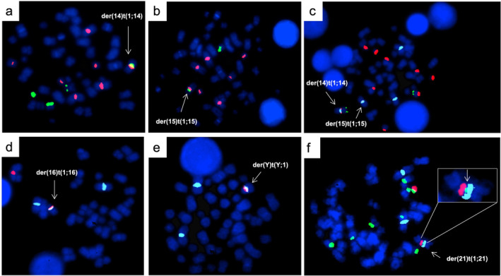
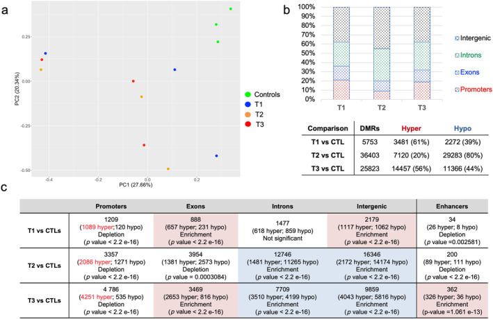
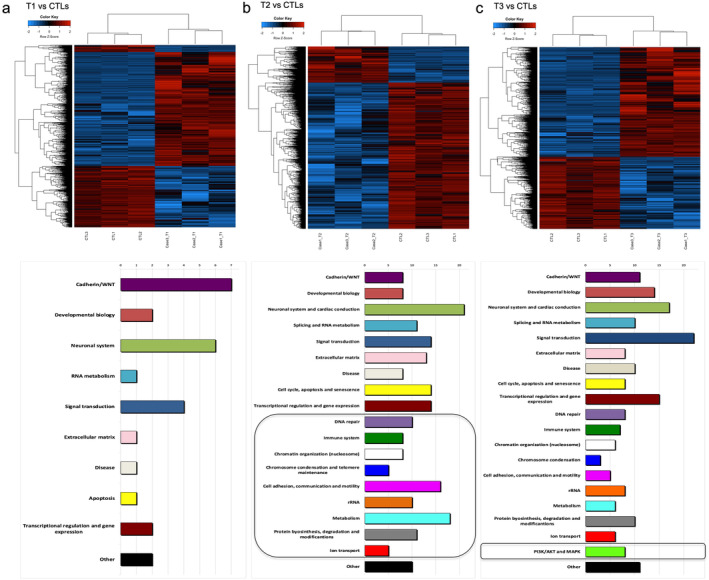
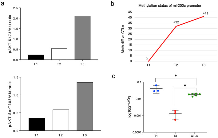
Jumping translocations (JT) are rare cytogenetic abnormalities associated with progression in myelodysplastic syndromes (MDS) and acute myeloid leukemia (AML). Typically, a tri-tetra-somic 1q chromosome is translocated to two or more recipient chromosomes. In multiple myeloma JT were shown to originate after DNA demethylation and decondensation. Using epigenomics, we investigated sequential samples in an SRSF2-mutated MDS and AML cohort with normal karyotype at diagnosis and 1qJT at disease evolution after 5'-azacytidine (AZA). 1qJT breakpoints fell within repetitive DNA at both 1q12 and the translocation partners, namely acrocentrics n. 14, 15, 21, and 22, chromosome 16, and chromosome Y. The global methylome at diagnosis showed hypermethylation at 61% of the differentially methylated regions (DMRs), followed by hypomethylation at 80% of DMRs under AZA, mostly affecting pathways related to immune system, chromatin organization, chromosome condensation, telomere maintenance, rRNA, and DNA repair. At disease evolution, a shift toward hypermethylation, intronic enhancers enrichment and epigenetic involvement of the PI3K/AKT and MAPK signaling emerged. In particular, AKT1 phosphorylation behaved as a hallmark of the progression. Overall, we provided new insights on the characterization of 1qJT in SRSF2-mutated myeloid neoplasms and first showed that epigenetics is a powerful tool to investigate the molecular landscape of repetitive DNA rearrangements.

Keywords: 5‐azacytidine; DNA methylation; MDS/AML; jumping 1q.


Conflict of interest statement

The authors declare no conflicts of interest.

Figures

FIGURE 1
FIGURE 1
FISH characterization of the three patients. Case 1 (a–c). Hybridization of the Sat.2 oligo probe for 1q12 sat‐II region (orange) and D14Z1 centromeric probe for chromosome 14 (green) (panel a); and D15Z3 centromeric probe for chromosome 15 (green) (panel b); tricolor FISH showing der(14) and der(15) in the same metaphase with pUC1.77 for 1q12 heterochromatin (aqua), RP5‐1174A5 probe for NORs (orange) in acrocentrics and RP11‐431B1 probe for 14q32 (green), used as a reference for chromosome 14 (panel c). Case 2 (d and e). Hybridization of Sat.2 oligo probe for 1q12 sat‐II region (aqua) and D16Z3 probe for pericentromeric heterochromatin of chromosome 16 (orange) (panel d); and DYZ1 probe for sat‐III of chromosome Y (orange) (panel e). Case 3 (f). Hybridization of pUC1.77 for 1q12 heterochromatin (aqua), RP5‐1174A5 probe for NORs (green) in acrocentrics and whole chromosome paint probe for chromosome 21 (orange). On the top right the insert showing a reduction of the signal for RP5‐1174A5 probe for NORs (green) in the der(21).
FIGURE 2
FIGURE 2
Summary of DMRs annotations. (a) Unsupervised principal component analysis (PCA) from mERRBS data separating leukemic samples from controls. (b) Upper, histograms showing DMRs distribution at each timepoint when compared to controls (FDR < 0.1; differential methylation > 25). Bottom, table reporting the total number DMRs with hyper‐ and hypo‐methylated DMRs at each timepoint. (c) Genomic localization of the identified DMRs. Statistical results for Binomial enrichment/depletion test were conduct on R, using 95% confidence interval and percentage of background annotation for each category (promoters, exons, introns, intergenic, and active enhancer) as true probability of success. As background, we used total identified DMRs without filters.
FIGURE 3
FIGURE 3
Summary of DNA methylation and pathways analysis. Supervised heatmaps (top) and enriched pathways (bottom) identified from DMRs annotation in the 1qJT cases analyzed versus controls at T1 (panel a), at T2 (panel b), and at T3 (panel c). Patients in the heatmaps are represented in the columns and CpGs in the rows. Blue and red heatmap colors refers to hypo‐ and hyper‐methylation, respectively as indicated in the top left color key legend. x‐axis on pathway histograms represented the number of pathways in each category, as reported by Arabic numbers on the top; black squares underline categories that emerged as new at each timepoint.
FIGURE 4
FIGURE 4
Results from AKT1 activation. (a) pAkt S473/Akt (upper) and pAkt T308/Akt (bottom) ratios of scanning densitometry from immunoblot analysis from case 1 at each timepoint (T1, T2, T3). (b) Methylation status of the mir200c promoter region at the three timepoints from mERRBS data. y‐axis indicates the methylation difference identified from mERRBS. (c) Significance for mir200c expression by qRT‐PCR (Mann–Whitney test, *p < 0.05); values are expressed as means ± SD.

References

    1. Allshire R. C. and Madhani H. D., “Ten Principles of Heterochromatin Formation and Function,” Nature Reviews Molecular Cell Biology 19, no. 4 (2018): 229–244. - PMC - PubMed
    1. Janssen A., Colmenares S. U., and Karpen G. H., “Heterochromatin: Guardian of the Genome,” Annual Review of Cell and Developmental Biology 34 (2018): 265–288. - PubMed
    1. Couture T., Amato K., DiAdamo A., and Li P., “Jumping Translocations of 1q in Myelodysplastic Syndrome and Acute Myeloid Leukemia: Report of Three Cases and Review of Literature,” Case Reports in Genetics (2018): 1–5. - PMC - PubMed
    1. Borri D., Ommati L. V. M., and Velloso E. D. R., “Jumping Translocation: An Unusual Cytogenetic Finding in Myeloid Neoplasm,” Hematology, transfusion and Cell Therapy 44, no. 4 (2022): 612–613. - PMC - PubMed
    1. Sawyer J. R., Tian E., J. D. Shaughnessy, Jr. , et al., “Hyperhaploidy Is a Novel High‐Risk Cytogenetic Subgroup in Multiple Myeloma,” Leukemia 31, no. 3 (2017): 637–644. - PMC - PubMed