. 2025 Jan;55(1):e202451142.
doi: 10.1002/eji.202451142. Epub 2024 Nov 27.

Distinct Requirements for CD4+ T Cell Help for Immune Responses Induced by mRNA and Adenovirus-Vector SARS-CoV-2 Vaccines

Affiliations

Distinct Requirements for CD4+ T Cell Help for Immune Responses Induced by mRNA and Adenovirus-Vector SARS-CoV-2 Vaccines

Lyn Yong et al. Eur J Immunol. 2025 Jan.

Abstract

CD4+ T cells have been established as central orchestrators of cellular and humoral immune responses to infection or vaccination. However, the need for CD4+ T cell help to generate primary CD8+ T cell responses is variable depending on the infectious agent or vaccine and yet consistently required for the recall of CD8+ T cell memory responses or antibody responses. Given the deployment of new vaccine platforms such as nucleoside-modified mRNA vaccines, we sought to elucidate the requirement for CD4+ T cell help in the induction of cellular and antibody responses to mRNA and adenovirus (Ad)-vectored vaccines against SARS-CoV-2. Using antibody-mediated depletion of CD4+ T cells in a mouse immunization model, we observed that CD4+ T cell help was dispensable for both primary and secondary CD8+ T cell responses to the BNT162b2 and mRNA-1273 mRNA vaccines but required for the AZD1222 Ad-vectored vaccine. Nonetheless, CD4+ T cell help was needed by both mRNA and Ad-vectored vaccine platforms for the generation of antibodies, demonstrating the centrality of CD4+ T cells in vaccine-induced protective immunity against SARS-CoV-2. Ultimately, this highlights the shared and distinct regulation of humoral and cellular responses induced by these vaccine platforms.

Keywords: CD4 T cell help; CD8 T cells; T cell memory; T cell priming; adenovirus vaccines; antibodies; mRNA vaccines; vaccination.


Conflict of interest statement

The authors note the following conflicts of interest: N.M.P. has received consulting fees from Infinitopes. P.K. has received consulting fees from UCB, Biomunex, AstraZeneca, and Infinitopes. E.B. consults for AstaZeneca, Roche, and Vaccitech and has patents in ChAdOx1 HBV and HCV vaccines.

Figures

FIGURE 1
FIGURE 1
CD4+ T cell help is dispensable for generation of antigen‐specific CD8+ T cell responses to mRNA‐1273. (A) A schematic diagram illustrating the experimental setup: Anti‐CD4 antibody was administered intraperitoneally 1 day prior to each dose of mRNA‐1273. Cells from blood and spleen were isolated for analysis of the cellular responses at 20 days post‐prime (d20), and 7‐ or 21 days post‐boost (d21 + 7 and d21 + 21, respectively). (B) Antigen‐specific CD8+ T cells were stained with a Kb/S539–546 tetramer, and the frequencies of Kb/S539–546 + CD8+ T cells were expressed as a percentage of CD8+ T cells. (C) The number of Kb/S539–546 + CD8+ T cells in the spleen. (D–F) Expression of KLRG1+CD127 effector phenotype (D), CD62LCD127+ effector memory phenotype (E), and CD62L+CD127+ central memory phenotype (F) were quantified as a percentage of Kb/S539–546 + CD8+ T cells (Tet+ CD8+ T cells). All bar graphs show the mean ± SEM. For all bar plots, each dot represents an animal, and data are from the number of mice as specified in (A; = 9–10). Data are pooled from two independent experiments. Experimental groups were compared to each other by Student's t‐test; *p < 0.05.
FIGURE 2
FIGURE 2
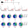
Polyfunctionality of CD8+ T cell responses to mRNA‐1273 is unimpaired without CD4+ T cell help. (A) A schematic diagram illustrating the experimental setup: Anti‐CD4 antibody was administered intraperitoneally 1 day prior to each dose of mRNA‐1273, where the boost was given 21 days post‐prime. Cells from blood and spleen were isolated for analysis of cellular responses at 21 days post‐prime and post‐boost (d21 and d21 + 21, respectively). Intracellular cytokine staining after stimulation with the immunodominant Kb‐restricted S539–546 peptide and flow cytometry were performed. (B–E) The frequency of CD107a+ (B), IFNγ+ (C), TNF+ (D), and IL‐2+ (E) CD8+ T cells was quantified as a percentage of CD8+ T cells. All bar graphs show the mean ± SEM. For all bar plots, each dot represents an animal, and data are from the number of mice as specified in (A; = 7–8). Data are pooled from two independent experiments. Experimental groups were compared to each other by Student's t‐test; *p < 0.05. (F–G) Polyfunctional cytokine profile at d21 (F) and d21 + 21 (G) of CD107a, IFNγ, TNF, and IL‐2 expression as a proportion of cytokine+ CD8+ T cells.
FIGURE 3
FIGURE 3
CD4+ T cell help is dispensable for generation of antigen‐specific CD8+ T cell responses to BNT162b2. (A) A schematic diagram illustrating the experimental setup: Anti‐CD4 antibody was administered intraperitoneally 1 day prior to each dose of BNT162b2. Cells from blood and spleen were isolated for analysis of the cellular responses at 20 days post‐prime (d20), and 7‐ or 21 days post‐boost (d21 + 7 and d21 + 21, respectively). (B) Antigen‐specific CD8+ T cells were stained with a Kb/S539–546 tetramer, and the frequencies of Kb/S539–546 + CD8+ T cells were expressed as a percentage of CD8+ T cells. (C) The number of Kb/S539–546 + CD8+ T cells in the spleen. (D–F) Expression of KLRG1+CD127 effector phenotype (D), CD62LCD127+ effector memory phenotype (E), and CD62L+CD127+ central memory phenotype (F) were quantified as a percentage of Kb/S539‐546 + CD8+ T cells (Tet+ CD8+ T cells). All bar graphs show the mean ± SEM. For all bar plots, each dot represents an animal, and data are from the number of mice as specified in (A; = 8). Data are pooled from two independent experiments. Experimental groups were compared to each other by Student's t‐test; *p < 0.05; ***p < 0.001.
FIGURE 4
FIGURE 4
Polyfunctionality of CD8+ T cell responses to BNT162b2 is unimpaired without CD4+ T cell help. (A) A schematic diagram illustrating the experimental setup: Anti‐CD4 antibody was administered intraperitoneally 1 day prior to each dose of BNT162b2, where the boost was given 21 days post‐prime. Cells from blood and spleen were isolated for analysis of cellular responses at 21 days post‐prime and post‐boost (d21 and d21 + 21, respectively). Intracellular cytokine staining after stimulation with the immunodominant Kb‐restricted S539–546 peptide and flow cytometry were performed. (B–E) The frequency of CD107a+ (B), IFNγ+ (C), TNF+ (D), and IL‐2+ (E) CD8+ T cells was quantified as a percentage of CD8+ T cells. All bar graphs show the mean ± SEM. For all bar plots, each dot represents an animal, and data are from the number of mice as specified in (A; = 8). Data are pooled from two independent experiments. Experimental groups were compared to each other by Student's t‐test; *p < 0.05. (F–G) Polyfunctional cytokine profile at d21 (F) and d21 + 21 (G) of CD107a, IFNγ, TNF, and IL‐2 expression as a proportion of cytokine+ CD8+ T cells.
FIGURE 5
FIGURE 5
Antigen‐specific CD8+ T cell responses to Ad‐vectored vaccines are dependent on CD4+ T cell help. (A) A schematic diagram illustrating the experimental setup: Anti‐CD4 antibody was administered intraperitoneally 1 day prior to each dose of AZD1222 and 14 days post‐prime. Animals were then boosted at 28 days post‐prime. Cells from blood and/or spleen were isolated for analysis of the cellular responses at 14‐ or 27 days post‐prime (d14 and d27, respectively), and 14 days post‐boost (d28 + 14). (B) Antigen‐specific CD8+ T cells were stained with a Kb/S539–546 tetramer, and the frequencies of Kb/S539–546 + CD8+ T cells were expressed as a percentage of CD8+ T cells. (C) The number of Kb/S539–546 + CD8+ T cells in the spleen. (D–F) Expression of KLRG1+CD127 effector phenotype (D), CD62LCD127+ effector memory phenotype (E), and CD62L+CD127+ central memory phenotype (F) were quantified as a percentage of Kb/S539–546 + CD8+ T cells (Tet+ CD8+ T cells). All bar graphs show the mean ± SEM. For all bar plots, each dot represents an animal, and data are from the number of mice as specified in (A; = 7–8). Data are pooled from two independent experiments. Experimental groups were compared to each other by Student's t‐test; *p < 0.05; **p < 0.01; ***p < 0.001. Ad, adenovirus.
FIGURE 6
FIGURE 6
Polyfunctionality of CD8+ T cell responses to AZD1222 is perturbed without CD4+ T cell help. (A) A schematic diagram illustrating the experimental setup: Anti‐CD4 antibody was administered intraperitoneally 1 day prior to each dose of AZD1222 and 14 days post‐prime. Animals were then boosted at 28 days post‐prime. Cells from blood and/or spleen were isolated for analysis of cellular responses at 14‐ and 27‐days post‐prime, and 14 days post‐boost (d14, d27, and d28 + 14, respectively). Intracellular cytokine staining after stimulation with the immunodominant Kb‐restricted S539–546 peptide and flow cytometry were performed. (B–E) The frequency of CD107a+ (B), IFNγ+ (C), TNF+ (D), and IL‐2+ (E) CD8+ T cells was quantified as a percentage of CD8+ T cells. All bar graphs show the mean ± SEM. For all bar plots, each dot represents an animal, and data are from the number of mice as specified in (A; = 9–10). Data are pooled from two independent experiments. Experimental groups were compared to each other by Student's t‐test; *p < 0.05; **p < 0.01; ***p < 0.001; ****p < 0.0001. (F) Polyfunctional cytokine profile of the blood at the different timepoints for CD107a, IFNγ, TNF, and IL‐2 expression as a proportion of cytokine+ CD8+ T cells.
FIGURE 7
FIGURE 7
CD4+ T cell help is essential in the induction of antibody responses irrespective of vaccine platforms. (A–C) Antibody responses in the sera were quantified as reciprocal dilutions by anti‐spike IgG ELISA post‐prime and post‐boost for each vaccine: d21 and d21 + 21 for mRNA‐1273 (A) and BNT162b2 (B), and d14 and 28 + 14 for AZD1222 (C). (D–I) Cells from inguinal lymph nodes were processed and stained for flow cytometry. The frequencies of GC B cells (Fas+ PNA+ IgD) following one dose of mRNA‐1273 (D) and BNT162b2 (E) or one and two doses of AZD1222 (F) were quantified as a percentage of B cells. The frequency of spike‐specific (S1+) B cells in response to mRNA‐1273 (G), BNT162b2 (H), and AZD1222 (I) was quantified similarly. All bar graphs show the mean ± SEM. For all bar plots, each dot represents an animal, and data are pooled from two independent experiments (= 7–8). Experimental groups were compared to each other by Student's t‐test; **p < 0.01; ***p < 0.001; ****p < 0.0001. ELISA, enzyme‐link immunosorbent assay; GC, germinal center.

References

    1. Swain S. L., McKinstry K. K., and Strutt T. M., “Expanding Roles for CD4+ T Cells in Immunity to Viruses,” Nature Reviews Immunology 12, no. 2 (2012): 136–148, https://www.nature.com/articles/nri3152. - PMC - PubMed
    1. Laidlaw B. J., Craft J. E., and Kaech S. M., “The Multifaceted Role of CD4+ T Cells in CD8+ T Cell Memory,” Nature Reviews Immunology 16, no. 2 (2016): 102–111, https://www.nature.com/articles/nri.2015.10. - PMC - PubMed
    1. Bennett S. R. M., Carbone F. R., Karamalis F., Flavell R. A., Miller J., and Heath W. R., “Help for Cytotoxic‐T‐Cell Responses Is Mediated by CD40 Signalling,” Nature 393, no. 6684 (1998): 478–480, https://www.nature.com/articles/30996. - PubMed
    1. Schoenberger S. P., Toes R. E. M., Van Dervoort E. I. H., Offringa R., and Melief C. J. M., “T‐Cell Help for Cytotoxic T Lymphocytes Is Mediated by CD40–CD40L Interactions,” Nature 393, no. 6684 (1998): 480–483, https://www.nature.com/articles/31002. - PubMed
    1. Castellino F., Huang A. Y., Altan‐Bonnet G., Stoll S., Scheinecker C., and Germain R. N., “Chemokines Enhance Immunity by Guiding Naive CD8+ T Cells to Sites of CD4+ T Cell–dendritic Cell Interaction,” Nature 440, no. 7086 (2006): 890–895, https://www.nature.com/articles/nature04651. - PubMed