. 2024 Nov 27;15(1):10309.
doi: 10.1038/s41467-024-54698-w.

Remodelling of Rea1 linker domain drives the removal of assembly factors from pre-ribosomal particles

Affiliations

Remodelling of Rea1 linker domain drives the removal of assembly factors from pre-ribosomal particles

Johan Busselez et al. Nat Commun. .

Abstract

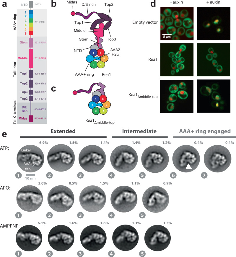
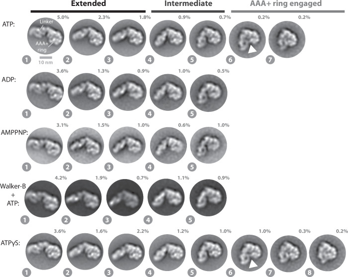
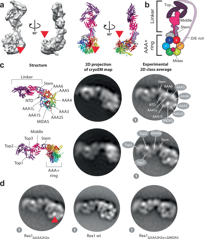
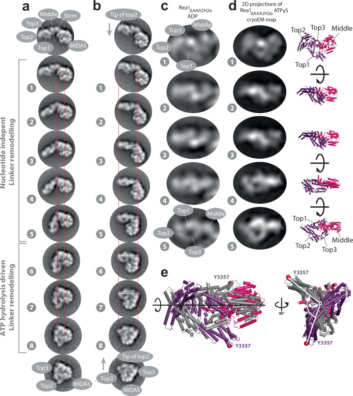
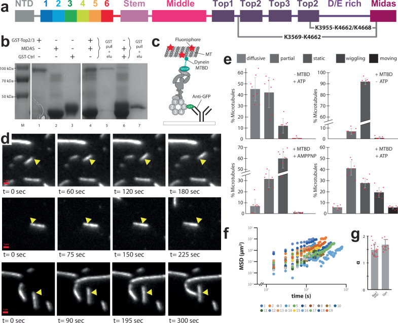
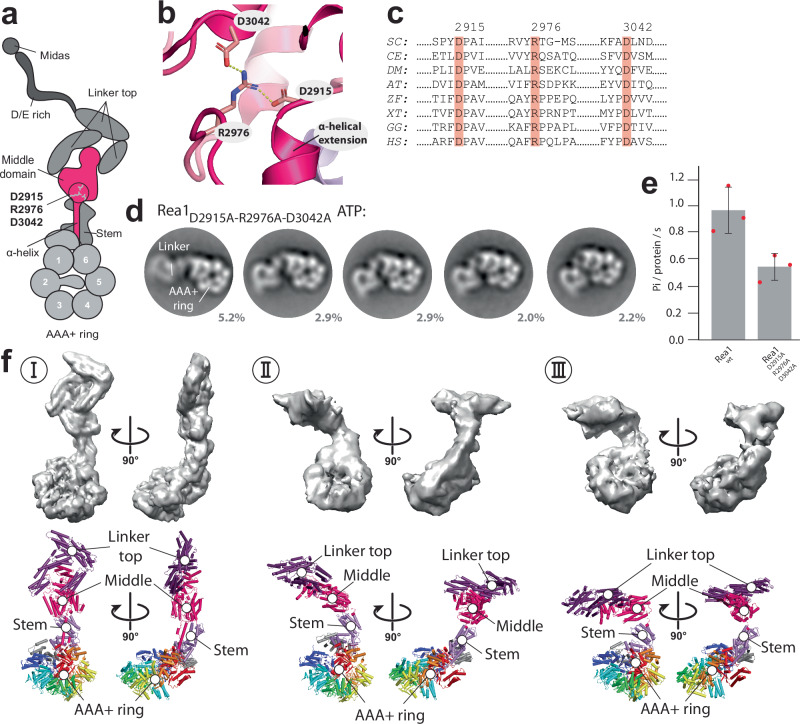
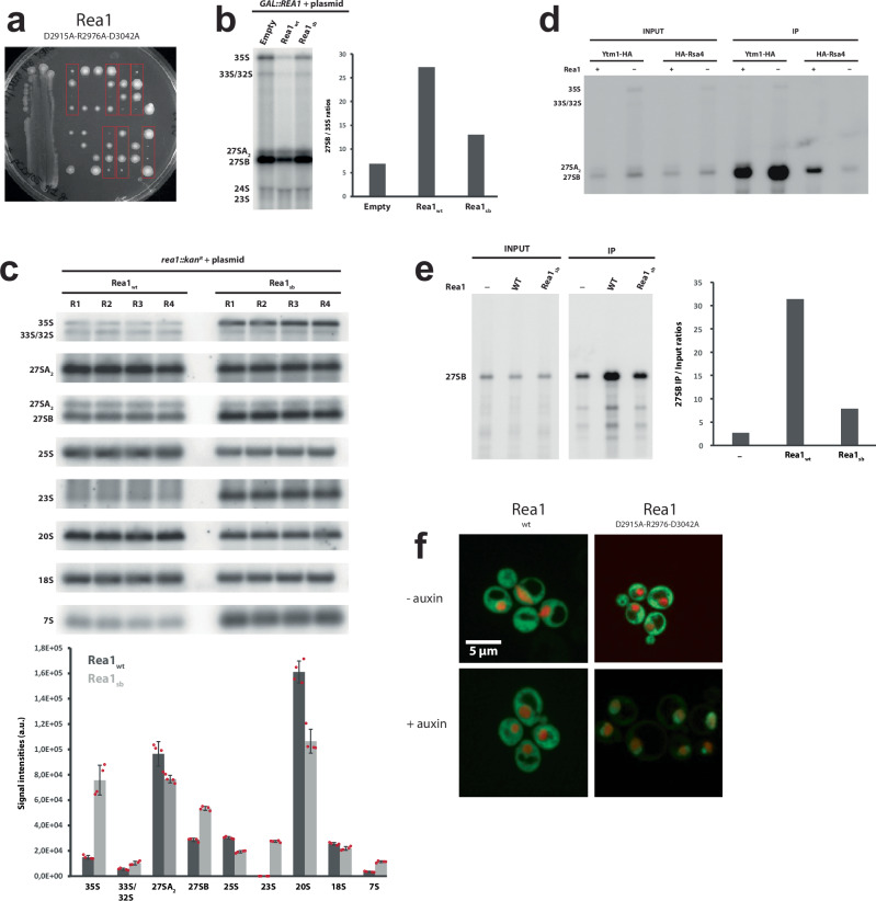
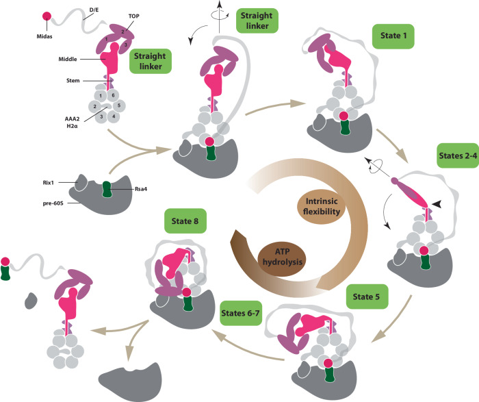
The ribosome maturation factor Rea1 (or Midasin) catalyses the removal of assembly factors from large ribosomal subunit precursors and promotes their export from the nucleus to the cytosol. Rea1 consists of nearly 5000 amino-acid residues and belongs to the AAA+ protein family. It consists of a ring of six AAA+ domains from which the ≈1700 amino-acid residue linker emerges that is subdivided into stem, middle and top domains. A flexible and unstructured D/E rich region connects the linker top to a MIDAS (metal ion dependent adhesion site) domain, which is able to bind the assembly factor substrates. Despite its key importance for ribosome maturation, the mechanism driving assembly factor removal by Rea1 is still poorly understood. Here we demonstrate that the Rea1 linker is essential for assembly factor removal. It rotates and swings towards the AAA+ ring following a complex remodelling scheme involving nucleotide independent as well as nucleotide dependent steps. ATP-hydrolysis is required to engage the linker with the AAA+ ring and ultimately with the AAA+ ring docked MIDAS domain. The interaction between the linker top and the MIDAS domain allows direct force transmission for assembly factor removal.


Conflict of interest statement

Competing interests: The authors declare no competing interests.

Figures

Fig. 1
Fig. 1. The Rea1 linker is a functionally important structural element and shows nucleotide independent as well as nucleotide dependent steps of remodelling.
a Domain organization of Rea1. b Schematic cartoon representation of Rea1. The helix 2 α-helical insertion of the AAA2 domain (AAA2H2α) sits in the central pore of the AAA+ ring. Adapted from ref. . c Schematic cartoon representation of a construct lacking the linker middle and top domains. The flexible D/E rich region with the substrate binding MIDAS domain are directly fused to the linker stem domain. Adapted from Sosnowski et al.. Elife 7, 10.7554/eLife.39163 (2018) under a CC BY license: https://creativecommons.org/licenses/by/4.0/. d Yeast nuclear export assay of pre-ribosomal particles. The pre-ribosomal particle marker Rpl25 is fused to GFP. Histone-mCherry marks the nucleus. The endogenous Rea1 is under the control of the auxin degron system. Upper panels: The addition of auxin leads to the accumulation of GFP fluorescence in the nucleus indicating a pre60S nuclear export defect due to degraded endogenous Rea1. Middle panels: The export defect can be rescued by a plasmid harbouring a Rea1wt copy. Lower panels: Providing a plasmid harbouring the construct in C. does not rescue the export defect, suggesting the linker middle and top domains are functionally important. Experiments were repeated three times. e Negative stain 2D classes representing AAA+ ring top views of Rea1wt in the presence of ATP, absence of nucleotide as well as in the presence of AMPPNP. States 1 – 5 represent the extended and intermediate linker conformations, which do not require nucleotide. In contrast to states 1 – 5, the AAA+ ring engaged states 6 and 7 require ATP hydrolysis. The white arrow head highlights a connection between the linker top and the AAA+ ring. Percentage numbers indicate how many particles of the corresponding data set sorted into the displayed 2D class averages.
Fig. 2
Fig. 2. Linker remodelling in Rea1ΔAAA2H2α.
The analysis of the ATP, ADP, AMPPNP and ATPγS data sets indicates that linker remodelling in Rea1ΔAAA2H2α is highly similar to linker remodelling in Rea1wt. Like in the case of Rea1wt, ATP-hydrolysis is required to engage the linker with the AAA+ ring. The latter point is also supported by the Rea1ΔAAA2H2α Walker-B mutant, which is impaired in ATP-hydrolysis activity and does not show the AAA+ ring engaged 2D class averages. Unlike Rea1wt, Rea1ΔAAA2H2α is able to sample state 8 in the presence of the slowly hydrolysable ATP analogue ATPγS. White arrow heads highlight a connection between the linker top and the AAA+ ring. Percentage numbers indicate how many particles of the corresponding data set sorted into the displayed 2D class averages.
Fig. 3
Fig. 3. Domain assignments in negative stain 2D class averages of Rea1ΔAAA2H2α.
a CryoEM map (left panels) and cartoon representation (right panels) of Rea1ΔAAA2H2α in the presence of ATPγS. The red arrow heads highlight the AAA+ ring docked MIDAS domain. b Schematic cartoon representation of the structure in a. The α-helical extension of AAA2 normally occupying the centre of the pore (compare Fig. 1b) has been deleted, which allows the MIDAS domain to dock onto the AAA+ ring. The D/E rich region connecting the MIDAS domain to the linker top is flexible and not visible in the structure. Adapted from Sosnowski et al.. Elife 7, 10.7554/eLife.39163 (2018) under a CC BY license: https://creativecommons.org/licenses/by/4.0/. c Upper panels: A 2D projection of the cryoEM map in A. low pass filtered to 25 Å shows a good match for the AAA+ ring region in the state 1 negative stain 2D class average. The projection allows the assignment of the NTD, AAA1–AAA6, the linker stem and the MIDAS domain. In contrast to the AAA+ ring region, the linker top and middle domains adopt a different conformation from the one seen in state 1. Lower panels: With a different 2D projection of the low pass filtered cryoEM map, a good match for the linker region in the state 1 negative stain 2D class average can be produced, allowing the assignment of the linker top1, top2 and top3 domains as well as the linker middle domain. The AAA+ ring does not match up with the AAA+ ring in the state 1 negative stain 2D class average. This mismatch indicates that -compared to the cryoEM structure in A. - the linker top and middle domains in state 1 have moved with respect to the AAA+ ring. d The assignment of the MIDAS domain in state 1 of Rea1ΔAAA2H2α (red arrow head, left panel) is further supported by comparisons with state 1 of Rea1wt, where the MIDAS domain is absent from the AAA+ ring (middle panel, compare also Supplementary Fig. 11) as well as analysis of the Rea1ΔAAA2H2α+ΔMIDAS double mutant (right panel).
Fig. 4
Fig. 4. The Rea1 linker pivots and rotates during remodelling.
a States 1 – 8 of Rea1ΔAAA2H2α ATPγS aligned on the linker stem and MIDAS domains (red lines). The linker middle and top domains swing towards the AAA+ docked MIDAS domain (compare also Supplementary Movie 1). The region between the linker stem and middle domains acts as pivot point. b States 1–8 of Rea1ΔAAA2H2α ATPγS aligned on long linker axis (tip of linker top2 domain and linker stem domain, white and red lines). The linker top and middle domains rotate during the pivot swing (compare also movie S2). The tip of the linker top2 domain points downwards in state 1 (grey arrow) but upwards in state 8 (grey arrow) highlighting the rotation. In the final linker remodelling conformation, state 8, the linker top2 and top3 domains as well as the MIDAS domain are in close proximity. States 1 -5 were also observed under APO conditions (compare Fig. 1e) indicating that large parts of the linker swing and the linker rotation are nucleotide independent and are part of the intrinsic conformational flexibility of the linker. The engagement of the rotated linker with the AAA+ ring during states 6–8 requires ATP hydrolysis. c Enlarged views of linker region in states 1–5 of Rea1ΔAAA2H2α ADP. d Left panels: Series of 2D projections of the linker top-middle part of the Rea1ΔAAA2H2α ATPγS cryoEM map low pass filtered to 25 Å and rotated around the long linker axis. Right panels: corresponding structures. The rotation of the linker in a and b can be approximated by a rigid-body rotation of the linker top and middle domains around the long linker axis. Additional internal rearrangements of the linker middle and top domains with respect to each other cannot be excluded. e Aligning states 1 (color coded) and 5 (grey) of d on the long linker axis indicates a total rotation angle of ≈100 °. Equivalent Cα atoms are shown as red spheres.
Fig. 5
Fig. 5. The linker top interacts with the MIDAS domain and linker remodelling is a force producing event.
a Crosslinks supporting state 8 detected in the Rea1ΔAAA2H2α ATPγS data set. The K3569-K4662 and K3955-K4662/K4668 crosslinks suggest an interaction between the linker top2 domain and a conserved MIDAS domain loop. b GST pulldown experiments provide further support for the interaction between the MIDAS and linker top2/3 domains. The samples were run on a SDS gel and stained with Coomassie blue. The GST-linker top2/3 fragment pulls down the MIDAS domain (lane 5). M: marker, lanes 1, 2 and 3: purified linker top2/3 construct, MIDAS domain and GST control. Lane 4, 5: GST-linker top2/3 + MIDAS domain input and pulldown. Lane 6, 7: GST-control + MIDAS domain input and pulldown. The pulldown experiment was carried out once. c Microtubule gliding assays provide evidence that Rea1 linker remodelling produces force. The dynein microtubule binding domain (cyan) was fused to the linker top2 domain using the spycatcher/spytag approach. GFP was fused to AAA5. The construct was anchored to a cover slide via GFP-antibodies and fluorescently labelled microtubules we applied. Adapted from Sosnowski et al.. Elife 7, 10.7554/eLife.39163 (2018) under a CC BY license: https://creativecommons.org/licenses/by/4.0/. d Movie frames of microtubule gliding events. Yellow arrow heads mark the microtubule position at the beginning of the movie (Supplementary Movies 3–5). The gliding events suggest that linker remodelling with respect to the AAA+ ring is able to produce mechanical force. e Statistical analysis of diffusive, partially bound, static, wiggling and moving microtubules under different conditions (n = 8). Moving microtubules are only observed in the presence of the dynein microtubule domain (MTBD) and ATP. f, g The microtubule gliding events result from directed movement. f Log mean squared displacement vs log time. g Slopes of the events in f, which represent the anomalous coefficient α, were averaged (n = 19). The average α of 1.49 ± 0.22 indicates that the events are not caused by a random diffusion process (α = 1). The motor protein dynein, known to power directed microtubule gliding, has a comparable α (1.65 ± 0.18, n = 11). Error bars show the standard deviation. Source data are provided as a Source Data file.
Fig. 6
Fig. 6. A highly conserved salt-bridge network in the linker middle domain has essential functions during linker remodelling.
a Schematic representation of the highly conserved linker middle domain salt-bridge network D2915-R2976-D3042. It is located above the α-helical extension of the middle domain and at the interface between the linker middle and stem domains. Adapted from Sosnowski et al.. Elife 7, 10.7554/eLife.39163 (2018) under a CC BY license: https://creativecommons.org/licenses/by/4.0/. b The D2915-R2976-D3042 salt-bridge network in the near-atomic resolution model of the S. cerevisiae Rea1 linker (PDB-ID: 6hyd). The dashed yellow lines indicate hydrogen bonds with a distance of 2.7 Å. c The D2915-R2976-D3042 salt-bridge network is conserved in Rea1/Midasin of SC: Saccharomyces cerevisiae, CE: Caenorhabditis elegans, DM: Drosophila melanogaster, AT: Arabidopsis thaliana, ZF: Zebrafish, XT: Xenopus tropicalis GG: Gallus gallus, HS: Homo sapiens. The S. cerevisiae Rea1 amino-acid residue numbering is shown at the top of the multiple sequence alignment. d Disrupting the D2915-R2976-D3042 salt-bridge network results in altered linker remodelling pathways. The linker is still able to swing towards the AAA+ ring but does not rotate around the long linker axis, which prevents the engagement of the linker with the AAA+ ring (compare also Supplementary Movie 6). e The inability of the Rea1 linker top and middle domains to correctly engage with the Rea1 AAA+ ring affects the ATPase activity. Disrupting the highly conserved D2915-R2976-D3042 salt-bridge network in the linker middle domain reduces the Rea1 ATPase activity by ≈50% (n = 3). The compromised ATPase activity indicates that the AAA+ ring engagement of the linker top and middle stimulates the Rea1 ATPase activity. Error bars show the standard deviation. f Three cryoEM structures of the Rea1D2915A-R2976A-D3042A mutant in the presence of ATP. The linker conformation I is highly similar to the linker conformation of Rea1ΔAAA2H2α ATPγS (compare Fig. 3a and Supplementary Fig. 8a, e). In conformations II and III the linker top and middle domains have swung towards the AAA+ ring (compare also Supplementary Fig. 18). Consistent with our previous domain assignments (compare Supplementary Movies 1 and 2), the region between the linker middle and stem domain acts as pivot point. Source data are provided as a Source Data file.
Fig. 7
Fig. 7. Functional impact of mutations in the conserved salt-bridge network D2915-R2976-D3042 of the Rea1 middle domain.
a A heterozygous REA1/rea1::kanR diploid strain was transformed with a plasmid directing expression of the Rea1D2915A-R2976A-D3042A mutant. After sporulation of the resulting strains, tetrads were dissected and haploid spores were spotted in rows on YPD medium. Spores containing the Rea1D2915A-R2976A-D3042A mutant could support viability (red rectangles), although growth was strongly impaired. b Impact on pre-rRNA processing. Left panel: Northern analyses of pre-rRNA processing in GAL::rea1 cells expressing Rea1D2915A-R2976A-D3042A (“Rea1sb”) or wild-type Rea1 (Rea1wt) from plasmids or transformed with an empty vector (empty), shifted 24 hours on glucose-containing medium. Right panel: Quantification of 27SB/35S ratio. The Rea1D2915A-R2976A-D3042A mutants displays reduced ratios compared to the Rea1wt control indicative of pre60S maturation defects. Experiment was done once. c Upper panel: Detailed northern pre-rRNA processing analysis of rea1::kanR strain expressing Rea1D2915A-R2976A-D3042A (“Rea1sb”) from a plasmid. Four wt strains and four Rea1D2915A-R2976A-D3042A expressing strains were analysed in parallel. Detection of the indicated (pre-)rRNAs by northern hybridization with anti-sense oligonucleotide probes. Lower panel: levels of the indicated (pre-)rRNAs in the Rea1D2915A-R2976A-D3042A mutant or wild-type (n = 4). Error bars show the standard deviation. d Rea1 depletion affects the association of Rsa4 with pre60S particles. REA1 (Rea1 +) or GAL::rea1 (Rea1 -) strains expressing HA-tagged Ytm1 or Rsa4 were grown in glucose-containing medium. Immunoprecipitation experiments were then carried out with anti-HA agarose beads and the indicated pre-rRNAs in the input and immunoprecipitated (IP samples) were detected by northern analyses. The experiment was repeated twice. e Rea1D2915A-R2976A-D3042A (“Rea1sb”) affects the association of Rsa4 with pre60S particles. Left panel: Analysis as in d, except that a GAL::rea1 strain expressing HA-tagged Rsa4 and Rea1D2915A-R2976A-D3042A or wild-type Rea1 (Rea1wt) from plasmids was used. -: strain transformed with an empty vector. Detection of 27SB pre-rRNA by northern hybridization. Right panel: ratios of the levels of precipitated 27SB pre-rRNA over input levels. Experiment was done once. f Disrupting the D2915-R2976-D3042 salt-bridge network leads to nuclear pre60S particle export defects. Experiments were repeated three times. Source data are provided as a Source Data file.
Fig. 8
Fig. 8. Model for the Rea1 mechanism.
Rea1 in the absence of pre60S particles exists in an autoinhibited form with the AAA2H2α insert occupying the central pore of the AAA+ ring and the linker in the straight conformation. The binding of Rea1 to pre60S particles relocates AAA2H2α towards the pre60S particles, which allows the MIDAS domain to dock onto the AAA+ ring to engage with its assembly factor substrate (here Rsa4). The linker remains in the straight conformation. The linker subsequently rotates and pivots towards the plane of the AAA+ ring to reach state 1. From here the linker middle and top domains rotate around the long linker axis and swing towards the AAA+ ring. The region between the linker middle and stem domains acts as pivot point. In state 5, the linker middle and top domains are fully rotated and in close proximity to the AAA+ ring. In states 6-8, the linker top engages with the AAA+ ring and in the final remodelling step state 8 the linker top2/3 domains interact with the MIDAS domain to allow the transmission of force for assembly factor removal. Up to state 5, linker remodelling is nucleotide independent and driven by the intrinsic conformational flexibility of the linker. States 6–8 require ATP hydrolysis. Cartoon adapted from Sosnowski et al. Elife 7, 10.7554/eLife.39163 (2018) under a CC BY license: https://creativecommons.org/licenses/by/4.0/.

References

    1. Thomson, E., Ferreira-Cerca, S. & Hurt, E. Eukaryotic ribosome biogenesis at a glance. J. Cell Sci.126, 4815–4821 (2013). - PubMed
    1. Kressler, D., Hurt, E. & Bassler, J. Driving ribosome assembly. Biochim Biophys. Acta1803, 673–683 (2010). - PubMed
    1. Harnpicharnchai, P. et al. Composition and functional characterization of yeast 66S ribosome assembly intermediates. Mol. Cell8, 505–515 (2001). - PubMed
    1. Venema, J. & Tollervey, D. Ribosome synthesis in Saccharomyces cerevisiae. Annu Rev. Genet33, 261–311 (1999). - PubMed
    1. Tang, L., Sahasranaman, A., Jakovljevic, J., Schleifman, E. & Woolford, J. L. Jr Interactions among Ytm1, Erb1, and Nop7 required for assembly of the Nop7-subcomplex in yeast preribosomes. Mol. Biol. Cell19, 2844–2856 (2008). - PMC - PubMed

Publication types

MeSH terms

Substances

LinkOut - more resources