. 2024 Nov 27;14(1):29501.
doi: 10.1038/s41598-024-81350-w.

18F-NaF uptake in skull-base bone as a predictor of treatment response in advanced nasopharyngeal carcinoma

Affiliations

18F-NaF uptake in skull-base bone as a predictor of treatment response in advanced nasopharyngeal carcinoma

Xingyu Mu et al. Sci Rep. .

Abstract

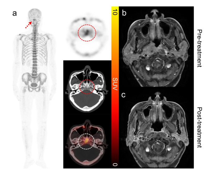
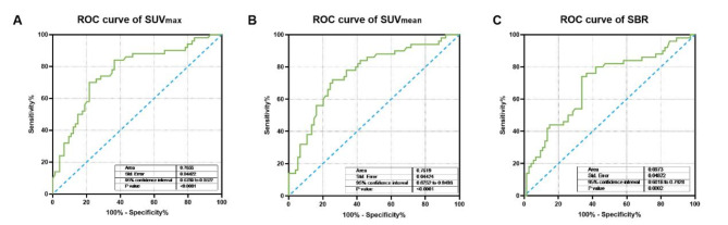
This study investigates the utility of 18F-sodium fluoride (18F-NaF) positron emission tomography/computed tomography (PET/CT) in assessing skull-base bone invasion (SBBI) and predicting treatment response in advanced nasopharyngeal carcinoma (NPC). A retrospective analysis was conducted on 142 patients with newly diagnosed advanced NPC who underwent 18F-NaF PET/CT for initial staging from December 2020 to December 2023. 18F-NaF PET/CT scans were analyzed for uptake values at the skull-base bone, and these were correlated with treatment outcomes of primary tumor using the Response Evaluation Criteria in Solid Tumors (RECIST) 1.1. Statistical analyses involved Mann-Whitney U tests for group comparisons and logistic regression for evaluating risk factors. Higher 18F-NaF uptake at the skull-base bone was significantly associated with advanced T stages (p < 0.0001) and the presence of bone metastases (p = 0.01). Patients exhibiting complete response (CR) to treatment had significantly lower skull-base 18F-NaF uptake compared to those with non-CR (p < 0.001). Receiver operating characteristic (ROC) analysis identified an SUVmax > 10.0 and SUVmean > 5.2 as predictive of non-CR, with AUC values of 0.77 and 0.76, respectively. Univariate and multivariable analysis confirmed SUVmax as a significant predictor of treatment response (OR = 7.03, 95% CI: 1.97-25.13, p < 0.05). Elevated 18F-NaF uptake at the skull-base bone is predictive of poorer treatment outcomes, highlighting its potential as a prognostic biomarker in advanced NPC. This study demonstrates that 18F-NaF PET/CT is a valuable imaging modality for evaluating SBBI in NPC, offering metabolic information that complements the anatomical findings from MRI.

Keywords: 18F-NaF; Nasopharyngeal carcinoma; PET/CT; Skull-base bone invasion; Treatment response.


Conflict of interest statement

Competing interests: The authors declare no competing interests. Ethical approval and consent to participate: This retrospective study received ethical clearance and the requirement to obtain informed consent was waived by the institutional review boards of Affiliated Hospital of Guilin Medical University (ID: 2023QTLL-16). The study protocol adheres to the tenets of the 1964 Declaration of Helsinki.

Figures

Fig. 1
Fig. 1
The flowchart of this study.
Fig. 2
Fig. 2
Distribution of 18F-NaF Uptake at Skull Base Across Tumor Stages in Patients with advanced NPC. (A) The SUVmax for each stage. (B) The SUVmean for each stage. (C) SBR for each stage. Each data point represents an individual patient measurement, overlaid on bars indicating the median value per stage.
Fig. 3
Fig. 3
A 57-y-old man with newly diagnosed locally advanced nasopharyngeal carcinoma who underwent 18F-NaF PET/CT and MRI at pre-treatment and MRI at post-treatment. (A) Maximum-intensity projection (MIP) and Axial images from 18F-NaF PET/CT: MIP illustrates a tracer accumulation at the base of the skull (red dashed arrow). Axial images demonstrate high 18F-NaF uptake in the lesion (dashed circle), with a SUVmax of 7.9. (B) Pre-treatment MRI: Axial contrast-enhanced T1-weighted MRI reveals an enhancing mass at the nasopharyngeal mass associated with bone invasion and soft-tissue extension. (C) Post-treatment MRI: Axial contrast-enhanced T1-weighted MRI taken after therapy shows significant reduction in the size, indicating a complete response to treatment.
Fig. 4
Fig. 4
A 37-year-old man with newly diagnosed locally advanced nasopharyngeal carcinoma who underwent 18F-NaF PET/CT and MRI at pre-treatment and MRI at post-treatment. (A) Maximum-intensity projection (MIP) and Axial images from 18F-NaF PET/CT: MIP image from an 18F-NaF PET/CT scan demonstrating a hypermetabolic lesion in the skull base (red dashed arrow). Axial images demonstrate high 18F-NaF uptake in the lesion (red dashed circle), with a SUVmax of 41.1. (B) Pre-treatment MRI: Axial contrast-enhanced T1-weighted MRI reveals an enhancing mass at the nasopharyngeal mass associated with bone invasion and soft-tissue extension. (C) Post-treatment MRI: Axial contrast-enhanced T1-weighted MRI taken after therapy shows no significant reduction in the size, indicating a stable disease after treatment.
Fig. 5
Fig. 5
ROC Curves of 18F-NaF uptake for predicting non-CR response to treatment.

Similar articles

Cited by

References

    1. Bray, F. et al. Global cancer statistics 2022: GLOBOCAN estimates of incidence and mortality worldwide for 36 cancers in 185 countries. CA Cancer J. Clin.74, 229–263 (2024). - PubMed
    1. Tang, L. L. et al. The Chinese Society of Clinical Oncology (CSCO) clinical guidelines for the diagnosis and treatment of nasopharyngeal carcinoma. Cancer Commun. (Lond).41, 1195–1227 (2021). - PMC - PubMed
    1. Lee, A. W., Ma, B. B., Ng, W. T. & Chan, A. T. Management of nasopharyngeal Carcinoma: Current practice and future perspective. J. Clin. Oncol.33, 3356–3364 (2015). - PubMed
    1. Pan, J. J. et al. Proposal for the 8th edition of the AJCC/UICC staging system for nasopharyngeal cancer in the era of intensity-modulated radiotherapy. Cancer.122, 546–558 (2016). - PMC - PubMed
    1. Wu, Z. et al. Characteristics of local extension based on tumor distribution in nasopharyngeal carcinoma and proposed clinical target volume delineation. Radiother. Oncol.183, 109595 (2023). - PubMed

MeSH terms

LinkOut - more resources