. 2024 Nov 27;14(1):29512.
doi: 10.1038/s41598-024-80931-z.

Preclinical evaluation of polymer encapsulated carbon-based nano and microparticles for sentinel lymph node tattooing

Affiliations

Preclinical evaluation of polymer encapsulated carbon-based nano and microparticles for sentinel lymph node tattooing

Marta Baselga et al. Sci Rep. .

Abstract

Selective sentinel lymph node biopsy (SNLB) is the standard method for detecting regional metastases in breast cancer patients. Identifying affected axillary lymph nodes before neoadjuvant treatment is crucial, as such treatment may alter drainage pathways and lymph node morphology, hindering the identification of sentinel lymph nodes. The use of carbon-based tattooing on sentinel lymph nodes (SLN) has been employed as a permanent tattooing method in clinical studies of Targeted Axillary Dissection (TAD), aiding in the SLN identification during surgery. Our study introduces a new method of lymph node tattooing based on poly lactic-co-glycolic (PLGA) particles with encapsulated carbon. This strategy substantially improves tattooing efficiency over single carbon suspensions currently used in clinical studies. We synthesized and characterized carbon-loaded PLGA micro- and nanoparticles, experimentally assessing their biological impact on porcine lymph nodes. The effect of particles' size and concentration was evaluated over time (from 1 to 16 weeks). Light and electron microscopy studies were conducted to characterize the cellular effects induced by the presence of these particles. Our findings reveal that the diverse physicochemical parameters of the particles interact differently with the lymphatic tissue, influencing their biodistribution within the lymph nodes and the intensity of the inflammatory response.

Keywords: Breast cancer; Carbon; Carbon-tattooing; Nanoparticles; Sentinel lymph node biopsy; Targeted-axillary dissection; Ultrastructure.


Conflict of interest statement

Declarations. Ethics approval and consent to participate: This research was approved by the Animal Research Ethics Committee of the University of Zaragoza under reference PI09/20. Competing interests: The authors declare no competing interests.

Figures

Fig. 1
Fig. 1
Brief outline of the methods used for the synthesis and preparation of the vectors. (a) Emulsion-solvent evaporation method. (b) Electrospraying method. (c) Dispersion of the resulting vectors in methylcellulose medium.
Fig. 2
Fig. 2
Macroscopic variables analyzed: (a) Ease of identification scale. (b) Estimation of the tattooed nodal area. (c) Morphometry of the particles clusters. (d) Extension measurement of the inflammatory response.
Fig. 3
Fig. 3
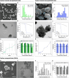
Characterization of the polymeric particles containing C-NPs synthesized (af) by emulsion (CS) and (gl) by electrospraying (CE), and (m-p) carbon nanoparticles (C-NPs). (a, g, m) Scanning electron micrographs of the particles. The carbon nanoparticles have been artificially coloured in blue to make them more visible in the micrographs. (b, h, o) Histograms of the size distributions. (c, i, n) Transmission electron micrographs of the particles, where carbon is clearly distinguished by its higher electron density. (d, j) Thermogravimetric curves. The solid blue line represents the mass loss of the micro and nanoparticles, the dashed black line represents the weight loss of empty PLGA, the solid black line represents the weight loss of carbon and the dashed blue line represents the weight loss of the stabilizer, in the case of the nanoparticles obtained by emulsion. (e, k) Curves of the release kinetics of the encapsulated carbon nanoparticles from the polymeric nano- and microparticles in PBS 1x medium. (f, l, p) Cell viability in the fibroblast (NHDF-Ad), macrophages (J774), and human breast adenocarcinoma (MDA-MB-231) cell lines. Untreated control cells were assigned as 100% viability. Statistical analyses are referred to untreated control cells (*p < 0.05, **p < 0.01, ***p < 0.001, ****p < 0.0001).
Fig. 4
Fig. 4
Macroscopic and microscopic variables analyzed. (a) Identification rates of CE20 (colored in blue and purple) and CS20 (colored in shades of green) tattoos. (b) Frequency of dissected lymph nodes showing difficult (1), moderate (2) or easy (3) identification. (c) Percentage of tattooed area over time for CS20 and CE20 vectors.
Fig. 5
Fig. 5
(a) Particles localization in the three different nodal regions (pericapsular region, adjacent tissue and inner region). (b) Particle’s location in CS20 and CE20 tattooed nodes. (c) Clusters size in CS20 and CE20-tattoed nodes. (d) Extension of the inflammatory tissue in CS and CE20 tattooed nodes. (eg) Inflammatory reaction associated with (e) CE20 and (f, g) CS20 vectors.
Fig. 6
Fig. 6
Structure and ultrastructural features of macrophages. (a) Macrophage with numerous lysosomes (arrows) in its cytoplasm. (b) Lamellipodia (arrowhead) and filopodia (arrow) emerging from the surface of a macrophage.
Fig. 7
Fig. 7
Different types of particles phagocytosed by macrophages. (a, b) Microparticles (CE). (c, d) Nanoparticles (CS). (e, f) Non-encapsulated carbon nanoparticles (C-NPs).
Fig. 8
Fig. 8
Effect of particles on macrophages. (a) Detail of the cytoplasm of a macrophage showing lysosomes with carbon particles (C-NPs) inside. (b) Carbon particles rupture the membrane of phagolysosomes (arrowheads), releasing lysosomal hydrolases that rupture the cell plasma membrane (arrows). (c) Plasma membrane of a macrophage lysed by spilled hydrolases. The rupture of the phagosome membrane (arrowheads) and the plasma membrane (arrows) is observed. (d) After rupture of the plasma membrane, the carbon particles remain intact in the extracellular medium. (e) The organelles spilled into the extracellular space preserve their structure.
Fig. 9
Fig. 9
Particle isolation in GMC and particle interaction with macrophages in the long term. (a) Carbon particles (C-NPs) internalized in a GMC. (b) Highly particle-laden macrophage (after 16 weeks). (c) The polymer of CS and CE particles is degraded, and free C-NPs appear inside macrophages. (d) Particle-laden macrophage is isolated within a matrix of collagen fibers.
Fig. 10
Fig. 10
Overview of the lymph node tattooing technique and its biological effect on the lymph node. Created with Scienfy®.

References

    1. Turner, R., Olilla, D., Krasne, D. & Giuliano, A. Histopathologic validation of the Sentinel Lymph Node hypothesis for breast carcinoma. Ann. Surg.226, 271–276 (1997). - PMC - PubMed
    1. Giuliano, A., Kirgan, D., Guenther, J. & Morton, D. Lymphatic mapping and Sentinel Lymphadenectomy for breast Cancer. Ann. Surg.220, 391–398 (1994). - PMC - PubMed
    1. Boughey, J. et al. Sentinel Node surgery after Neoadyuvant Chemotherapy in patients with node-positive breast Cancer: the American College of Surgeons Oncology Group. Z1071 clinical trial. JAMA310, 1455–1461 (2013). - PMC - PubMed
    1. Kuehn, T. et al. Sentinel-Lymph-Node Biopsy in patients with breast Cancer before and after Neoadjuvant Chemotherapy (SENTINA): a prospective, Multicenter Cohort Study. Lancet Oncol.14, 608–618 (2013). - PubMed
    1. Boileau, J. et al. Sentinel Node Biopsy after Neoadyuvant Chemotherapy in Biopsy-Proven node-positive breast Cancer: the SN FNAC Study. J. Clin. Oncol.20, 2588–2564 (2015). - PubMed

LinkOut - more resources