. 2024 Nov 27;14(1):29506.
doi: 10.1038/s41598-024-79667-7.

Photoacoustic polydopamine-indocyanine green (PDA-ICG) nanoprobe for detection of senescent cells

Affiliations

Photoacoustic polydopamine-indocyanine green (PDA-ICG) nanoprobe for detection of senescent cells

Muhamad Hartono et al. Sci Rep. .

Abstract

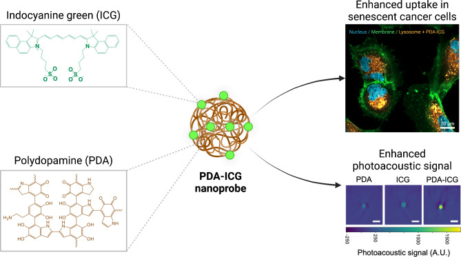
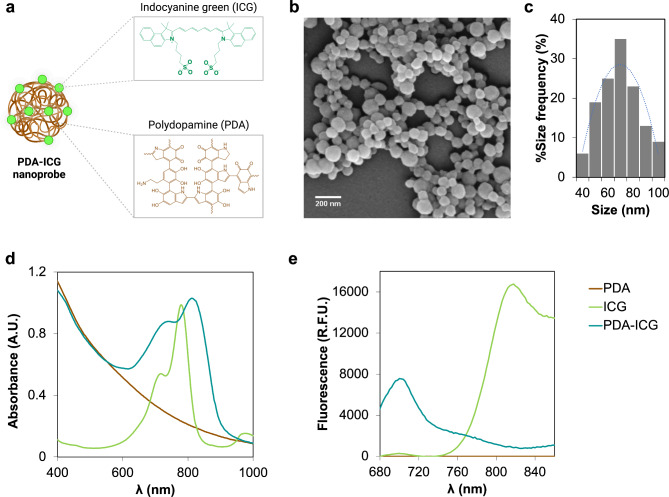
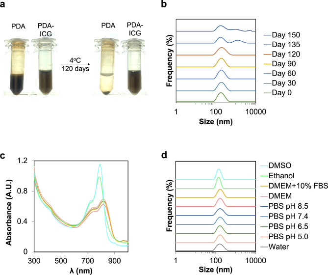
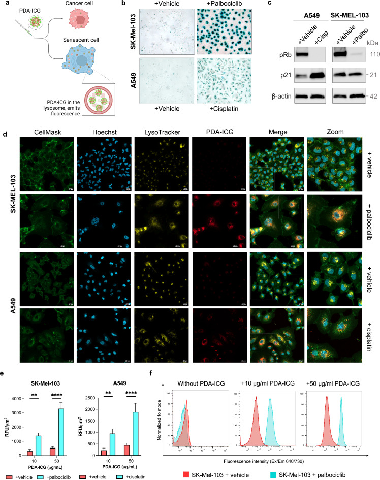
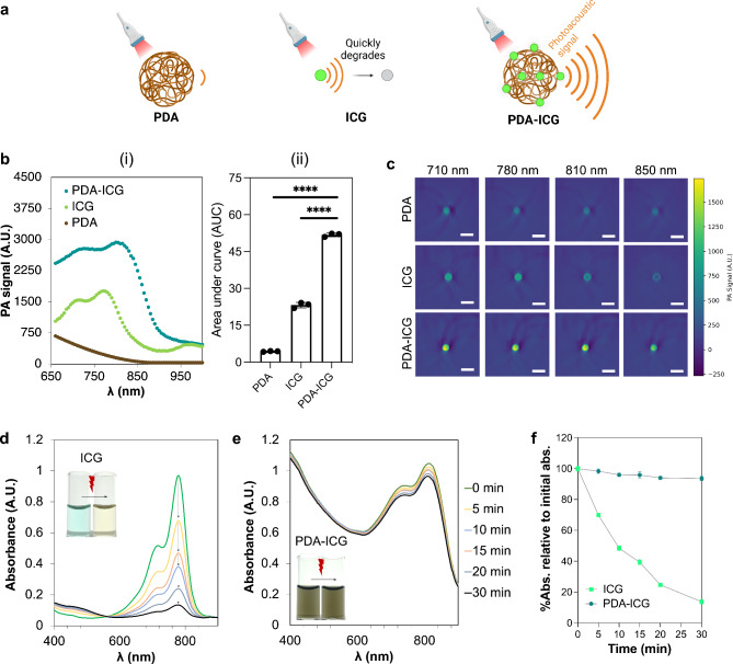
Cellular senescence is considered an important tumour suppression mechanism in response to damage and oncogenic stress in early lesions. However, when senescent cells are not immune-cleared and persist in the tumour microenvironment, they can drive a variety of tumour-promoting activities, including cancer initiation, progression, and metastasis. Additionally, there is compelling evidence demonstrating a direct connection between chemo(radio)therapy-induced senescence and the development of drug resistance and cancer recurrence. Therefore, detection of senescent cells in tissues holds great promise for predicting cancer occurrence earlier, assessing tumour progression, aiding patient stratification and prognosis, and informing about the efficacy of potential senotherapies. However, effective detection of senescent cells is limited by lack of biomarkers and readout strategies suitable for in vivo clinical imaging. To this end, a nanoprobe composed of biocompatible polydopamine (PDA) nanoparticle doped with FDA-approved indocyanine green (ICG) dye, namely PDA-ICG, was designed as a contrast agent for senescence detection using photoacoustic imaging (PAI). In an in vitro model of chemotherapy-induced senescence, PDA-ICG nanoprobe showed an elevated uptake in senescent cells relative to cancer cells. In addition to its improved photostability, 2.5-fold enhancement in photoacoustic signal relative to ICG was observed. Collectively, the results indicate that the PDA-ICG nanoprobe has the potential to be used as a contrast agent for senescence detection of chemotherapy-induced senescence using PAI.

Keywords: Cancer; Detection; ICG; Photoacoustic; Polydopamine; Senescence.


Conflict of interest statement

Declarations. Competing interests: Ljiljana Fruk, Daniel Munoz Espin and Andrew G Baker are co-founders of Senesys Bio, a company improving the formulation of senolytics and declare competing interests. Other authors (MH, TRE, ASE and SEB) declare no competing interests.

Figures

Fig. 1
Fig. 1
Schematic layout of present study. Photoacoustic-active PDA-ICG nanoprobe showing enhanced uptake in senescent cells. The nanoprobe was prepared by loading ICG dye into PDA nanoparticles.
Fig. 2
Fig. 2
Synthesis and characterization of PDA-ICG nanoprobe. a. Schematic illustration of PDA-ICG molecular structures. b. SEM photograph showing spherical PDA-ICG, with corresponding (c) size distribution (n = 200 nanoparticles) with an average size of 75 ± 8 nm. d. Absorbance and (e) fluorescence emission spectra of bare PDA, ICG and PDA-ICG suspended in water (lexcitation = 640 nm). Compared to free ICG, the fluorescence emission of PDA-ICG shifts to lower wavelength (700 nm) and was quenched.
Fig. 3
Fig. 3
PDA-ICG exhibits excellent colloidal and long-term stability. a. Images of PDA and PDA-ICG suspended in water at the same concentration (0.5 mg/ml), before and after being stored at 4°C for 120 days. b. Corresponding DLS measurements of PDA-ICG in water over different period of storage. Conjugating PDA with ICG improves nanoparticles colloidal stability. Colloidal stability of PDA-ICG suspended in deionized water, biological media and organic solvents measured with UV-Vis spectroscopy (c) and DLS (d). Measurement in biological media were taken after 24 h incubation at 37°C, while in other media were taken 5 min after the nanoprobe was suspended. PDA-ICG was stable in biological media but were broken apart in organic solvents.
Fig. 4
Fig. 4
Study of cellular uptake and intracellular localization of PDA-ICG nanoprobe on SK-MEL-103 and A549 cells using confocal fluorescence microscopy and flow cytometry. a. Schematic illustration showing the cellular uptake of PDA-ICG by either cancer cells or senescent cells. Cells were first validated for senescence 10 days following treatments with either chemotherapeutic drugs or vehicles. b. Representative images of b-galactosidase staining of fixed control (vehicle-treated) cells, palbociclib-treated SK-MEL-103 cells and cisplatin-treated A549 cells. c. Western blot analysis of the expression of relevant senescence markers: phosphorylated retinoblastoma (pRb), and cell cycle inhibitor p21 with β-actin as reference. d. Confocal microcopy images of cells following 24-h incubation with 10 µg/ml PDA-ICG nanoparticles. Cell membranes, nucleus and lysosomes were stained with CellMask (green), Hoechst 33,342 (blue) and LysoTracker (yellow). PDA-ICG was observed in the Alexa Fluor 700 channel (red). Scale bar = 20 μm. e. Corresponding fluorescence intensity for nanoprobe-treated cells, showing senescent cells having higher PDA-ICG fluorescence intensity compared to cancer cells (mean ± SD, n = 3, ** for p ≤ 0.01, **** for p < 0.0001). f. Histogram showing mean fluorescence values of SK-MEL-103 cells after incubation with PDA-ICG; PDA-ICG fluorescence was measured from the Alexa Fluor 700 channel that corresponded to the fluorescence of PDA-ICG nanoparticles.
Fig. 5
Fig. 5
Photoacoustic properties and photostability of PDA-ICG nanoprobe. a) Schematic illustration of the comparison among photoacoustic signals generated by PDA, ICG and PDA-ICG. b) (i) Photoacoustic signal in phantoms generated by bare PDA, free ICG and PDA-ICG (left). (ii). Area under the curve (AUC) measured from (i) that represents the integrated photoacoustic mean pixel intensities (mean ± SD, n = 3, **** for p < 0.0001, ordinary one-way ANOVA). c) Phantom photoacoustic images of bare PDA, free ICG and PDA-ICG at different excitation wavelengths. Absorbance spectra of free ICG (d) and PDA-ICG (e) taken immediately after irradiation with a 740 nm laser. Insets are photographs of ICG and PDA-ICG solution taken before and after laser irradiation. f. Percentage of absorbance over time relative to the initial absorbance of ICG versus PDA-ICG, wherein the absorbance of ICG is constantly declining over time.

References

    1. Muñoz-Espín, D. & Serrano, M. Cellular senescence: from physiology to pathology. Nat. Rev. Mol. Cell Biol.15, 482–496 (2014). - PubMed
    1. Schmitt, C. A., Wang, B. & Demaria, M. Senescence and cancer — role and therapeutic opportunities. Nat. Rev. Clin. Oncol.19, 619–636 (2022). - PMC - PubMed
    1. Coppé, J. P., Desprez, P. Y., Krtolica, A. & Campisi, J. The Senescence-Associated Secretory phenotype: the Dark side of Tumor suppression. Annu. Rev. Pathol. Mech. Dis.5, 99–118 (2010). - PMC - PubMed
    1. Di Micco, R., Krizhanovsky, V. & Baker, D. d’Adda Di Fagagna, F. Cellular senescence in ageing: from mechanisms to therapeutic opportunities. Nat. Rev. Mol. Cell. Biol.22, 75–95 (2021). - PMC - PubMed
    1. Chaib, S., Tchkonia, T. & Kirkland, J. L. Cellular senescence and senolytics: the path to the clinic. Nat. Med.28, 1556–1568 (2022). - PMC - PubMed

LinkOut - more resources