Review
. 2024 Nov 27;11(1):48.
doi: 10.1186/s40580-024-00456-z.

Manganese oxide nanomaterials: bridging synthesis and therapeutic innovations for cancer treatment

Affiliations
Review

Manganese oxide nanomaterials: bridging synthesis and therapeutic innovations for cancer treatment

Sandip Gangadhar Balwe et al. Nano Converg. .

Abstract

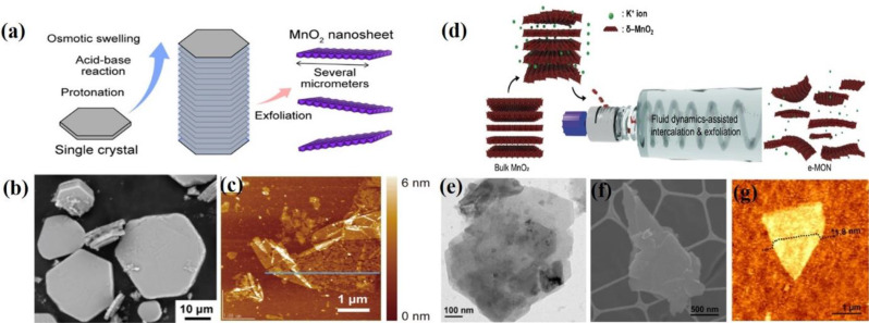
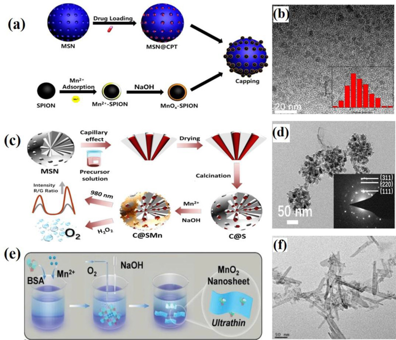
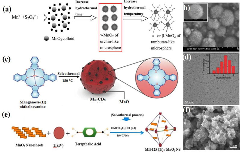
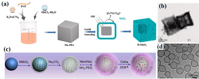
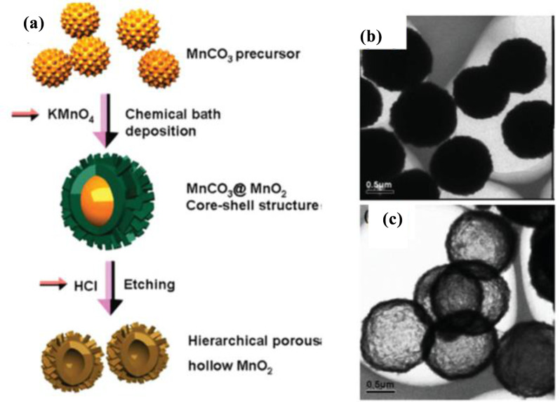
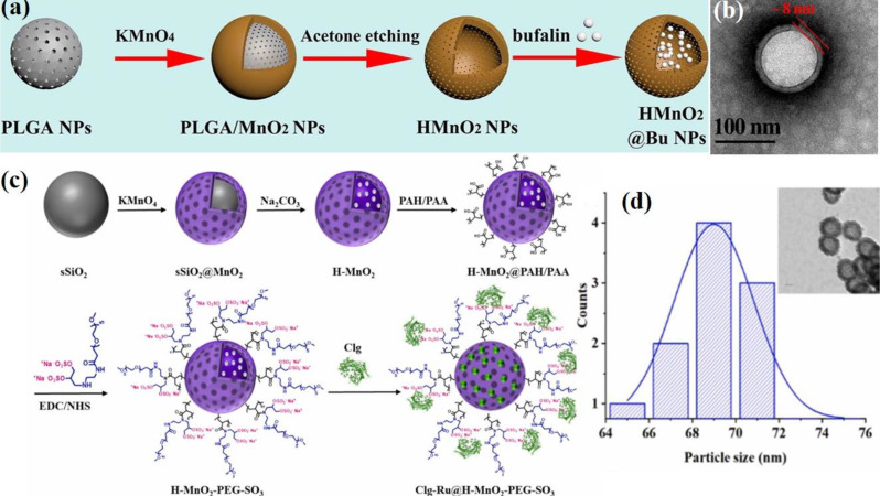
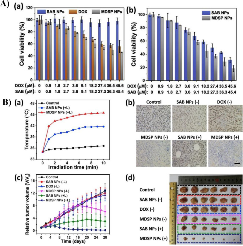
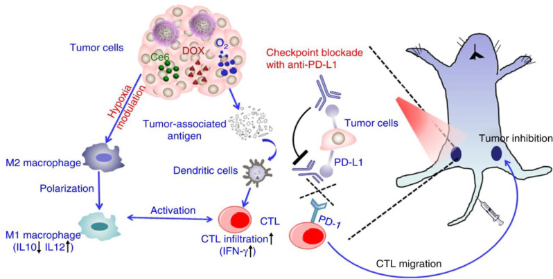
The advent of precision medicine in oncology emphasizes the urgent need for innovative therapeutic strategies that effectively integrate diagnosis and treatment while minimizing invasiveness. Manganese oxide nanomaterials (MONs) have emerged as a promising class of nanocarriers in biomedicine, particularly for targeted drug delivery and the therapeutic management of tumors. These nanomaterials are characterized by exceptional responsiveness to the tumor microenvironment (TME), high catalytic efficiency, favorable biodegradability, and advanced capabilities in magnetic resonance imaging. These attributes significantly enhance drug delivery, facilitate real-time bioimaging, and enable early tumor detection, thereby improving the precision and effectiveness of cancer therapies. This review highlights the significant advancements in the synthesis and therapeutic applications of MONs, beginning with a comprehensive overview of key synthetic methods, including thermal decomposition, potassium permanganate reduction, exfoliation, adsorption-oxidation, and hydro/solvothermal techniques. We delve into the preparation of MONs and H-MnO₂-based nanomaterials, emphasizing their chemical properties, surface modifications, and toxicity profiles, which are critical for their clinical application. Moreover, we discuss the notable applications of H-MnO₂-based nanomaterials in pH-responsive drug release, overcoming multidrug resistance (MDR), immunotherapy, and the development of nanovaccines for synergistic cancer treatments. By addressing the current challenges in the clinical translation of MONs, we propose future research directions for overcoming these obstacles. By underscoring the potential of MONs to transform cancer treatment paradigms, this review aims to inspire further investigations into their multifunctional applications in oncology, thus ultimately contributing to more effective and personalized therapeutic strategies.

Keywords: Anticancer material; Hypoxia; Immunotherapy; MONs; Nanovaccine; TME.


Conflict of interest statement

Declarations. Competing interests: The authors declare that they have no known competing financial interests or personal relationships that could have appeared to influence the work reported in this paper.

Figures

Fig. 1
Fig. 1
Synthesis methods for MONs. (a) Schematic diagram illustrating the synthesis procedure for MnOx nanocrystals via thermal decomposition. (b) X-ray powder diffraction (XRD) pattern of 7 nm diameter MnO nanocrystals. (c) TEM image of MnO nanocrystals. Reproduced with permission [39]. (d) Schematic illustration of the synthetic procedure for MnO₂ NSs via a multi-step or single-step exfoliation method. (e) SEM image of a freeze-dried aggregate of MnO₂ NSs. (f) Tapping-mode AFM image of MnO2 NSs. Reproduced with permission [56]
Fig. 2
Fig. 2
Exfoliation strategies for MnO₂ nanosheets (NSs). (a) Schematic diagram illustrating the synthesis procedure for MnO2 NSs. (b) SEM images of MnO2 NSs (c) AFM image of MnO2 NSs obtained from the protonated single crystals. Reproduced with permission [57]. (d) Schematic illustration of the synthesis of e-MON using a fluid-dynamic assisted intercalation and exfoliation method. (e) HR-TEM image of e-MON. (f) SEM image of e-MON. (g) AFM image of e-MON. Reproduced with permission [58]
Fig. 3
Fig. 3
Schematic illustration of synthesis procedures using the adsorption–oxidation method. (a) Synthesis of MnOx-SPION capped MSN. (b) TEM images of MnOx-SPION. Reproduced with permission [59]. (c) Synthesis process of C@SMn. (d) TEM images of C@SMn. Reproduced with permission [60]. (e) Wet-chemical synthesis of enzymatic 2D MnO₂ nanosheets (NSs). (f) TEM images of MnO₂ NSs. Reproduced with permission [61]
Fig. 4
Fig. 4
Schematic illustration of synthesis procedures using the hydro/solvothermal method. (a) Hydrothermal synthesis of α or β-MnO₂ microspheres. (b) SEM images of MnO2 microspheres. Reproduced with permission [63]. (c) Solvothermal method for the synthesis of Mn-CD. (d) TEM images of Mn-CDs. Reproduced with permission [65]. (e) Synthesis of MIL-125 (Ti)/MnO₂ NSs through a solvothermal process. (f) SEM images of MIL-125(Ti)/MnO2 NSs. Reproduced with permission [67]
Fig. 5
Fig. 5
Schematic illustration of the facile synthesis of MnO₂ hollow nanostructures via a self-templated method. (a) Diagram of the fabrication of MnO₂ hollow nanostructures (i: MnCO3, ii: MnO2 shell structures with MnCO3 cores, iii: MnO2 hollow morphologies). (b, c) SEM images of MnO2 hollow microspheres and microcube. Reproduced with permission [70]. (d) Formation process of hollow β-MnO₂ microspheres. (e) SEM image of hollow β-MnO2 microspheres. Reproduced with permission [71]. (f) Synthesis of 3D α-MnO₂ PHMSs. (g) SEM image of α-MnO2 PHMSs. Reproduced with permission [74]
Fig. 6
Fig. 6
Schematic illustration of the synthesis of H-MnO₂ nanostructures: (a) Synthesis process of H–MnO₂ nanozymes via a self-template sacrifice and redox strategy. Reproduced with permission [75]. (b) TEM image of H-MnO₂. (c) Synthesis process of H-MnO₂-PEG nanoshells via a hard templating method; (d) TEM image of H-MnO₂ nanoshells. Reproduced with permission [46]
Fig. 7
Fig. 7
(a) Schematic illustration of the facile synthesis of hierarchical hollow MnO₂ spheres. (b) FESEM image of MnCO₃ sphere used as a precursor. (c) TEM image of ε-MnO₂ hollow sphere. Reproduced with permission [79]
Fig. 8
Fig. 8
Schematic illustration depicting the step-by-step synthesis process for H-MnO₂ nanocomposites. (a) Preparation process of H-MnO₂@Bu NPs, using poly(lactic-co-glycolic acid) (PLGA) as a hard template. (b) TEM image of H-MnO2 NPs. Reproduced with permission [83]. (c) Synthesis process for Clg-Ru@H–MnO₂–PEG–SO₃ using solid silica (sSiO₂) as a hard template. (d) TEM image and particle size distribution of H–MnO2–PEG–SO3 NPs Reproduced with permission [80]
Fig. 9
Fig. 9
(a–f) Comparison of Ru penetration with collagenase and antiangiogenic agent. (a) Schematic of Clg-Ru@H–MnO₂–PEG–SO₃ and Soraf-Clg-Ru@H–MnO₂–PEG–SO₃ preparation. (b1-b3) 3D images of Ru in tumor tissues after Clg-Ru@H–MnO₂–PEG–SO₃ injection. (c1-c3) 3D images after Soraf-Clg-Ru@H–MnO₂–PEG–SO₃ injection. (d-e) 3D reconstructions showing Ru penetration distribution. (f1-f2) Graphs showing Ru distribution based on distance from blood vessels. (g-j) Comparison of apoptosis induced by Soraf-Clg-Ce6@H–MnO₂–PEG–SO₃ vs. SorafCe6@H–MnO₂–PEG–SO₃, with 3D images and graphs showing apoptotic cell distribution. Reproduced with permission [80]
Fig. 10
Fig. 10
(A) The in vitro cell viability test of HCT-116 cells. (a) Cell viability of HCT-116 after different treatments without xenon laser irradiation. (b) Cell viability of HCT-116 after different NP treatments with xenon laser irradiation. B) The in vivo test of tumor bearing mice. (a) Temperature of tumor under irradiation after i.v. injection of saline and NPs. (b) Immunostaining images of HIF-1α in tumor tissue after different treatments. (c) Relative tumor volume according to different injection groups of mice. (d) Comparison of size of tumor extirpated from mice of different injection groups. Reproduced with permission [91]
Fig. 11
Fig. 11
Schematic diagrams of assembly pathway of BSA-MnO₂-DOX nanoparticles and pathway of drug release and the inhibition of multidrug resistance (MDR). Reproduced with permission [92]
Fig. 12
Fig. 12
Schematic diagrams of the mechanism of anti-tumor immune responses induced by the combination of H-MnO₂-PEG/C&D and anti-PD-L1 Ab therapy. Reproduced with permission [46]
Fig. 13
Fig. 13
Schematic representation of the synthesis process and various functions of H-M-pp/C&D + 3 NPs loaded with functional genes, serving as synergistic nanovaccines for cancer immunotherapy. Reproduced with permission [12]
Fig. 14
Fig. 14
(a) CLSM images of A549 cells after treatment with H-M-pp/C&D + 3 nanoparticles. (b) The cell toxicities of H-M-pp, H-M-pp/C&D, and H-M-pp/C&D + miR-145 by flow cytotometry analysis. (c) TNF-α induced by DC2.4 cells incubated with PBS, H-M, and H-M-pp/C&D + CPG was determined by ELISA. (d) Cell viability at different concentrations of H-M-pp, (e) the effect of PDT on cell viability, and (f) cell viability under combined treatment with H-M-pp/C&D + 3 nanoparticles. Reproduced with permission [12]

References

    1. H. Sung, J. Ferlay, R.L. Siegel, M. Laversanne, I. Soerjomataram, A. Jemal, F. Bray, Global cancer statistics 2022: GLOBOCAN estimates of incidence and mortality worldwide for 36 cancers in 185 countries. Cancer J. Clin. 71, 209 (2021). 10.3322/caac.21834 - PubMed
    1. J.A. Kemp, Y.J. Kwon, Cancer nanotechnology: current status and perspectives. Nano Converg. 8, 34 (2021). 10.1186/s40580-021-00282-7 - PMC - PubMed
    1. A.L. Harris, Hypoxia — a key regulatory factor in tumour growth. Nat. Rev. Cancer. 2, 38 (2002). 10.1038/nrc704 - PubMed
    1. J. Lim, H. Ching, J.-K. Yoon, N.L. Jeon, Y.T. Kim, Microvascularized tumor organoids-on-chips: advancing preclinical drug screening with pathophysiological relevance. Nano Converg. 8, 12 (2021). 10.1186/s40580-021-00261-y - PMC - PubMed
    1. E.B. Rankin, A.J. Giaccia, Hypoxic control of metastasis. Science. 352, 175 (2016). 10.1126/science.aaf4405 - PMC - PubMed

LinkOut - more resources