. 2025 Jan;637(8046):716-725.
doi: 10.1038/s41586-024-08257-4. Epub 2024 Nov 27.

Cancer cells impair monocyte-mediated T cell stimulation to evade immunity

Affiliations

Cancer cells impair monocyte-mediated T cell stimulation to evade immunity

Anais Elewaut et al. Nature. 2025 Jan.

Abstract

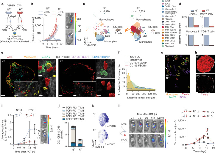
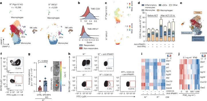
The tumour microenvironment is programmed by cancer cells and substantially influences anti-tumour immune responses1,2. Within the tumour microenvironment, CD8+ T cells undergo full effector differentiation and acquire cytotoxic anti-tumour functions in specialized niches3-7. Although interactions with type 1 conventional dendritic cells have been implicated in this process3-5,8-10, the underlying cellular players and molecular mechanisms remain incompletely understood. Here we show that inflammatory monocytes can adopt a pivotal role in intratumoral T cell stimulation. These cells express Cxcl9, Cxcl10 and Il15, but in contrast to type 1 conventional dendritic cells, which cross-present antigens, inflammatory monocytes obtain and present peptide-major histocompatibility complex class I complexes from tumour cells through 'cross-dressing'. Hyperactivation of MAPK signalling in cancer cells hampers this process by coordinately blunting the production of type I interferon (IFN-I) cytokines and inducing the secretion of prostaglandin E2 (PGE2), which impairs the inflammatory monocyte state and intratumoral T cell stimulation. Enhancing IFN-I cytokine production and blocking PGE2 secretion restores this process and re-sensitizes tumours to T cell-mediated immunity. Together, our work uncovers a central role of inflammatory monocytes in intratumoral T cell stimulation, elucidates how oncogenic signalling disrupts T cell responses through counter-regulation of PGE2 and IFN-I, and proposes rational combination therapies to enhance immunotherapies.


Conflict of interest statement

Competing interests: The laboratories of A.C.O. and J.Z. received research support and funding from Boehringer Ingelheim. J.Z. is a founder and scientific advisor of Quantro Therapeutics. G.C. has received grants from Celgene, Boehringer-Ingelheim, Roche, BMS, Iovance Therapeutics, and Kite Pharma. The institution that G.C. is affiliated with has received fees for his participation on an advisory board or for presentation at a company-sponsored symposium from Genentech, Roche, BMS, AstraZeneca, NextCure, Geneos Tx, and Sanofi/Avensis. G.C. has patents in the domain of antibodies and vaccines targeting the tumour vasculature, as well as technologies related to T cell expansion and engineering for T cell therapy. G.C. has received royalties from the University of Pennsylvania regarding CAR T cell technology. G.C. is a named inventor on patent applications filed by the Ludwig Institute for Cancer Research pertaining to the subject matter disclosed herein. D.D.L. has received a grant from Hoffmann-La Roche. All other authors declare no competing interests.

Figures

Fig. 1
Fig. 1. Tumour-infiltrating T cells are restimulated in permissive TMEs.
a, Schematic of subcutaneous injection of YUMM1.7OVA NTT cells or RTT cells in Rag2–/– mice and OT-1Luc ACT through intravenous (i.v.) injection. b, Left, ACT responses of NTT and RTT tumours (n = 5 mice per group). Right, representative bioluminescence imaging (BLI) pictures of T cells 96 h after ACT; key indicates radiance (photons per second that leave a square centimeter of tissue and radiate into a solid angle of one steradian). c, Uniform manifold approximation and projection (UMAP) maps of scRNA-seq of CD45+ cells in NTT and RTT tumours 72 h after ACT (n = 4 tumours pooled per group). d, Relative cell frequencies from scRNA-seq. e, Representative multiparameter IF images of YUMM1.7OVA NTT and RTT tumours 48 h after ACT (n  = 3 tumours per group). Scale bar, 400 µm (zoom-ins, 50 µm and 10 µm). f, Relative T cell frequency and distance to the next immune cell in NTT tumours (n = 3 tumours per group). g, Representative IF image of Nur77–GFP OT-1 cells in YUMM1.7OVA NTT and RTT tumours 48 h after ACT (n = 3 tumours per group). Scale bar, 10 µm. Arrows indicate Nur77+ OT-1 cells. h, Representative IF image of YUMM1.7OVA NTT and RTT tumours 72 h after ACT. Dashed lines depict the tumour border (n = 2 tumours per group). Scale bar, 500 µm. i, Left, quantification of T cells by BLI over time after intratumoral (i.t.) ACT (mean ± s.e.m., n = 4 mice per group). Two-way analysis of variance (ANOVA) with Sidak’s multiple comparisons test. Right, representative BLI image 96 h after ACT (key as in b). j, T cell states by flow cytometry 120 h after i.t. ACT (mean ± s.e.m., n = 5 NTT, n = 4 RTT tumours). k, Effector memory T cell signature on scRNA-seq of T cells (n = 14 NTT, n = 18 RTT pooled tumours). l, Left, representative BLI images of T cells in NTT and contralateral (CL) RTT tumours after i.t. ACT (key as in b). Right, growth curves (mean ± s.e.m., n = 6 mice per group). Arrows in b and l indicate day of ACT. Source Data
Fig. 2
Fig. 2. Inflammatory monocytes restimulate T cells within the TME.
a, Left, ACT response of YUMM1.7OVA NTT tumours in Rag2–/– mice (n = 4 mice) and Batf3–/–Rag2–/– (n = 5 mice). Right, representative BLI image of T cells 96 h after ACT (key as in Fig. 1b). b, Gene expression in individual clusters from scRNA-seq from Fig. 1c. c, Scoring of an ISG signature on our scRNA-seq data from Fig. 1c. Mod., module. d, Regulon specificity score of monocytes calculated using SCENIC in NTT tumours. e, Left, representative histograms depicting CFSE intensity. Right, quantification of T cell proliferation after 72 h of co-culture of naive CFSE-labelled T cells and inflammatory (Ly6A+) or non-inflammatory monocytes (Ly6A) isolated from NTT tumours in Rag2–/– mice (n = 4 tumours). Two-tailed unpaired Student’s t-test. f, Left, schematic of injection of YUMM1.7OVA NTT CTRL or B2m KO cells in BALB/c mice. Right, representative histograms depicting H2-Kb levels. g, Left, schematic of injection of NTT CTRL or NTT B2m KO tumours in Rag2–/– mice and Batf3–l–Rag2–/– mice. Middle, quantification of T cell infiltration by BLI. Right, representative BLI image of T cell infiltration 96 h after ACT (n = 5 NTT in Rag2–/– and n = 6 mice for other groups) (key as in Fig. 1b). One-way ANOVA with Tukey’s multiple comparisons test. h, Left, UMAP of human inflammatory macrophages from a melanoma scRNA-seq myeloid dataset. Right, enrichment score (ES) values of the inflammatory monocyte gene signature for each cell cluster. i, Top, representative field of view (FOV) of human metastatic melanoma (n = 2 of 72 FOVs) analysed by CosMx spatial transcriptomics profiling. Bottom, Pearson’s correlation values between cell types across FOVs (n = 72) were determined and displayed as a heatmap (n = 34 melanoma samples). The white arrow depicts activated CD8+ T cells, the black arrow depicts CXCL9+CXCL10+C1QC+ macrophages, and the red arrow CXCL9+CXCL10+ macrophages and DCs. Mac, macrophage. Bar graphs depict the mean ± s.e.m. Source Data
Fig. 3
Fig. 3. Cancer cells produce PGE2 and downregulate IFN-I responses to confer immunotherapy resistance.
a, Pathway enrichment analysis of differential gene expression in cancer cells isolated from YUMM1.7OVA NTT tumours and RTT tumours (n = 3 tumours per group; Supplementary Table 3). Adjusted P values were computed using the Benjamini–Hochberg correction. b, PGE2 ELISA of YUMM1.7OVA tumours in Rag2–/– mice (n = 10 NTT, n = 11 RTT CTRL, n = 7 RTT Ptgs1/2 KO over 2 independent experiments). c, Top, representative BLI image of T cells 96 h after ACT (key as in Fig. 1b). Bottom, BLI quantification (n = 4 mice NTT, n = 6 mice RTT CTRL and RTT Ptgs1/2 KO). d, Left, response of YUMM1.7OVA tumours in Rag2–/– mice to ACT (n = 7 mice per group). Right, YUMM3.3 in C57BL/6 mice (n = 5 mice per group). e, IFNβ ELISA of supernatants from YUMM1.7OVA NTT and RTT cells (n = 5 replicates per group over 2 independent experiments). f, Top, representative BLI images of T cells in YUMM1.7OVA RTT CTRL and RTT IRF3/7 tumours in Rag2–l– mice 96 h after ACT (key as in Fig. 1b). Bottom, BLI quantification (n = 6 mice per group). g, Left, response of YUMM1.7OVA tumours in Rag2–/– mice to ACT (n = 4 mice RTT CTRL, n = 5 mice RTT IRF3/7). Right, YUMM3.3 in C57BL/6 mice (n = 6 mice per group). h, Response to ACT of YUMM1.7OVA RTT Ptgs2 KO (left) and RTT IRF3/7 tumours (right) in Rag2–/– (n = 4 mice per group) and Batf3–/–Rag2–/– mice (n = 5 mice per group). i, PGE2 and IFNβ ELISAs of NTT and RTT A375 human melanoma (n = 2 replicates per group for PGE2, n = 3 tumours per group for IFNβ). Arrows in d, g and h indicate day of ACT. Bar graphs depict the mean ± s.e.m. Statistical analysis was performed with a two-tailed unpaired Student’s t-test (e,f,i) or one-way ANOVA with Tukey’s multiple comparisons test (b,c). Source Data
Fig. 4
Fig. 4. PGE2 and IFN-I responses determine myeloid cell abundance and their functional inflammatory state in the TME.
a, UMAP of scRNA-seq of CD45+ cells in YUMM1.7OVA RTT Ptgs1/2 KO and RTT IRF3/7 tumours 72 h after ACT (n = 3 tumours pooled per group). See Fig. 1c for cell cluster annotation. b, Scoring of the TME-COX and TME-IRF3/7 signatures (Supplementary Table 4) in myeloid fractions of responder (n = 6) and non-responder (n = 7) patients before TIL infusion. c, Scoring of an ISG signature in the scRNA-seq from a. d, Flow cytometry quantification of myeloid populations normalized to the CD45+ fraction from YUMM1.7OVA NTT tumours in Rag2–/– mice treated with anti-IFNAR1 or anti-IFNγ (n = 5 tumours per group, except n = 4 in anti-IFNAR1 + ACT and ant-IFNγ + ACT). e, RNA velocity of the MoMac compartment from RTT Ptgs1/2 KO tumours. f, Representative contour plots of Ly6C+ monocytes depicting Ly6A expression 72 h after i.t. transfer into NTT and RTT tumours in Rag2–/– mice (n = 5 tumours per group). g, Left, BLI quantification of T cells 96 h after ACT of YUMM1.7OVA RTT cells into Cd11ccre-Ptger2–/–Ptger4fl/fl mice (EP2/EP4 KO) or Cd11ccre (CTRL) mice (n = 7 mice per CTRL group and n = 8 mice per EP2/EP4 KO group); two-tailed Mann–Whitney U-test. Right, representative BLI image (key as in Fig. 1b). h, Representative contour plots of Ly6C+ monocytes depicting expression of Ly6A 48 h after treatment with CM from cancer cells with or without a COX1/2i (indomethacin) and with or without anti-ΙFNAR1 or isotype (n = 3 biological replicates). i, Heatmap of scaled ISG expression in BMDCs exposed to CM from NTT or RTT cells and with or without a COX1/2i in the presence of anti-ΙFNAR1 or isotype (n = 4 technical replicates) measured by RT–qPCR. j, Heatmap of scaled ISG expression in BMDCs treated with IFNβ and PGE2 measured by RT–qPCR (n = 4 technical replicates). Bar graphs depict the mean ± s.e.m. Source Data
Fig. 5
Fig. 5. Pharmacological modulation of PGE2 and IFN-I responses reinstates an immune-permissive TME and immunotherapy response.
a, Forest plot of pooled odds ratios and 95% CIs across clinical studies for overall response rates in patients receiving ICB with or without NSAID co-medication (n = 722 patients over 8 independent cohorts; Extended Data Fig. 9a and Supplementary Table 5). Statistical analysis was performed with a random effects model and data are presented as the mean ± 95% CI. b, UMAP of scRNA-seq of CD45+ cells of RTT CTRL and COX2i-treated YUMM1.7OVA RTT tumours 72 h after ACT (n = 3 tumours pooled per condition). See Fig. 1c for cell cluster annotation. c, Relative frequency of cell types across conditions as assessed by scRNA-seq. d, Left, treatment schedule of YUMM1.7OVA RTT tumours in Rag2–/– mice with celecoxib (COX2i) in combination with FLT3L or 5-AZA. Right, BLI quantification. Top, representative BLI images 72 h after ACT for vehicle (n = 8 mice), COX2i (n = 9 mice), COX2i + 5-AZA (n = 6 mice) and COX2i + FLT3L (n = 8 mice) groups (key as in Fig. 1b). Bar graphs depict the mean ± s.e.m. One-way ANOVA with Tukey’s multiple comparisons test. e, Left, Survival of Rag2–/– mice bearing YUMM1.7OVA RTT tumours treated with ACT and vehicle (n = 8 mice), COX2i (n = 9 mice), COX2i + 5-AZA (n = 7 mice) or COX2i + FLT3L (n = 8 mice). Right, survival of C57BL/6 mice bearing YUMM3.3 RTT tumours treated with an anti-PD1 and anti-CTLA4 with vehicle (n = 6 mice), COX2i (n = 8 mice), COX2i + FLT3L (n = 9 mice) or COX2i + 5-AZA (n = 9 mice). Log-rank Mantel–Cox test. Source Data
Extended Data Fig. 1
Extended Data Fig. 1. Characterization of immune permissive versus suppressive TMEs in mouse melanoma models.
a, Representative images of bioluminescent live imaging (BLI) of T cell infiltration after adoptive CD8+ OT-1Luc T cell transfer (ACT) in YUMM1.7OVA NTT and RTT tumors. b, Scheme outlining the in vitro killing of NTT and RTT cells with activated CD8+ OT-1 T cells (1:1 ratio) (left), histograms (flow cytometry) displaying MHCI levels of NTT and RTT cells (middle) and normalized ratio of 50:50 NTT/RTT cells mixed cultures in an in vitro killing assay with CD8+ OT-1 T cells. Normalization was performed with NTT and RTT percentages from the same 50:50 mixed cultures without CD8+ OT-1 T cells to account for proliferation differences (n = 3 biological replicates) (right). Two-tailed unpaired Student’s t-test. c, UMAP of scRNA-seq of CD45+ cells of YUMM1.7OVA NTT and RTT tumors in Rag2−/− before ACT (n = 3 tumors pooled/group). d, Relative frequency of cell types across conditions in YUMM1.7OVA tumors post-ACT (n = 4 tumors pooled/group). e, Expression of myeloid cell markers in each cluster (scRNA-seq). f, Heatmap of scaled gene expression (scRNA-seq) across cell clusters between NTT and RTT YUMM1.7OVA tumors post-ACT. g, Response to immune checkpoint blockade (ICB, anti-PD1/CTLA-4) of YUMM3.3 NTT and RTT tumors (n = 6 NTT, n = 7 NTT + ICB, n = 8 RTT, n = 9 RTT + ICB). Two-way ANOVA with Tukey’s multiple comparison. h, UMAP of scRNA-seq of CD45+ cells of YUMM3.3 NTT and RTT (n = 1 tumor/group). i, Relative frequency of cell types across conditions. j, Heatmap of scaled gene expression (scRNA-seq) across cell clusters between YUMM3.3 NTT and RTT. Bar graphs and growth curves depict the mean ± s.e.m. ns = not significant. Source Data
Extended Data Fig. 2
Extended Data Fig. 2. Permissive TMEs act as local reservoirs for T cell expansion.
a, Representative immunofluorescence (IF) of YUMM1.7OVA NTT and RTT tumors 48 h post-ACT(n = 3 tumors/group). Scale bars depicted in image. b, Quantification of immune populations based on IF staining (n = 3 tumors/group). c, Quantification of relative T cell frequency and distance to the tumor periphery in NTT and RTT tumors. Percentages of T cells found in the tumor border or tumor center are depicted (n = 3 tumors per condition). d, Quantification of Nur77+ OT-1 cells in YUMM1.7OVA NTT and RTT tumors 48 h post-ACT (n = 6 tumors for NTT, n = 5 for RTT). e, Average distances between Nur77+ OT-1 cells and cDC1s or monocytes based on IF stainings (n = 6 tumors for NTT, n = 5 for RTT). f, Quantification of CD8+ OT-1Luc T cell infiltration by BLI post-ACT in YUMM1.7OVA NTT and RTT in Rag2−/− mice treated with FTY720 (n = 5 mice/group). g, Quantification of T cell proliferation by CFSE dilution via flow cytometry 72 h post intratumoral (i.tu.) T cell injection into NTT and RTT tumors in Rag2−/− (n = 5 mice/group). h, Relative frequencies of T cell phenotypes (flow cytometry) after activation in vitro prior to ACT (n = 1, experiment repeated 3 times). i, Gating strategy for the phenotypic characterization of T cells. j, Relative frequencies of CD8+ T cell populations based on defined T cell signatures of i.tu. injected T cells isolated and subjected to scRNA-seq 5 days post-transfer (n = 14 NTT, n = 18 RTT pooled tumors). k, Projection of a progenitor exhausted T cell signature score and Cxcr6 gene expression on scRNA-seq from intratumoral T cells. l, Scheme outlining the contralateral (CL) injection of tumors in opposite flanks of the same mouse followed by intravenous (i.v) or i.tu. ACT in one of the tumors. m, Growth curves depicting the response to i.v ACT of primary or CL tumor in the same mouse (n = 4 mice NTT/NTT, n = 3 mice RTT/RTT and n = 6 mice NTT/RTT). n, Quantification of T cell infiltration by BLI in mice harboring CL NTT/NTT tumors (n = 6 mice), RTT/RTT tumors (n = 3 mice) or an NTT and RTT tumor (n = 10 mice) after i.v ACT. o, Representative BLI images from Extended Data Fig. 2m, n. p, Flow cytometry characterization of the TME of tumors in the CL setting (n = 3 mice NTT/NTT, n = 3 mice RTT/RTT, n = 5 mice NTT/RTT tumors). q, Quantification of T cell infiltration by BLI post-ACT in NTT and RTT in Rag2−/− mice treated with FTY720 (n = 7 mice/group). r, Response to ACT of mice bearing CL tumors (NTT/RTT) and treated with FTY720 or untreated (CTRL), (n = 7 mice/group). s, Response to ACT of Rag2−/− mice bearing CL tumors (NTT/NTT) with one of the NTT tumors injected i.tu (n = 6 mice). t, Response to ACT of Rag2−/− mice bearing CL tumors (NTT/RTT) with RTT tumors injected i.tu (n = 6 mice). u, Representative BLI images of T cell infiltration in mice harboring NTT/NTT, RTT/ RTT or RTT/ NTT tumors, with one of the tumors receiving i.tu. T cell transfer (n = 6 mice). Bar graphs and growth curves depict the mean ± s.e.m. Error bars in s and t not visible due to synchronous tumor regression. Statistical analysis was performed with a two-tailed unpaired student’s t-test in b, c, d, and g. A one-way ANOVA with Tukey’s multiple comparisons test in f, n, p and q. Two-way ANOVA with Tukey’s multiple comparisons test in m and r on day 17 pi. ns = not significant. Arrow indicates day of ACT. Source Data
Extended Data Fig. 3
Extended Data Fig. 3. Inflammatory monocytes cross-dress with cancer-derived pMHCI complexes.
a, Scheme outlining the dendritic cell (DC) vaccination of YUMM1.7OVA RTT tumors with bone marrow-derived cDC1s (BM-DCs) injected i.tu. and treated with ACT. b, Left, representative BLI images and right, quantification of T cell infiltration by BLI (n = 6 mice CTRL and n = 7 mice for DC vaccination). c, Response to ACT of RTT tumors vaccinated with BM-DCs followed by ACT. Black arrow indicates the day of ACT and gray arrows the days of cDC1 vaccination (n = 6 mice CTRL and n = 7 mice for DC vaccination). d, Representative IF staining of YUMM1.7OVA NTT and RTT tumors in Batf3−/ − Rag2−/− mice, 48 h post ACT, scale bar in NTT = 500 µm, zoom-ins=20 µm and 10 µm, scale bar in RTT = 1000 µm, (n = 3 tumors/group). e, Quantification of relative T cell frequency and distance to the tumor periphery in NTT and RTT tumors injected in Rag2−/− Batf3−/− mice. Percentages of T cells found in the tumor border or tumor center are depicted (n = 3 tumors/group). f, Quantification of relative T cell frequency and distance to the next immune cell in NTT tumors in Rag2−/− Batf3−/− mice (n = 3 tumors). g, Scheme outlining the depletion of dendritic cells with diphtheria toxin (DT) in NTT tumors in Rag2−/− mice reconstituted with bone marrow from Rag2−/− Zbtb46-DTR mice. DT was administered every 3 days starting on the day of tumor injection for the duration of the experiment. h, Contour plots depicting the frequency of dendritic cells (left) and flow quantification (flow cytometry) (n = 3 tumors/group). i, Representative BLI images (left) and quantification of T cell infiltration (n = 5 mice for untreated, n = 6 DT treated) (right). j, Scoring of a monocyte derived dendritic cell (MonoDC) signature in the YUMM1.7OVA scRNA-seq. k, Scoring of the Bosteels et al. ISG signature (Supplementary Table 2) in the YUMM1.7OVA scRNA-seq. l, Quantification of Nur77+ or Nur77- OT-1 T cells interacting with Ly6c+ monocytes based on IF staining of NTT tumors 48 h post-ACT (n = 6 tumors). m, Median fluorescence intensity (MFI) quantification of SIINFEKL-H2Kb staining (n = 6 mice/group) (left) and histograms (right). n, MFI quantification of NTT-derived H2-Kb on BALB/c pMHC-I cross-dressed monocytes (n = 6 mice/group) in NTT and NTT B2m KO. o, Scheme of the process of cross-dressing and in vitro T cell activation (left) and quantification of OT-1 T cell proliferation after 72 h of co-culture with monocytes FACS-sorted from YUMM1.7OVA NTT tumors in BALB/c mice (n = 3 technical replicates). Bar graphs depict the mean ± s.e.m. Statistical analysis was performed with a two-tailed unpaired student’s t-test in b, e, h, i, l, m, n and o. ns = not significant. Source Data
Extended Data Fig. 4
Extended Data Fig. 4. Inflammatory monocytes correspond to ISG+ and CXCL9+ macrophages in human data-sets.
a, Inflammatory monocyte and Monocyte 1 gene signature projection on a UMAP, and enrichment scores (ES) calculated with Gene Set Variance Analysis (GSVA) for different myeloid populations in a human melanoma scRNA-seq myeloid data-set (MEL) (Cheng et al.). b, Inflammatory monocyte and Monocyte 1 gene signature projection on a UMAP, and ES calculated with GSVA for different myeloid populations, in human non-small cell lung cancer (NSCLC) scRNA-seq myeloid data-set (Cheng et al.). c, Inflammatory monocyte and Monocyte 1 gene signature projection on a UMAP, and ES calculated with GSVA for different myeloid populations, in human melanoma scRNA-seq myeloid data-set (Barras et al.). See also Supplementary Table 2.
Extended Data Fig. 5
Extended Data Fig. 5. Oncogenic MAPK signaling counter-regulates the production of PGE2 and IFN-I.
a, Targeted metabolomic analysis of eicosanoids in YUMM1.7OVA NTT and RTT tumors isolated at day 10 post-injection from Rag2−/− mice (n = 3 tumors/group). b, Tumor growth of non-ACT RTT CTRL and RTT Ptgs1/2 KO (n = 7 mice/group) in Rag2−/− mice. c, Scheme outlining the injection of YUMM1.7OVA RTT Ptgs2 KO into Rag2−/− mice followed by ACT. d, Quantification of PGE2 by ELISA from YUMM1.7OVA RTT Ptgs2 KO tumors isolated at day 10 post-injection (n = 4 for RTT CTRL and n = 6 for RTT Ptgs2 KO, over 2 independent experiments). e, Survival of Rag2−/− mice harboringYUMM1.7OVA RTT CTRL or RTT Ptgs2 KO tumors upon ACT treatment (n = 5 mice/group). f, Quantification of PGE2 by ELISA from YUMM3.3 NTT and RTT tumors (n = 2 biological replicates). g, Tumor growth of YUMM3.3 RTT CTRL and RTT Ptgs2 KO (n = 5 mice/group) in Rag2−/− mice. h, Quantification of IFN-β by ELISA from YUMM1.7OVA NTT, RTT and RTT IRF3/7 tumors grown in Rag2−/− mice normalized to tumor weight (n = 4 tumors/group, pooled from 2 independent experiments). i, Upstream regulator analysis (Ingenuity) of differentially expressed genes in cancer cells sorted from RTT vs. NTT YUMM1.7OVA tumors grown in Rag2−/− mice. Benjamini-Hochberg correction for multiple testing. j, Tumor growth of non-ACT treated YUMM1.7OVA RTT CTRL (n = 5 mice) and RTT IRF3/7 (n = 4 mice) in Rag2−/− mice. k, Quantification of IFN-β by ELISA from YUMM3.3 NTT and RTT tumors grown in C57BL/6 mice normalized to tumor weight (n = 3 tumors/group). l, Heatmap of scaled ISG expression in YUMM1.7OVA RTT upon treatment with MEK inhibitor (MEKi) for 48 h. m, Western blot of RTT YUMM1.7OVA depicting COX2 protein levels upon treatment with RAF inhibitor (RAFi) or MEKi for 48 h. For gel source data see Supplementary Fig. 1. Experiment repeated 2 independent times. n, Heatmap of scaled gene expression in YUMM1.7OVA RTT cancer cells upon treatment with MEKi in vivo (n = 5 tumors per condition). o, ISG expression in YUMM1.7OVA RTT Ptgs2 KO cell line compared to NTT and RTT CTRL by RT-qPCR (n = 4 technical replicates). All expression values are depicted as the log2FC of the expression of each gene compared to NTT. p, Quantification of IFN-β by ELISA from YUMM1.7OVA RTT CTRL and RTT Ptgs2 KO tumors grown in Rag2−/− mice normalized to tumor weight (n = 6 tumors/group). q, Quantification of PGE2 by ELISA from YUMM1.7OVA RTT CTRL and RTT IRF3/7 tumors grown in Rag2−/− mice (n = 3 tumors/group). r, Quantification of IFN-β and PGE2 by ELISA from NTT and RTT variants of M249, LOX and H358 cell lines. For IFN-β analysis, tumors were grown in NSG mice and normalized to tumor weight (n = 3 tumors/group). For PGE2, supernatants from in vitro cell lines were analyzed (n = 2 biological replicates/group). s, Western blot of RTT, NTT and NTT NRAS variants of A375 human melanoma cell lines depicting COX2 protein levels upon treatment with RAFi for 48 h. For gel source data see Supplementary Fig. 1. Experiment performed once. Bar graphs depict the mean ± s.e.m. Statistical analysis was performed with a two-tailed student’s t-test in a, d, k, p, q and r and one-way ANOVA with Tukey’s multiple comparison in h. p-value in e was calculated using a Log-rank Mantel Cox test. ns = not significant. ND = Not detected. Source Data
Extended Data Fig. 6
Extended Data Fig. 6. High PGE2 and low IFN-I instruct an immune evasive TME.
a, Gating strategy for the identification of intratumoral myeloid populations. b, Flow cytometry myeloid characterization pre and 72 h post-ACT (n = 6 for RTT ± ACT, IRF37 RTT ± ACT, n = 5 for NTT ± ACT, RTT Ptgs2 KO + ACT, n = 4 RTT Ptgs2 KO). c, Flow cytometry characterization of dendritic cell populations pre and 72 h post-ACT, (n = 5 for NTT, RTT Ptgs2 KO ± ACT, IRF37 RTT + ACT, n = 6 for NTT + ACT, RTT ± ACT, IRF37 RTT). d, Relative frequency of cell types across conditions in YUMM1.7OVA (scRNA-seq), for RTT CTRL the RTT mCherry sample was used. e, Heatmap of scaled gene expression (scRNA-seq) between NTT, RTT CTRL, RTT Ptgs1/2 KO and RTT IRF3/7 for individual cell clusters. f, Flow cytometry characterization of SIINFEKL peptide on MHCI of dendritic cells isolated from tumors and pulsed ex vivo with SIINFEKL peptide (n = 6 tumors/group) and MFI quantification. g, Scheme outlining in vivo NK cell depletion in Rag2−/− mice harboring YUMM1.7OVA RTT Ptgs2 KO tumors. h, Representative plot depicting NK1.1+ cells in NK cell-depleted vs CTRL RTT Ptgs2 KO tumors measured by flow cytometry (left) and quantification of cDCs (n = 4 CTRL and n = 3 anti-NK1.1 treated tumors) (right). i, Representative BLI images (left) and quantification of CD8+ OT-1Luc T cell infiltration by BLI (right), (n = 3 mice for NTT, n = 5 mice for all other groups). j, Tumor response to ACT (n = 5 mice/group). Arrow indicates day of ACT. Bar graphs and growth curves depict the mean ± s.e.m. Data in i depicts the mean ± s.e.m. Statistical analysis was performed with a two-tailed unpaired student’s t-test in h. A one-way ANOVA with Tukey’s multiple comparison in f. Two-way ANOVA with Tukey’s multiple comparison in i. ns = not significant. Source Data
Extended Data Fig. 7
Extended Data Fig. 7. Type I and type II IFNs determine the inflammatory state.
a, b, Overall survival of melanoma patients stratified according to the TME-COX and TME-IRF3/7 signatures (Supplementary Table 4) (left) and correlation of intratumoral CD8+ T cell signature (right) in the Gide et al. RNA-seq data-set. PCC=Pearson correlation coefficient. c, TME-COX and TME-IRF3/7 signatures projected on the myeloid UMAP from baseline tumors (split for responder and non-responders to TIL therapy) from the Barras et al. patient cohort (above) and individual score across different myeloid populations (below). d, Gene set enrichment analysis (GSEA) of NTT and RTT tumors pre and post-ACT based on scRNA-seq analysis. Normalized enrichment score (NES). Multiple correction testing with false discovery rate. e, Pathway enrichment analysis of the top variable genes in the immune tumor microenvironment (TME) from NTT and RTT tumors pre and post-ACT analyzed by scRNA-seq. 1, 2 and 3 depict each gene cluster identified. Benjamini-Hochberg correction for multiple testing. f, TME characterization of YUMM1.7OVA RTT Ptgs2 KO tumors pre and 72 h post-ACT upon αIFN-γ and αIFNAR1 treatment (n = 5 tumors/group, except n = 4 for isotype and αIFNAR + ACT). g, Trajectory analysis and pseudotime of the MoMac compartment of YUMM1.7OVA NTT and RTT tumors. h, Density plots of F4/80+ macrophages after exposure of BM-derived monocytes to PGE2 for 48 h (left) and quantification (right) (n = 3 replicates per condition). i, Scheme outlining the intratumoral (i.tu.) transfer of CD45.1+ BM-derived monocytes into NTT and RTT tumors injected into CD45.2+ Rag2−/− mice (left) and inflammatory monocytes frequencies 3 days after transfer (right) (n = 5 tumors/group). j, Scheme outlining the knockout of EP2 (Ptger2) and EP4 (Ptger4) in OT-1 T cells and i.v. injection into YUMM1.7OVA RTT tumor-bearing Rag2−/− mice (left) and tumor growth (n = 6 mice/group) (right). Arrow indicates the day of ACT. k, Expression of Ptger2 and Ptger4 across populations from the YUMM1.7OVA scRNA-seq. Bar graphs depict the mean ± s.e.m. Statistical analysis was performed with a two-tailed unpaired student’s t-test in h and i. p-value in a and b was calculated using a Cox’s proportional hazards model (survival) and with a two-sided Pearson’s correlation (correlations). Source Data
Extended Data Fig. 8
Extended Data Fig. 8. PGE2 suppresses myeloid responsiveness to IFN-I.
a, Tumor growth of YUMM1.7 (left) and YUMM3.3 (right) RTT in Itgax-Cre (CTRL) (n = 7 mice for YUMM3.3 and n = 8 for YUMM1.7) or CD11cCre(Itgax-Cre)Ptger2−/−/Ptger4fl/fl treated with anti-CD8 to deplete CD8+ T cells (n = 5 mice for YUMM3.3 and n = 6 for YUMM1.7) or isotype control (n = 7 mice for YUMM3.3 and n = 10 for YUMM1.7). b, TME characterization (flow cytometry) of RTT tumors in CD11cCre-Ptger2−/−Ptger4fl/fl mice (KOEP2/EP4) or CD11cCre control mice (CTRL) (n = 8 tumors/group). c, Quantification of Ly6A+ cells after exposing BM-derived Ly6C+ monocytes to conditioned media (CM) from cancer cell lines ± COX1/2 inhibitor (COX1/2i, indomethacin) during media conditioning ± αΙFNAR1 during 48 h of culture (n = 3 biological replicates per condition). d, MFI quantification of AXL, MHC-I and CD86 (flow cytometry) of BM-DCs treated with fresh media (C) or CM from NTT, RTT and RTT IRF3/7 cells for 24 h ± αΙFNAR1 or isotype control (n = 3 biological replicates per condition). e, Heatmap of scaled expression of ISGs in BM-DCs exposed to CM from RTT IRF3/7 treated with COX1/2i (indomethacin) or untreated, in the presence of αΙFNAR1 or isotype control measured by RT-qPCR (n = 4 technical replicates). f, Heatmap of scaled ISG expression in BM-DCs exposed to CM from NTT and RTT treated with MEKi or untreated, in the presence of αΙFNAR1 or isotype control (n = 4 technical replicates) measured by RT-qPCR. g, Scheme outlining the culture of human monocyte models to CM from cancer cell lines ± COX1/2i during media conditioning (left) and heatmaps of scaled ISG expression (right). h, Quantification of AXL+ BM-DCs treated with IFN-β and PGE2 (flow cytometry) (left) (n = 3 biological replicates) and heatmap of scaled ISG expression measured by RT-qPCR (right) (n = 4 technical replicates). Bar graphs and growth curves depict the mean ± s.e.m. Statistical analysis were performed with a two-way ANOVA with Tukey’s multiple comparisons test in a, a two-tailed unpaired student’s t-test (APCs, cDC1 and infl.monocytes) and two-tailed Mann Whitney U (total cDCs and infl.cDC2s) in b, one-way ANOVA with Tukey’s multiple comparison in c, d and h. ns = not significant. Source Data
Extended Data Fig. 9
Extended Data Fig. 9. Meta-analysis of retrospective clinical studies.
a-c, Forest plot of pooled odds ratios (ORs) and hazard ratios (HRs) with 95% confidence intervals (CI) across included studies of patients receiving ICB with and without concomitant non-steroidal anti-inflammatory drugs (NSAIDs) medication. Number of participants are included in each figure panel. a, Odds ratios comparing overall response rates. b, Hazard ratios comparing progression-free survival. c, Hazard ratios comparing overall survival. I2: I-squared; P: probability. d, PRISMA flow diagram of literature search and study selection process for meta-analysis. Statistical analysis was performed with a random effects model, data are presented as mean values ± 95% confidence interval. See also Supplementary Tables 5 and 6.
Extended Data Fig. 10
Extended Data Fig. 10. Pharmacological inhibition of PGE2 and type I IFN modulators resensitize cross-resistant tumors to immunotherapy.
a, Relative frequency of each cell type across conditions (scRNA-seq). b, Scoring of the Duong et al. inflammatory signature in RTT COX2i-treated tumors. c, Heatmap of scaled gene expression (scRNA-seq) in YUMM1.7 OVA RTT vehicle-treated tumors (CTRL) and RTT celecoxib (COX2i)-treated tumors (n = 3 tumors were pooled/group). d, Response of YUMM1.7OVA RTT tumors to vehicle + adoptive T cell transfer (ACT) (n = 7 mice), celecoxib (n = 4 mice), celecoxib + ACT (n = 6 mice) and etoricoxib ± ACT (n = 7 mice/group). Red arrow and box indicate COX2i treatment stop. e, UMAP of scRNA-seq of CD45+ cells from YUMM1.7OVA RTT mice treated with vehicle or COX2i + 5-AZA isolated 72 h post-ACT (n = 3 tumors pooled/group). f, Relative frequency of each cell type across conditions in e (scRNA-seq). g, Scoring of the Duong et al. inflammatory signature in COX2i + 5-AZA treated tumors. h, Response of YUMM1.7OVA RTT tumors to 5-azacytidine (5-AZA) (n = 4 mice), 5-AZA + ACT (n = 7 mice), vehicle+ACT (n = 8 mice), Flt3L+ACT (n = 7 mice), COX2i+ACT (n = 9 mice), COX2i + 5-AZA + ACT (n = 7 mice) and COX2i+Flt3L+ACT (n = 8 mice). i, Representative image of IF staining of vehicle-treated and COX2i+Flt3L RTT YUMM1.7OVA tumors 96 h post-ACT (n = 2 tumors per condition). Scale bar = 1000 µm, zoom-in = 50 µm. Black arrows indicates day of ACT. Source Data
Extended Data Fig. 11
Extended Data Fig. 11. Combination therapies in melanoma, lung, pancreatic and colorectal mouse cancer models.
a, Scheme outlining the re-injection of YUMM1.7OVA RTT cells into tumor-free responder mice from Fig. 5e (left) and tumor growth of re-challenged mice (n = 4 mice for COX2i + Ftl3L, n = 3 COX2i and n = 2 COX2i + 5-AZA) or naive control mice (n = 5 mice). b, Scheme outlining the injection of YUMM3.3 RTT tumors into C57BL/6 mice and the treatment regimen with COX2i, Flt3L, 5-AZA and anti-PD1/CTLA-4 (ICB) or isotype control. c, Response to control vehicle treatment or vehicle + ICB treatment of YUMM3.3 RTT tumors (n = 6 mice). d, Response to single treatments and ICB combination treatments of YUMM3.3 RTT tumors. COX2i, Flt3L, 5-AZA + ICB, COX2i+ICB and Flt3L+ICB (n = 8 mice), COX2i + 5-AZA + ICB and COX2i+Flt3L+ICB (n = 10 mice). e, Survival plot. f, Scheme outlining the injection of CT-26 RTT tumors into BALB/c mice and the treatment regimen with COX2i, Flt3L, 5-AZA and anti-PD1 (ICB) or isotype control. g, Response to treatment of CT26 RTT tumors. Vehicle and Vehicle+ICB (n = 8 mice), COX2i + 5-AZA + ICB and COX2i+Flt3L+ICB (n = 9 mice). h, Survival plot. i, Scheme outlining the intrapancreatic injection of EPP2Luc cells into C57BL/6 mice and the treatment regimen with COX2i, Flt3L and anti-PD1 (ICB) or isotype control. j, Left, representative BLI images of EPP2Luc tumor burden and right, quantification of control (n = 5 mice), anti-PD1 (n = 6 mice), anti-PD1 + COX2i + Ftl3L (n = 6 mice). k, Survival plot of j. l, Scheme outlining the injection of KPAR tumors into C57BL/6 mice and the treatment regimen with COX2i, Flt3L and anti-PD1 (ICB) or isotype control. m, Response to treatment of KPAR tumors, vehicle ± ICB (n = 6 mice) and ICB + COX2i + Ftl3L (n = 8 mice). n, Survival plot of m. o, Scheme summarizing the role of inflammatory monocytes in T cell restimulation and the functional convergence of PGE2 and IFN-I to determine an inflammatory TME, T cell restimulation and immunotherapy response. Bar graphs depict the mean ± s.e.m. Statistical analysis was performed with a one-way ANOVA with Tukey’s multiple comparisons test in j. Log-rank Mantel Cox test in h, k, n. ns = not significant. Source Data

References

    1. Binnewies, M. et al. Understanding the tumor immune microenvironment (TIME) for effective therapy. Nat. Med.24, 541–550 (2018). - PMC - PubMed
    1. Spranger, S. & Gajewski, T. F. Impact of oncogenic pathways on evasion of antitumour immune responses. Nat. Rev. Cancer18, 139–147 (2018). - PMC - PubMed
    1. Di Pilato, M. et al. CXCR6 positions cytotoxic T cells to receive critical survival signals in the tumor microenvironment. Cell184, 4512–4530.e22 (2021). - PMC - PubMed
    1. Magen, A. et al. Intratumoral dendritic cell–CD4+ T helper cell niches enable CD8+ T cell differentiation following PD-1 blockade in hepatocellular carcinoma. Nat. Med.10.1038/s41591-023-02345-0 (2023). - PMC - PubMed
    1. Chen, J. H. et al. Human lung cancer harbors spatially organized stem-immunity hubs associated with response to immunotherapy. Nat. Immunol.25, 644–658 (2024). - PubMed

MeSH terms

LinkOut - more resources