

Ultrasound diagnosis of first trimester umbilical cord entanglement in monochorionic monoamniotic twins - case report and review of the literature
- PMID: 39604909
- PMCID: PMC11603871
- DOI: 10.1186/s12884-024-06962-6
Ultrasound diagnosis of first trimester umbilical cord entanglement in monochorionic monoamniotic twins - case report and review of the literature
Abstract
Background: Diagnosis of umbilical cord entanglement (UCE) by ultrasound (US) in monochorionic monoamniotic (MCMA) twins in the second and third trimesters is common. However, only a few cases have been reported on the diagnosis of UCE as early as the first trimester. Herein, we report a case of the earliest-ever sonographic diagnosis of UCE and demonstrate the feasibility of its diagnosis by US.
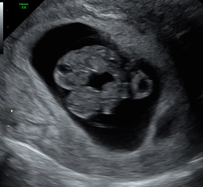
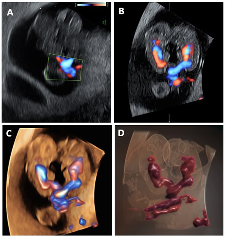
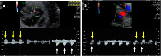
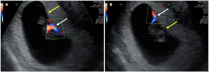
Case presentation: A 32-year-old gravida 2 para 1 woman conceived after assisted reproductive technology (ART) treatment. In transvaginal US examination at 8.5 gestational weeks, two embryos with regular heartbeats in the same amniotic sac and with only one yolk sac were demonstrated. The fetal crown-rump lengths were 20 and 21 mm, appropriate for 8.4 and 8.5 gestational weeks, respectively. HD-flow power Doppler 2D and 3D US demonstrated two tightly entangled umbilical cords of the two fetuses. Spectral Doppler US showed two different heart rates (162 and 167 beats per minute) and blood flow in opposite directions from the point of entanglement of the two umbilical cords. This was consistent with a diagnosis of a first-trimester MCMA pregnancy with UCE. Missed abortion of the two embryos was diagnosed by US examination at 10.5 weeks, and the pregnancy was terminated by dilatation and curettage without further complications.
Conclusions: UCE in the first trimester may occur as early as eight gestational weeks, and its diagnosis by ultrasound is feasible. UCE diagnosed in the first trimester may be a poor prognostic factor.
Keywords: Entanglement; First trimester; Monochorionic-monoamniotic; Twin; Umbilical cord.
© 2024. The Author(s).
Conflict of interest statement
Declarations. Ethics approval and consent to participate: Not applicable. Consent for publication: Written informed consent was obtained from the patient for publication of this case report. Competing interests: The authors declare no competing interests.
Figures
References
-
- Van Mieghem T, Abbasi N, Shinar S, Keunen J, Seaward G, Windrim R, Ryan G. Monochorionic monoamniotic twin pregnancies. Am J Obstet Gynecol MFM. 2022;4(2S):100520. 10.1016/j.ajogmf.2021.100520. Epub 2021 Oct 30. PMID: 34728404. - PubMed
-
- Khairudin D, Khalil A. Monochorionic monoamniotic twin pregnancies. Best Pract Res Clin Obstet Gynecol. 2022;84:96–103. 10.1016/j.bpobgyn.2022.08.004. Epub 2022 Aug 24. PMID: 36123247. - PubMed
-
- Glinianaia SV, Rankin J, Khalil A, Binder J, Waring G, Sturgiss SN, Thilaganathan B, Hannon T. Prevalence, antenatal management and perinatal outcome of monochorionic monoamniotic twin pregnancy: a collaborative multicenter study in England, 2000–2013. Ultrasound Obstet Gynecol. 2019;53(2):184–92. Epub 2018 Dec 30. PMID: 29900612. - PubMed
-
- Hack KE, van Gemert MJ, Lopriore E, Schaap AH, Eggink AJ, Elias SG, van den Wijngaard JP, Vandenbussche FP, Derks JB, Visser GH, Nikkels PG. Placental characteristics of monoamniotic twin pregnancies in relation to perinatal outcome. Placenta. 2009;30(1):62–5. Epub 2008 Nov 17. PMID: 19010539. - PubMed
-
- Van Mieghem T, De Heus R, Lewi L, et al. Prenatal management of monoamniotic twin pregnancies. Obstet Gynecol. 2014;124:498–506. - PubMed
Publication types
MeSH terms
LinkOut - more resources
Full Text Sources
