

Head-to-head comparison of tau PET tracers [18F]PI-2620 and [18F]RO948 in non-demented individuals with brain amyloid deposition: the TAU-PET FACEHBI cohort
- PMID: 39605030
- PMCID: PMC11603960
- DOI: 10.1186/s13195-024-01622-5
Head-to-head comparison of tau PET tracers [18F]PI-2620 and [18F]RO948 in non-demented individuals with brain amyloid deposition: the TAU-PET FACEHBI cohort
Abstract
Background: Second-generation tau tracers for positron emission tomography (PET) show high affinity for paired helical filaments tau deposits characteristic of Alzheimer´s disease and low off-target binding. Differences in their chemical structure though may lead to variations in their regional tau uptake and off-target signal. In this work, we aimed to compare the in-vivo uptake of tau tracers [18F]PI-2620 and [18F]RO948 in the early stages of the AD continuum.
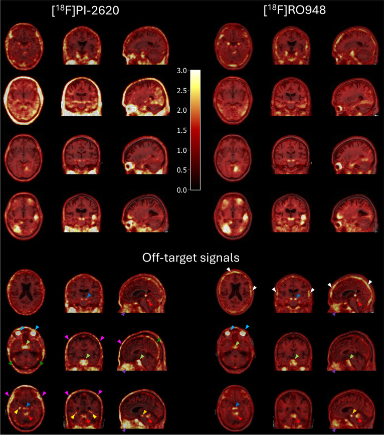
Methods: Data from the TAU-PET FACEHBI clinical trial (EUDRA-CT 2021-000473-83) were analyzed. All participants were non-demented and underwent tau imaging with [18F]PI-2620 and [18F]RO948 PET within 3 months, amyloid imaging with [18F]Florbetaben and brain magnetic resonance imaging. Tau PET standardized uptake values ratios (SUVR) were calculated in Braak and typical off-target regions using the inferior cerebellar cortex as a reference region.
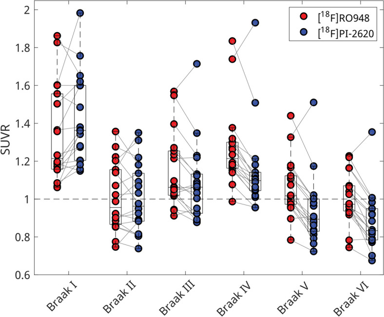
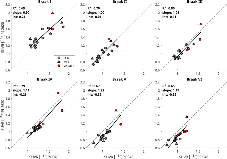
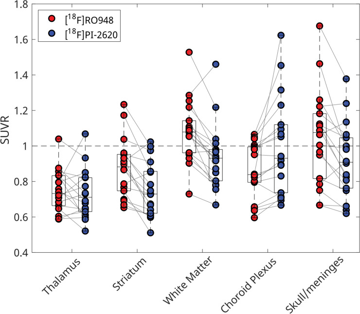
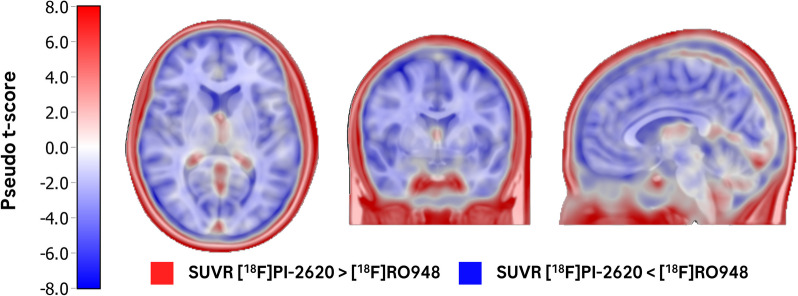
Results: The cohort consisted of 18 individuals with subjective cognitive decline (n = 13) and mild cognitive impairment (n = 5), with centiloid values ranging from 17 to 159. Both tau tracers showed similar tau pathology distribution but presented a distinct off-target signal pattern on visual read. SUVR measurements for [18F]PI-2620 and [18F]RO948 were highly correlated in all Braak regions (R2 range [0.65-0.80]). Regarding off-target signal, [18F]PI-2620 had higher SUVRs in vascular structures, and [18F]RO948 had higher SUVRs in the skull/meninges.
Conclusions: In a cohort of individuals at early stages of the AD continuum, tau PET tracers [18F]PI-2620 and [18F]RO948 showed similar in-vivo uptake in all Braak regions and distinct off-target signal. These preliminary results support the development of standardized quantification scales for tau deposition that are tracer-independent.
Trial registration: AEMPS EudraCT 2021-000473-83. Registered 30 December 2021.
Keywords: Alzheimer; FACEHBI; Mild cognitive impairment; Positron emission tomography; Subjective cognitive decline; Tau; [18F]PI-2620; [18F]RO948.
© 2024. The Author(s).
Conflict of interest statement
Declarations. Ethics approval and consent to participate: The study was conducted in accordance with the Declaration of Helsinki. The TAU-PET FACEHBI protocol received the approval from the Spanish Drug Agency (AEMPS for its initials in Spanish) and was registered as phase II clinical trial (CT) (EudraCT: 2021-000473-83, approval date December 15th 2021) and by the Ethics Committee of Hospital Universitari de Bellvitge in Hospitalet de Llobregat, Spain. The FACEHBI-2 protocol (which includes study v5) was approved by the Ethics Committee of Hospital Clínic i Provincial in Barcelona, Spain. Informed consent was obtained from all participants involved in the study. Consent for publication: The authors affirm that human research participants provided informed consent for publication of the images in Fig. 2. Competing interests: MB has consulted for Araclon, Avid, Grifols, Lilly, Nutricia, Roche, Eisai and Servier. She received fees from lectures and funds for research from Araclon, Biogen, Grifols, Nutricia, Roche and Servier. She reports grants/research funding from Abbvie, Araclon, Biogen Research Limited, Bioiberica, Grifols, Lilly, S.A, Merck Sharp & Dohme, Kyowa Hakko Kirin, Laboratorios Servier, Nutricia SRL, Oryzon Genomics, Piramal Imaging Limited, Roche Pharma SA, and Schwabe Farma Iberica SLU, all outside the submitted work. She has not received personal compensations from these organizations. AR is member of scientific advisory board of Landsteiner Genmed and Grifols SA. AR has stocks of Landsteiner Genmed. MM has consulted for F. Hoffmann-La Roche Ltd and is a member of the Scientific Advisory Board of Biomarkers of Araclon. MT, EB, CG, GK are full-time employees, and owns stock or stock options, of F. Hoffmann-La Roche Ltd. NRV, SB, EPM, ES are full-time employees of Life Molecular Imaging GmbH. The rest of authors declare that they have no competing interests.
Figures


References
-
- Braak H, Braak E. Neuropathological stageing of Alzheimer-related changes. Acta Neuropathol. 1991;82:239–59 Springer-Verlag. - PubMed
Publication types
MeSH terms
Substances
LinkOut - more resources
Full Text Sources
Research Materials
Miscellaneous
