[Preprint]. 2024 Nov 11:2024.11.11.621817.
doi: 10.1101/2024.11.11.621817.

In vivo Treatment of a Severe Vascular Disease via a Bespoke CRISPR-Cas9 Base Editor

Affiliations

In vivo Treatment of a Severe Vascular Disease via a Bespoke CRISPR-Cas9 Base Editor

Christiano R R Alves et al. bioRxiv. .

Update in

  • Treatment of a severe vascular disease using a bespoke CRISPR-Cas9 base editor in mice.
    Alves CRR, Das S, Krishnan V, Ha LL, Fox LR, Stutzman HE, Shamber CE, Kalailingam P, McCarthy S, Lino Cardenas CL, Fong CE, Imai T, Mitra S, Yun S, Wood RK, Benning FMC, Roh K, Lawton J, Kim N, Silverstein RA, Ferreira da Silva J, de la Cruz D, Richa R, Xie J, Gray-Edwards HL, Malhotra R, Chung DY, Chao LH, Tsai SQ, Maguire CA, Lindsay ME, Kleinstiver BP, Musolino PL. Alves CRR, et al. Nat Biomed Eng. 2025 Sep 11. doi: 10.1038/s41551-025-01499-1. Online ahead of print. Nat Biomed Eng. 2025. PMID: 40935887

Abstract

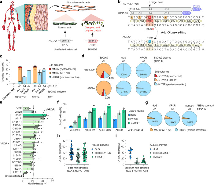
Genetic vascular disorders are prevalent diseases that have diverse etiologies and few treatment options. Pathogenic missense mutations in the alpha actin isotype 2 gene (ACTA2) primarily affect smooth muscle cell (SMC) function and cause multisystemic smooth muscle dysfunction syndrome (MSMDS), a genetic vasculopathy that is associated with stroke, aortic dissection, and death in childhood. Here, we explored genome editing to correct the most common MSMDS-causative mutation ACTA2 R179H. In a first-in-kind approach, we performed mutation-specific protein engineering to develop a bespoke CRISPR-Cas9 enzyme with enhanced on-target activity against the R179H sequence. To directly correct the R179H mutation, we screened dozens of configurations of base editors (comprised of Cas9 enzymes, deaminases, and gRNAs) to develop a highly precise corrective A-to-G edit with minimal deleterious bystander editing that is otherwise prevalent when using wild-type SpCas9 base editors. We then created a murine model of MSMDS that exhibits phenotypes consistent with human patients, including vasculopathy and premature death, to explore the in vivo therapeutic potential of this base editing strategy. Delivery of the customized base editor via an engineered SMC-tropic adeno-associated virus (AAV-PR) vector substantially prolonged survival and rescued systemic phenotypes across the lifespan of MSMDS mice, including in the vasculature, aorta, and brain. Together, our optimization of a customized base editor highlights how bespoke CRISPR-Cas enzymes can enhance on-target correction while minimizing bystander edits, culminating in a precise editing approach that may enable a long-lasting treatment for patients with MSMDS.

Keywords: ACTA2; CRISPR-Cas; MDMS; MSMDS; aortic aneurysm; base editing; bystander editing; genome editing; moyamoya disease; pediatric disease; vasculopathies.


Conflict of interest statement

Competing interests C.L.C., R.M., C.A.M., D.Y.C., B.P.K., M.E.L., and P.L.M. are inventors on a patent application filed by Mass General Brigham (MGB) that describes the development of genome editing technologies to treat MSMDS. C.R.R.A, R.A.S., J.F.dS., and B.P.K. are inventors on additional patents or patent applications filed by MGB that describe genome engineering technologies. S.Q.T. is an inventor on a patent covering CHANGE-seq. S.Q.T. is a member of the scientific advisory board of Prime Medicine and Ensoma. R.M., D.Y.C., C.A.M., B.P.K., M.E.L., and P.M.M received sponsored research support from Angea Biotherapeutics, a company developing gene therapies for vasculopathies. R.M. receives research funding from Amgen and serves as a consultant for Pharmacosmos, Myokardia/BMS, Renovacor, Epizon Pharma, and Third Pole and performs speaker bureaus through Vox Media, all of which are unrelated to the current work. C.A.M. has financial interests in Chameleon Biosciences, Skylark Bio, and Sphere Gene Therapeutics, companies developing Adeno Associated Virus (AAV) vector technologies for gene therapy applications. C.A.M. performs paid consulting work for all three companies. C.A.M.’s interests were reviewed and are managed by Massachusetts General Hospital and Mass General Brigham in accordance with their conflict-of-interest policies. B.P.K. is a consultant for EcoR1 capital, Novartis Venture Fund, and Jumble Therapeutics, and is on the scientific advisory boards of Acrigen Biosciences, Life Edit Therapeutics, and Prime Medicine. B.P.K. has a financial interest in Prime Medicine, Inc., a company developing therapeutic CRISPR-Cas technologies for gene editing. B.P.K.’s interests were reviewed and are managed by MGH and MGB in accordance with their conflict-of-interest policies. The other authors declare no competing interests.

Figures

Figure 1.
Figure 1.. Development of a bespoke adenine base editor to correct ACTA2 R179H.
a, Schematic of multisystemic smooth muscle dysfunction syndrome (MSMDS) caused by an ACTA2 R179H mutation. b, Schematic of the genomic region surrounding ACTA2 R179H with base editor guide RNA (gRNA) target sites shown; potential bystander edits are shown in orange boxes. c, A-to-G base editing to correct ACTA2 R179H in homozygous HEK 293T cells when using ABEs comprised of deaminase domains ABE8.20m and ABE8e fused to WT SpCas9 (with gRNA A8), or PAM variant SpCas9 enzymes SpG or SpCas9-VRQR (with gRNA A4). Base edited alleles assessed by targeted sequencing and analyzed via CRISPResso2. d, Fraction of reads with precise ACTA2 H179R correction with or without the M178V bystander edit, analyzed from data in panel c. e, Modified reads at the ACTA2 R179H target site in homozygous HEK 293T cells when using SpCas9-VRQR nuclease variant enzymes harboring amino acid substitutions to potentiate on-target activity, assessed by targeted sequencing. All conditions utilized ACTA2 R179H gRNA A4; eVRQR, enhanced SpCas9-VRQR. f, A-to-G base editing to correct ACTA2 R179H in homozygous HEK 293T cells when using ABEs including ABEmax,, ABE8.8m, ABE8.20m, or ABE8e fused to SpG, SpCas9-VRQR, or eVRQR when paired with ACTA2 R179H gRNA A4. g, Fraction of reads with precise ACTA2 H179R correction with or without the M178V bystander edit, analyzed from data in panel f. h,i, Summary of A-to-G base editing in HEK 293T cells with SpG, SpCas9-VRQR, or eVRQR ABE8e constructs when using gRNAs targeting sites with canonical PAMs (NGAN or NGNG; panel h) or non-canonical PAMs (NGBN or NGNH; panel I) for SpCas9-VRQR or eVRQR. B = C, G, or T; H = A, C, or T. For data in panels c-h, mean, s.e.m., and individual datapoints shown from experiments with between n = 3 to 6 independent biological replicates.
Figure 2.
Figure 2.. Analysis base editing specificity to correct ACTA2 R179H.
a, Schematic of potential ACTA2 bystander edits induced by adenine base editors (ABEs) paired with ACTA2 R179H gRNA A4. b, HDAC9 transcript levels assessed by RT-qPCR in human smooth muscle cells (SMCs) following transduction with lentiviral vectors that express ACTA2 variant cDNAs harboring each mutation indicated in panel A (see also Sup. Fig. 5a); wild-type (WT) ACTA2 cDNA in blue, MSMDS-causative ACTA2 R179H cDNA in red, and cDNAs encoding bystander edits in orange. c, Number of putative off-target sites in the human genome with up to 3 mismatches for the spacers of gRNAs A4 with NGAN and NGNG PAMs, A7 with NGNN PAMs, and A8 with NGGN, NAGN and NGAN PAMs, annotated by CasOFFinder. d, Total number of GUIDE-seq2-detected off-target sites when using wild-type (WT) SpCas9 nuclease with gRNA A8 or eVRQR with gRNA A4. e, Percentage of total GUIDE-seq reads attributable to the on-target site or cumulative off-target sites. f, Total number of CHANGE-seq-BE detected off-target sites with ABE8e-WT with gRNA A8 or ABE8e-eVRQR with gRNA A4, performed using genomic DNA from patients with MSMDS. g, Number of CHANGE-seq-BE identified off-target sites that account for greater than 1% of total reads and are common across experiments performed using genomic DNA extracted from fibroblasts from 3 independent patients with MSMDS. h, Percentage of CHANGE-seq-BE reads detected at the on-target site relative to the total number of reads in each experiment. i, Venn diagram of nominated off-target sites with eVRQR and gRNA A4, between in silico CasOFFinder nomination, GUIDE-seq2 (performed via plasmid expression of nucleases in cells), or CHANGE-seq-BE (performed in vitro using ABE8e-VRQR protein). j, Summary of the levels of on- and off-target base editing in homozygous HEK 293T ACTA2 R179H cells that were untreated (naïve) or treated with ABE8e-eVRQR and gRNA A4. Genomic DNA was subjected to rhAmpSeq for the on-target site and 121 off-target sites (nominated by CasOFFinder, GUIDE-seq2, or CHANGE-seq-BE assays) with data analysis via CRISPResso2 for n = 3 independent biological replicates; base editing efficiencies plotted for only the most edited base for each target site, typically an adenine in the middle of the edit window. For panels b and f-h, mean, s.e.m, and individual datapoints shown for n = 3 independent biological replicates.
Figure 3.
Figure 3.. Development and characterization of an Acta2 R179H mouse model.
a, Schematic of the Acta2 locus for wild-type mice, control mice (Acta2fl/+), and MSMDS mice (Acta2fl/+ / Myh11Cre+). Expression of Cre recombinase in smooth muscle cells of Acta2fl/+ mice crossed with Myh11-Cre mice via the Myh11 promoter excises the exon 5–9 and Neo’ cassette to enable expression of the mutant R179H allele. SA, splice acceptor; ex, exon; rBH pA, Rabbit β-globin polyadenylation signal. b-e, Characterization of phenotypes in MSMDS mice (Myh11-Cre:Acta2fl) when compared to control mice, including survival (panel b), weight gain (panel c), exercise performance as demonstrated by latency to fall via rotarod assay (panel d), and distance traveled in open field testing (panel e). f, Comparison of aortic diameter between MSMDS and control mice at 4–5 weeks of age. Log-rank test revealed a significant difference (P < 0.001) between treated and untreated mice in panel b. Repeated measurement ANOVA revealed significant difference (P < 0.01) between control and MSMDS mice in panels c-e. T-test revealed significant difference (***P <0.001 and ** P <0.01) in panel f. Mean and s.e.m. shown in panels c-f. Sample size indicated in panels c-f with indication of living mice under each time point, and individual datapoints shown in panel f.
Figure 4.
Figure 4.. In vivo correction of Acta2 R179H in MSMDS mice.
a, Schematic of plasmid transfection experiments in HEK 293T ACTA2 R179H cells to compare conventional ABE and gRNA expression plasmids to ITR-containing intein-split AAV production plasmids for ABE8e-eVRQR and gRNA A4. TadA8e, TadA domain from ABE8e; Npu(N), N-terminal DnaE intein from Nostoc punctiforme (Npu); Npu(C), C-terminal Npu intein; ITR, inverted terminal repeat; Cas9(S55R)(N), residues 1–573 of nSpCas9(D10A/S55R); VRQR(C), residues 574–1,368 of SpCas9-VRQR. b, A-to-G base editing to correct the ACTA2 R179H mutation in HEK 293T R179H cells via plasmid delivery. Editing assessed by targeted sequencing; mean, s.e.m., and individual datapoints shown for n = 3 independent biological replicates. c, Schematic of P3 intravenous (IV) injections of dual AAV-PR-ABE or AAV9-ABE vectors that express intein-split ABE8e-VRQR(S55R) and gRNA A4 into in MSMDS and control mice. Mice were sacrificed and tissues were harvested 7–8 weeks after injections. d-g, A-to-G editing to correct Acta2 R179H following IV injections of dual AAV9 or AAV-PR vectors encoding ABE8e-eVRQR with gRNA A4 (AAV-ABE), with analysis of editing in the liver (panel d), heart (panel e), brain vasculature (panel f), or aorta (panel g). h, Summary of bystander editing or insertion or deletion mutations (indels) following P3 IV injections of AAV-PR encoding ABE8e-eVRQR with gRNA A4 into MSMDS or control mice, with editing evaluated by targeted sequencing of genomic DNA from the liver, heart, brain vasculature, or aorta. Mean, s.e.m. and individual datapoints shown in panels d-h.
Figure 5.
Figure 5.. Phenotypic changes in MSMDS mice following AAV-mediated delivery of ABEs.
Analyses were performed with untreated control mice (Acta2fl/+), untreated MSMDS mice (Acta2fl/+ / Myh11Cre+), and MSMDS mice treated with AAV-PR or AAV9 to deliver ABE8e-eVRQR and gRNA-A4 (AAV-ABE) at P3. a,b, Survival of mice at 8 weeks of age (panel a) or until natural death (panel b). c-f, Characterization of body mass (panel c), rotarod performance (panel d), distance traveled in open field testing (panel e), and aortic diameter (panel f). g, Representative images of cerebral arteries from untreated control, MSMDS, and AAV-ABE treated MSMDS mice. Sections were immunostained for smooth muscle actin (SMA) using DAB staining to visualize vascular structures. Fixed tissues were sectioned and imaged with a Zeiss LSM 800 microscope. Scale bar: 20 μm. h-i, Calculation of the mean normalized tortuosity index (panel h) and SMA intensity (panel i) in cerebral arteries. For tortuosity, the inner and outer perimeters of vessels were outlined using ImageJ/FIJI, with the index determined by dividing the inner perimeter by the outer perimeter. SMA intensity was measured by selecting regions of interest (ROIs) and calculating the mean pixel intensity within these regions using ImageJ/FIJI to compare SMA labeling across samples. j, Evaluation of neurovascular anatomy via representative coronal sections from untreated control, mutant, and treated MSMDS mice immunostained for myelin basic protein (MBP) using DAB staining and visualized with a Zeiss LSM 800 microscope. Scale bar: 500 μm. k, Cortical myelination assessed by measuring the total area of the cortex and the area of myelinated cortex using ImageJ/FIJI. The proportion of myelinated cortex was calculated as the percentage of the myelinated area relative to the total cortical area. Log-rank test revealed a significant difference (P < 0.001) between treated and untreated mice in panels a-b. Repeated measures ANOVA revealed significant differences (P < 0.01) between treated and untreated MSMDS mice in panels c-e. One-way ANOVA followed by Fisher’s exact test revealed significant difference in panels f, h, i and k. *P <0.05, **P <0.01, ***P <0.001, ****P <0.0001. Mean and s.e.m. shown in panels c-f, h-i, and k. Sample size indicated in panels c-e, and individual datapoints shown in panels f, h-i and k.

References

    1. Cw T. et al. Heart Disease and Stroke Statistics-2023 Update: A Report From the American Heart Association. Circulation 147, (2023). - PMC - PubMed
    1. Ostrem B. E. L., Godfrey D., Caruso P. A. & Musolino P. L. Monogenic Causes of Cerebrovascular Disease in Childhood: A Case Series. Pediatr. Neurol. 149, 39–43 (2023). - PubMed
    1. Jankovic M. et al. The Genetic Basis of Strokes in Pediatric Populations and Insight into New Therapeutic Options. Int. J. Mol. Sci. 23, 1601 (2022). - PMC - PubMed
    1. Grossi A. et al. Targeted re-sequencing in pediatric and perinatal stroke. Eur. J. Med. Genet. 63, 104030 (2020). - PubMed
    1. Ilinca A. et al. A stroke gene panel for whole-exome sequencing. Eur. J. Hum. Genet. 27, 317–324 (2019). - PMC - PubMed

Publication types