[Preprint]. 2024 Nov 18:2024.11.15.623838.
doi: 10.1101/2024.11.15.623838.

Intravenous gene therapy improves lifespan and clinical outcomes in feline Sandhoff Disease

Affiliations

Intravenous gene therapy improves lifespan and clinical outcomes in feline Sandhoff Disease

Anne S Maguire et al. bioRxiv. .

Abstract

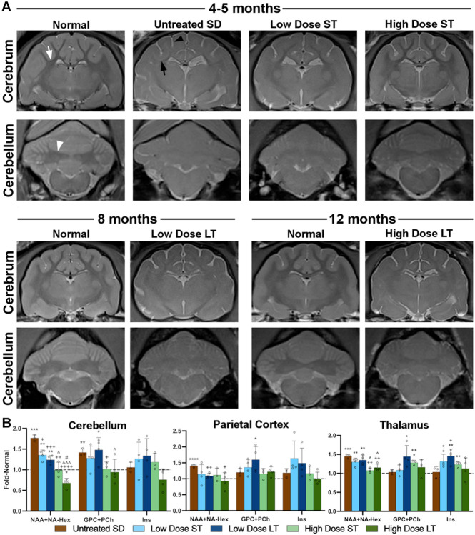
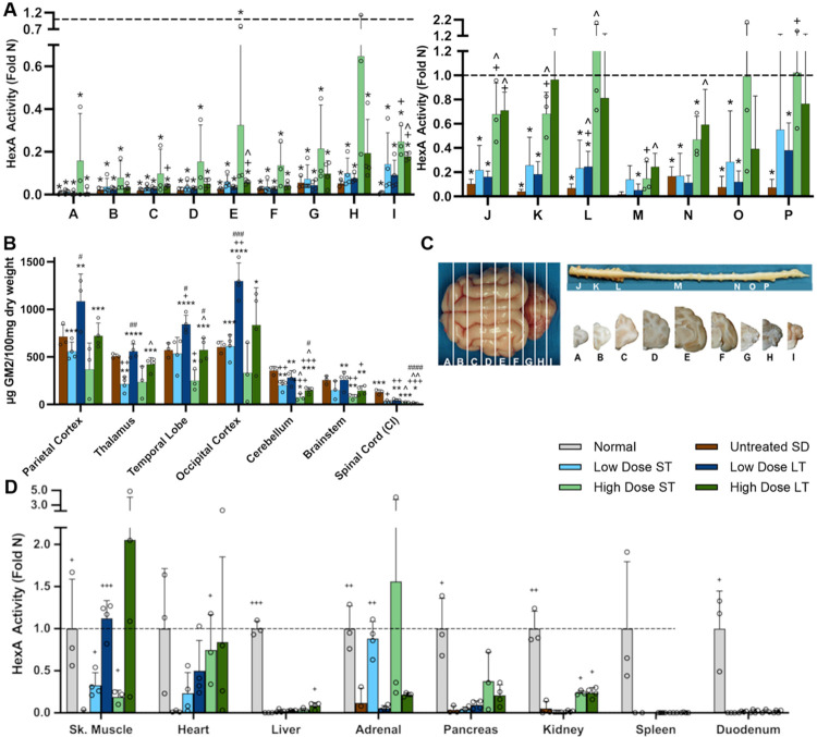
Sandhoff Disease (SD), a fatal neurodegenerative disorder, is caused by the absence of ß-hexosaminidase (Hex) and subsequent accumulation of GM2 ganglioside in lysosomes. Previous studies have led to adeno-associated virus (AAV) gene therapy for children with GM2 gangliosidosis in both expanded access and Phase I/II clinical trials via intracranial and/or cerebrospinal fluid-based delivery. The current study investigated intravenous (IV) gene therapy of SD cats, treated at one month of age with a bicistronic AAV vector. While untreated SD cats lived to 4.3±0.2 months, cats treated with low and high doses lived to 8.3±1.2 and 12.4±2.7 months, respectively. In-life assessments revealed clear clinical benefit of AAV treatment, with the most dramatic improvement seen in the reduction of overt full-body tremors. Cerebrospinal fluid levels of aspartate aminotransferase (AST) and lactate dehydrogenase (LDH) were decreased, indicating a reduction of cell damage within the central nervous system. Magnetic resonance imaging (MRI) and spectroscopy (MRS) acquired on a 7 Tesla scanner indicated that structural pathology and metabolite abnormalities are partially normalized by AAV treatment. Dose-dependent reduction of GM2 ganglioside storage and increases in Hex activity were most substantial in the caudal regions of the brain and in the spinal cord. Immunohistochemistry revealed reduction in neuroinflammatory cell populations and partial correction of myelin deficits. These results support the dose-dependent efficacy of AAV delivered IV for significant restoration of clinical metrics and Hex function in a feline model of SD.


Figures

Figure 1.
Figure 1.. Survival and quality of life improve with AAV treatment.
(A) Kaplan-Meier curve demonstrates improved survival with AAV treatment. The lifespan of cats treated with the low dose and high dose nearly doubled and tripled, respectively. +p<0.05 v. untreated SD, ^p<0.05 v. low dose. (B) Weights of treated cats were partially normalized. *p<0.05 untreated SD v. normal; +p<0.05 low dose v. normal; #p<0.05 high dose v. normal. (C) Neurological clinical rating scores (CRS) indicate dose-dependent delay of neurologic dysfunction. CRS for the high-dose cohort (green) artifactually increased at 13 months because only one cat with better-than-average treatment survived to this age.
Figure 2.
Figure 2.. Liver stiffness in SD cats is improved with high dose AAV treatment.
Shear wave ultrasound elastography demonstrated significant decreases in speed and elasticity at all ages of untreated and low dose treated SD cats compared to normal liver. In SD cats treated with the high dose, wave speed and elasticity returned to near-normal levels until 7 months of age, after which they decreased to untreated levels. *0.05>p>0.01, **0.01>p>0.001, ***0.001^p<0.05 versus low dose at same age.
Figure 3.
Figure 3.. AAV treatment partially normalizes 7T MR images and MRS metabolites.
(A) T2-weighted anatomical images. In normal cats, white matter is darker than, or hypointense to, gray matter in the corona radiata of the cerebrum (white arrow) and the DCN of the cerebellum (white arrowhead). The CSF signal (bright) around the meninges is minimal in normal cats. In untreated SD cats at humane endpoint, the DCN are isointense to (same shade as) surrounding gray matter and the corona radiata (black arrow) are hyperintense to (lighter than) gray matter due to hypomyelination and storage in neuron cell bodies. The increased amount of CSF signal (black arrowhead) in untreated SD cats is especially apparent surrounding the gyri and is attributed to generalized atrophy. Pathological MRI changes are mitigated by AAV treatment, with the most prominent preservation of brain architecture in the high-dose, short-term cohort. (B) MRS of 3 brain voxels: cerebellum, parietal cortex, thalamus. The spectroscopic peak of the neurometabolic marker NAA, which merges with the NA-Hex storage product in SD, is significantly increased in all regions of SD cat brains and normalized by the high dose of AAV in the cerebellum and thalamus. GPC+PCh, a measure of membrane turnover that correlates with demyelination in this model, is significantly increased in the cerebellum of untreated SD cats and normalized in the high-dose cohort at both short- and long-term timepoints. Myoinositol (Ins) is unchanged in untreated and treated SD cats, though variability is high in some groups. ST: Short-term, LT: Long-term. *0.05>p>0.01, **0.01>p>0.001, ***0.001+0.05>p>0.01, ++0.01>p>0.001, +++0.001<p<0.0001, ++++p<0.00001 vs. untreated SD cats; ^0.05>p>0.01, ^^^0.001<p<0.0001 vs. low dose at same age. #p<0.05 vs. short-term at same dose. Dashed horizontal line represents levels in age-matched normal cat brains, to which all SD data were standardized.
Figure 4.
Figure 4.. AAV treatment reduces CSF markers of cell damage and increases Hex activity.
(A, B) CSF levels of aspartate aminotransferase (AST) and lactate dehydrogenase (LDH) are significantly increased in SD cats, indicating cell damage within the central nervous system. AST and LDH are significantly reduced in cats treated with the high AAV dose at the short-term time point (5 months of age). AST and LDH rose between the short- and long-term time points as clinical disease progressed. (C) Specific activity of all hexosaminidase isozymes (Total Hex) is significantly decreased to background levels in the CSF of untreated SD cats. AAV treatment normalizes Total Hex levels across doses and age groups. ST, short-term; LT, long-term. *p<0.05 versus age-matched normal cats, +p<0.05 versus untreated SD cats, ^p<0.05 versus low dose at same age.
Figure 5.
Figure 5.. Hex activity increases and GM2 ganglioside storage decreases with AAV treatment.
(A) HexA activity (fold normal, or Fold N) in the CNS increases with AAV dose, with the cerebellum, cervical spinal cord, and lumbar spinal cord showing the best response. (B) GM2 ganglioside in untreated SD cats was significantly increased above normal (trace) levels in all CNS regions except spinal cord. AAV treatment reduced GM2 levels in certain regions of the CNS (depending on dose and time point) but only restored it to normal levels in the cervical intumescence (CI) of the spinal cord. (C) Brain and spinal cord block designations. Dorsal surface of the brain is shown on the left, with corresponding transverse hemi-sections on the right (below spinal cord). (D) HexA activity in peripheral tissues, including the legend for panels A-D. *p<0.05, **0.01>p>0.001, ***0.001+p<0.05, ++0.01>p>0.001, +++0.001<p<0.0001, ++++p<0.00001 vs. untreated SD cats; ^0.05>p>0.01, ^^0.01>p>0.001, ^^^0.001<p<0.0001 vs. low dose at same age. #p<0.05, ##0.01>p>0.001, ###0.001<p<0.0001, ####p<0.00001 vs. short term at same dose.
Figure 6.
Figure 6.. Astrocyte and microglia pathology is transiently normalized after AAV treatment.
SD cats treated with the high dose were evaluated short-term (16 weeks post-treatment or ~5 months of age) and long-term (humane endpoint at ~12 months of age). Brain sections were stained for (A) GFAP (astrocyte marker) or (B) Iba1 (microglia marker). Micrographs at the top of each panel show representative staining in the thalamus and demonstrate normalization of glial cell morphology in the short-term cohort but not in the long-term cohort. Graphs depict the percentage of positive staining for each marker standardized to that of normal cats (represented by dashed horizontal line). Scale bars: 20 μm. ST: short-term, LT: long-term, WM: white matter, GM: gray matter, DCN: deep cerebellar nuclei. *p<0.05, **0.01>p>0.001, ***0.001+p<0.05 vs. untreated SD cats .
Figure 7.
Figure 7.. Myelin deficits in SD cats are partially normalized through AAV treatment.
(A) Oligodendrocytes staining positive for Olig2 and (B) percentage of tissue staining positive for Luxol Fast Blue (LFB) are below normal levels in SD cats and partially restored in AAV-treated cats. Micrographs at 20x are from the parietal cortex white matter of representative cats. Scale bar: 20 μm. ST: short-term, LT: long-term, WM: white matter, GM: gray matter, DCN: deep cerebellar nuclei. *p<0.05, **0.01>p>0.001, ***0.001+p<0.05, ++0.01>p>0.001 vs. untreated SD cats; #p<0.05 vs. short term at same dose.

References

    1. Leal A. F. et al., GM2 Gangliosidoses: Clinical Features, Pathophysiological Aspects, and Current Therapies. Int J Mol Sci 21, (2020). - PMC - PubMed
    1. Bradbury A. M. et al., Therapeutic response in feline sandhoff disease despite immunity to intracranial gene therapy. Mol Ther 21, 1306–1315 (2013). - PMC - PubMed
    1. Bradbury A. M. et al., Biomarkers for disease progression and AAV therapeutic efficacy in feline Sandhoff disease. Exp Neurol 263, 102–112 (2015). - PMC - PubMed
    1. Cachon-Gonzalez M. B. et al., Effective gene therapy in an authentic model of Tay-Sachs-related diseases. Proc.Natl.Acad.Sci.U.S.A. 103, 10373–10378 (2006). - PMC - PubMed
    1. Cachon-Gonzalez M. B. et al., Gene transfer corrects acute GM2 gangliosidosis--potential therapeutic contribution of perivascular enzyme flow. Mol Ther 20, 1489–1500 (2012). - PMC - PubMed

Publication types

LinkOut - more resources