[Preprint]. 2024 Nov 21:2024.11.12.623252.
doi: 10.1101/2024.11.12.623252.

An Open Access Resource for Marmoset Neuroscientific Apparatus

Affiliations

An Open Access Resource for Marmoset Neuroscientific Apparatus

Isabela Zimmermann Rollin et al. bioRxiv. .

Update in

  • An open access resource for marmoset neuroscientific apparatus.
    Zimmermann Rollin I, Papoti D, Bishop M, Szczupak D, Corigliano MR, Hitchens TK, Zhang B, Pell SKA, Guretse SS, Dureux A, Murai T, Sukoff Rizzo SJ, Klassen LM, Zeman P, Gilbert KM, Menon RS, Lin MK, Everling S, Silva AC, Schaeffer DJ. Zimmermann Rollin I, et al. Imaging Neurosci (Camb). 2025 Feb 21;3:imag_a_00483. doi: 10.1162/imag_a_00483. eCollection 2025. Imaging Neurosci (Camb). 2025. PMID: 40800752 Free PMC article.

Abstract

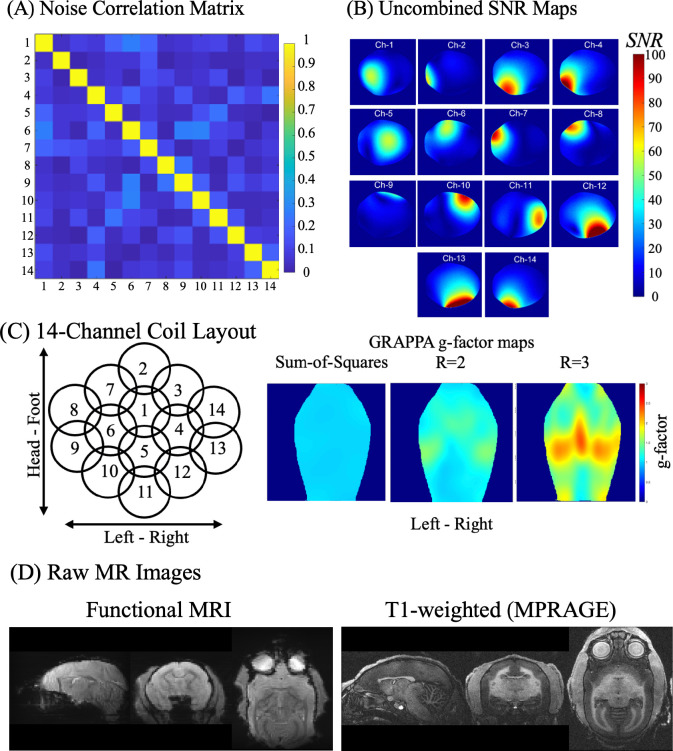
The use of the common marmoset (Callithrix jacchus) for neuroscientific inquiry has grown precipitously over the past two decades. Despite windfalls of grant support from funding initiatives in North America, Europe, and Asia to model human brain diseases in the marmoset, marmoset-specific apparatus are of sparse availability from commercial vendors and thus are often developed and reside within individual laboratories. Through our collective research efforts, we have designed and vetted myriad designs for awake or anesthetized magnetic resonance imaging (MRI), positron emission tomography (PET), computed tomography (CT), as well as focused ultrasound (FUS), electrophysiology, optical imaging, surgery, and behavior in marmosets across the age-span. This resource makes these designs openly available, reducing the burden of de novo development across the marmoset field. The computer-aided-design (CAD) files are publicly available through the Marmoset Brain Connectome (MBC) resource (https://www.marmosetbrainconnectome.org/apparatus/) and include dozens of downloadable CAD assemblies, software and online calculators for marmoset neuroscience. In addition, we make available a variety of vetted touchscreen and task-based fMRI code and stimuli. Here, we highlight the online interface and the development and validation of a few yet unpublished resources: Software to automatically extract the head morphology of a marmoset from a CT and produce a 3D printable helmet for awake neuroimaging, and the design and validation of 8-channel and 14-channel receive arrays for imaging deep structures during anatomical and functional MRI.

Keywords: CT; Electrophysiology; MRI; Marmoset; Open Science; PET.


Figures

Figure 1:
Figure 1:
(A) A screenshot of the home page of the Neuroscientific Apparatus Resource. (B) After selecting a design, the user is directed to a page where they can access a zoomable and rotatable 3D rendering along with photos and relevant design files.
Figure 2:
Figure 2:
(A) CT images available in an online viewer (also downloadable) across the age span for marmosets of different sexes and weights. (B) Examples of calculators available on the website, including a (i) medication calculator for doses of different agents from a single input (weight of animal), (ii) adeno-associated viral (AAV) injection dose calculator (ii), and (iii) Ernst Angle calculator for optimizing contrast with MR images. (C) Submission Page for groups interested in contributing to the resource.
Figure 3:
Figure 3:
Automated Marmoset Imaging Helmet Generation, an Open-Source Software (AMIHGOS). (A) The software home screen allows users to load NIFTI or DICOM format files as (B) raw CT image inputs. (C) In the ROI selection step, users select boundary points on the nose and just posterior to the glenohumeral joint (just caudal to the shoulder joint) and define the ROI to be included in the helmet generation. (D) Users then align their input image with the template image by selecting corresponding points on the ear canals, the top of the head, and the front of the orbitals in each image. (E) Registration is confirmed by dragging the “Image Z” or “Alpha” bars with the cursor to ensure the template and input images are aligned. (F) Users can adjust any necessary translational modifications and generate an STL file of the customized helmet, which can be sent for 3D printing.
Figure 4:
Figure 4:
(A) Comparison of the size and morphology of a rat and a marmoset skull. (B) Single-element electrical scheme for a coil, adapted from Papoti et al. 2017. (C) 8-channel coil layout and rendering on a marmoset skull. (D) 14-channel coil layout on a marmoset skull and photograph of assembled coil. (E) Assembly of all components needed for awake imaging with the 8-channel coil, including the coil itself, preamplifier box, body restrainer, chin plate and helmet.
Figure 5:
Figure 5:
(A) Restraint training progression over 14 days, showing the phases of adaptation to the MRI environment. (B) CT images were used to generate custom-fitted helmets tailored to each marmoset’s anatomy, ensuring an optimized fit for stable, awake PET ([¹⁸F] FDG, 80 MBq, 60-minute static acquisition), PET, and MRI scans, showing fit for small (325 g), medium (425 g), and large (500 g) adult marmosets. (C) Helmet motion comparison between a universal foam-lined helmet (1 mm wall thickness) and AMIHGOS-generated custom helmets.
Figure 6:
Figure 6:
(A) Noise correlation matrix showing the correlation levels between channels, with an average correlation coefficient of 10.7% across channels. (B) Uncombined SNR maps from the central coronal plane demonstrate individual coil element sensitivity. (C) Schematic of the 8-channel coil layout and GRAPPA g-factor maps. (D) Raw MR images from a marmoset brain, including fMRI and T2-weighted RARE sequences.
Figure 7:
Figure 7:
(A) Noise correlation matrix from a noise-only acquisition, showing low inter-channel correlations with an average of 8.3% and a maximum correlation of 27.2% between channels 13 and 14, indicating minimal residual coupling. (B) Uncombined SNR maps from a central coronal orientation, demonstrating individual coil element sensitivity. (C) Layout of the 14-channel coil array and GRAPPA g-factor maps. (D) Raw fMRI and T1-weighted MPRAGE MRI acquired from a marmoset.

References

    1. Gilbert K. M. et al. Open-source hardware designs for MRI of mice, rats, and marmosets: Integrated animal holders and radiofrequency coils. J Neurosci Methods 312, 65–72 (2019). - PubMed
    1. Kita Y. et al. Cellular-resolution gene expression profiling in the neonatal marmoset brain reveals dynamic species- and region-specific differences. Proceedings of the National Academy of Sciences 118, (2021). - PMC - PubMed
    1. Liu C. et al. Marmoset Brain Mapping V3: Population multi-modal standard volumetric and surface-based templates. Neuroimage 226, 117620 (2021). - PMC - PubMed
    1. Liu C. et al. A resource for the detailed 3D mapping of white matter pathways in the marmoset brain. Nat Neurosci 23, 271–280 (2020). - PMC - PubMed
    1. Liu C. et al. A digital 3D atlas of the marmoset brain based on multi-modal MRI. Neuroimage 169, 106–116 (2018). - PMC - PubMed

Publication types

LinkOut - more resources