[Preprint]. 2024 Nov 24:2024.11.23.624981.
doi: 10.1101/2024.11.23.624981.

MAT2a and AHCY inhibition disrupts antioxidant metabolism and reduces glioblastoma cell survival

Affiliations

MAT2a and AHCY inhibition disrupts antioxidant metabolism and reduces glioblastoma cell survival

Emma C Rowland et al. bioRxiv. .

Update in

Abstract

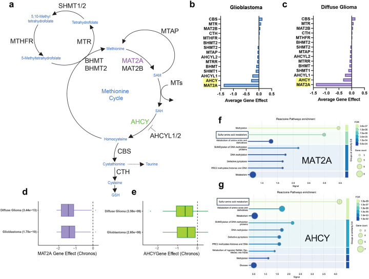
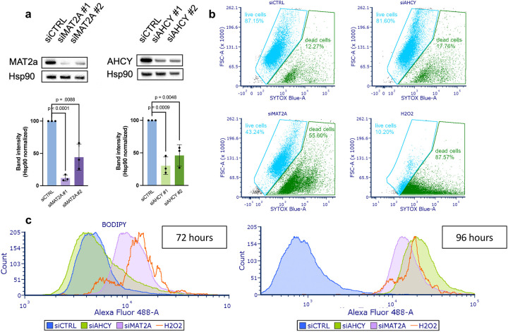
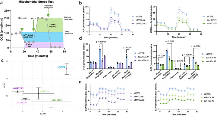
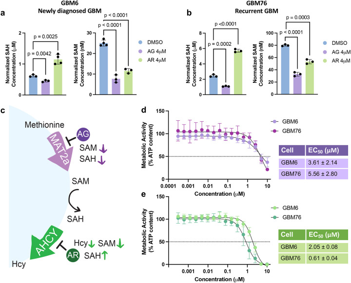
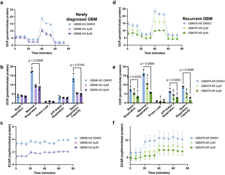
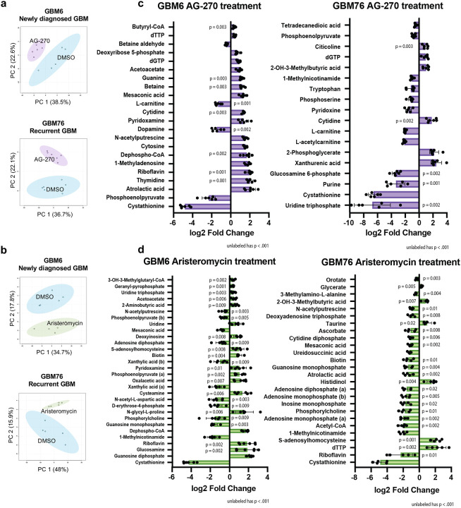
Glioblastoma (GBM) is a highly aggressive primary malignant adult brain tumor that inevitably recurs with a fatal prognosis. This is due in part to metabolic reprogramming that allows tumors to evade treatment. We therefore must uncover the pathways mediating these adaptations to develop novel and effective treatments. We searched for genes that are essential in GBM cells as measured by a whole-genome pan-cancer CRISPR screen available from DepMap and identified the methionine metabolism genes MAT2A and AHCY. We conducted genetic knockdown, evaluated mitochondrial respiration, and performed targeted metabolomics to study the function of these genes in GBM. We demonstrate that MAT2A or AHCY knockdown induces oxidative stress, hinders cellular respiration, and reduces the survival of GBM cells. Furthermore, selective MAT2a or AHCY inhibition reduces GBM cell viability, impairs oxidative metabolism, and changes the metabolic profile of these cells towards oxidative stress and cell death. Mechanistically, MAT2a or AHCY regulates spare respiratory capacity, the redox buffer cystathionine, lipid and amino acid metabolism, and prevents DNA damage in GBM cells. Our results point to the methionine metabolic pathway as a novel vulnerability point in GBM.

Keywords: glioblastoma; lipid peroxidation; metabolism; metabolomics; methionine; mitochondria; oxidative stress.


Conflict of interest statement

The authors declare that they have no conflicts of interest with the contents of this article.

Figures

Figure 1.
Figure 1.. Pooled CRISPR screen data reveals methionine metabolism gene essentiality in GBM.
a The methionine cycle contains multiple enzymes to generate methyl donor SAM, and downstream antioxidant substrates. b, c Average gene effect scores representative of DepMap pooled CRISPR screens for indicated genes involved in the methionine cycle in Glioblastoma and Diffuse Glioma lineages. Less than 0 indicates impaired cell growth and greater than 0 indicates enhanced cell growth. d, e Gene effect scores for both MAT2A and AHCY in Diffuse Glioma and Glioblastoma lineages represented as boxplots scores filtered using p-value <0.00005 according to DepMap significance. f, g Significantly enriched pathways from the Reactome database for both MAT2A and AHCY, reflecting high signal for sulfur amino acid metabolism, generated using the STRING database.
Figure 2.
Figure 2.. Genetic knockdown of MAT2A and AHCY induces cell death and lipid peroxidation in GBM.
a Gene knockdown in LN229 cells 48 hours post-transfection, with heat shock protein 90 (Hsp90) as the loading control. Blots quantified using ImageJ and normalized to siCTRL band intensity. b Cells stained with SYTOX Blue for flow cytometry analysis of cell death 96 hours post-transfection, with hydrogen peroxide as positive control. c BODIPY staining for flow cytometry analysis of lipid peroxidation 72 hours and 96 hours post-transfection as indicated on the graph. Cell counts were normalized to peak intensity, with hydrogen peroxide as positive control.
Figure 3.
Figure 3.. Genetic knockdown of MAT2A and AHCY induces mitochondrial dysfunction in GBM.
a Graphic of Mitochondrial Stress Test indicating changes in oxygen consumption rate (OCR) following electron transport chain inhibitor compound injections. Highlighted regions correlate to respiratory parameters by calculated differences between OCR values. b Mitochondrial Stress Test OCR with LN229 cells with genetic knockdown of either MAT2A or AHCY 72 hours post-transfection. c Energy map with mean maximal OCR and corresponding ECAR values of genetic knockdown LN229 cells, with top right indicating energetic, bottom right indicating glycolytic, top left indicating aerobic, and bottom left indicating quiescent phenotype. d Quantification of respiratory parameters for MAT2A and AHCY knockdown cells compared to control. Unpaired t-test with Holm-Šídák correction was performed. e Mitochondrial Stress Test ECAR with LN229 cells corresponding to OCR measurements, with top right corner indicating a more energetically active state and bottom left corner indicating a more energetically inactive state.
Figure 4.
Figure 4.. Selective MAT2A and AHCY inhibitors reduce cell viability in GBM.
a, b SAH and SAM levels in patient-derived primary GBM cells 24 hours after 4μM of Aristeromycin (AR), 4μM of AG-270 (AG) treatment or vehicle treatment. c Graphic of enzyme inhibition by each respective inhibitor compound and resulting changes in metabolite levels. d, e Dose-response curves for AG-270 (top) and Aristeromycin (bottom) 72 hours post-treatment with corresponding EC50 values in both GBM6 and GBM76 cells. ATP content was measured using CellTiterGlo to indicate cell viability.
Figure 5.
Figure 5.. MAT2A and AHCY enzyme inhibition disrupts mitochondrial function in GBM.
a Mitochondrial Stress Test OCR with newly diagnosed GBM6 cells treated with AG-270 72 hours prior. b OCR for recurrent GBM76 cells treated with Aristeromycin 72 hours prior. c, d Quantification of respiratory parameters corresponding to OCR measurements compared to control. Performed unpaired t-test with Holm-Šídák correction. e, f Mitochondrial Stress Test ECAR corresponding to OCR measurements.
Figure 6.
Figure 6.. GBM exhibits oxidative stress and compromised lipid metabolism upon MAT2A and AHCY inhibition.
a, b Principal component analysis of GBM6 and GBM76 cells grown in serum free media comparing DMSO to AG-270 (a) or Aristeromycin (b) treatment. Analyzed after 24 hours using LC-MS. Sample peak intensities were normalized using log transformation and Pareto scaling. c,d Significantly reduced and increased metabolites in GBM6 and GBM76 cells treated with AG-270 (c) and Aristeromycin (d) compared to control using unpaired t-test with a p-value of .05. (b) indicates the alkaline metabolite while (a) indicates the acidic metabolite upon ionization.

Similar articles

References

    1. Wen P. Y. et al. (2020) Glioblastoma in adults: a Society for Neuro-Oncology (SNO) and European Society of Neuro-Oncology (EANO) consensus review on current management and future directions. Neuro-Oncol. 22, 1073–1113 - PMC - PubMed
    1. Kotecha R., Odia Y., Khosla A. A. & Ahluwalia M. S. (2023) Key Clinical Principles in the Management of Glioblastoma. JCO Oncol. Pract. 19, 180–189 - PubMed
    1. Fernandes C. Costa A. Osório L. et al. (2017) Glioblastoma Chapter 11: Current Standards of Care in Glioblastoma Therapy. Codon Publications 197–241. doi:10.15586/codon.glioblastoma.2017.ch11 - DOI - PubMed
    1. Nguyen T. T. T., Shang E., Westhoff M.-A., Karpel-Massler G. & Siegelin M. D. (2022) Therapeutic Drug-Induced Metabolic Reprogramming in Glioblastoma. Cells 11, 2956. - PMC - PubMed
    1. Duraj T. et al. (2021) Beyond the Warburg Effect: Oxidative and Glycolytic Phenotypes Coexist within the Metabolic Heterogeneity of Glioblastoma. Cells 10, 202. - PMC - PubMed

Publication types

LinkOut - more resources