[Preprint]. 2024 Nov 22:2024.11.22.624763.
doi: 10.1101/2024.11.22.624763.

Electrical synapse molecular diversity revealed by proximity-based proteomic discovery

Affiliations

Electrical synapse molecular diversity revealed by proximity-based proteomic discovery

Jennifer Carlisle Michel et al. bioRxiv. .

Abstract

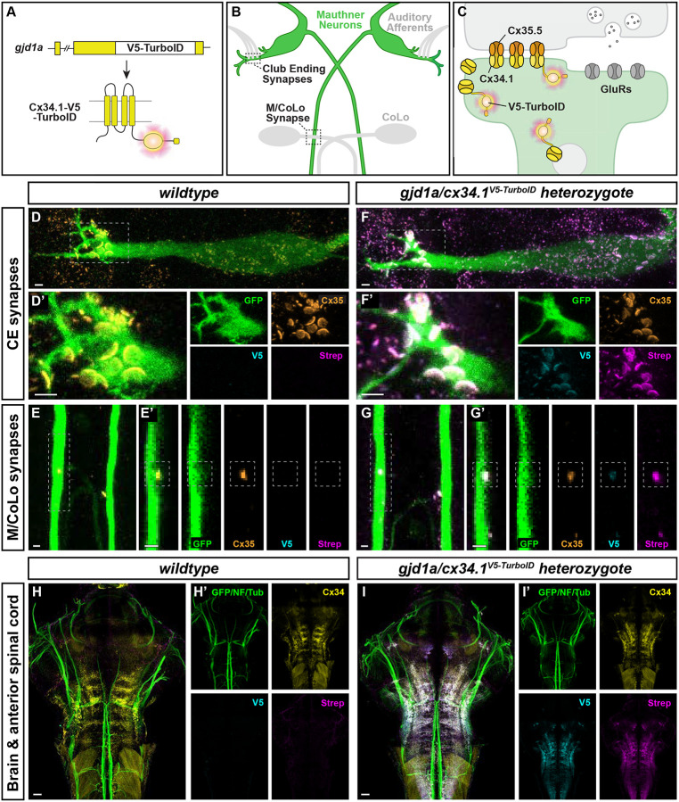
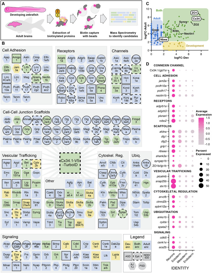
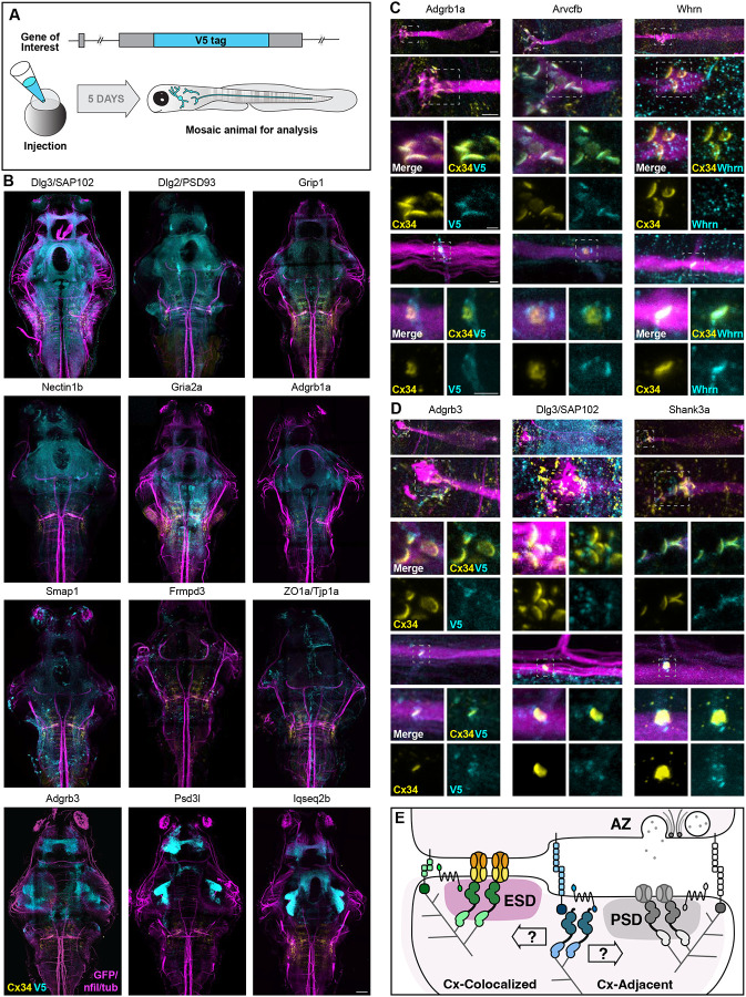
Neuronal circuits are composed of synapses that are either chemical, where signals are transmitted via neurotransmitter release and reception, or electrical, where signals pass directly through interneuronal gap junction channels. While the molecular complexity that controls chemical synapse structure and function is well appreciated, the proteins of electrical synapses beyond the gap-junction-forming Connexins are not well defined. Yet, electrical synapses are expected to be molecularly complex beyond the gap junctions. Connexins are integral membrane proteins requiring vesicular transport and membrane insertion/retrieval to achieve function, homeostasis, and plasticity. Additionally, electron microscopy of neuronal gap junctions reveals neighboring electron dense regions termed the electrical synapse density (ESD). To reveal the molecular complexity of the electrical synapse proteome, we used proximity-dependent biotinylation (TurboID) linked to neural Connexins in zebrafish. Proteomic analysis of developing and mature nervous systems identifies hundreds of Connexin-associated proteins, with overlapping and distinct representation during development and adulthood. The identified protein classes span cell adhesion molecules, cytoplasmic scaffolds, vesicular trafficking, and proteins usually associated with the post synaptic density (PSD) of chemical synapses. Using circuits with stereotyped electrical and chemical synapses, we define molecular sub-synaptic compartments of ESD localizing proteins, we find molecular heterogeneity amongst electrical synapse populations, and we examine the synaptic intermingling of electrical and chemical synapse proteins. Taken together, these results reveal a new complexity of electrical synapse molecular diversity and highlight a novel overlap between chemical and electrical synapse proteomes. Moreover, human homologs of the electrical synapse proteins are associated with autism, epilepsy, and other neurological disorders, providing a novel framework towards understanding neuro-atypical states.


Figures

Figure 1.
Figure 1.. Connexin-TurboID localizes to electrical synapses and biotinylates proteins in vivo.
(A) Schematic diagram of the gjd1a/cx34.1 gene locus modified by in-frame insertion of V5-TurboID. The horizontal black bar represents the DNA strand, a white box represents V5-TurboID, and yellow boxes represent individual gjd1a exons. Below, a cartoon of the Cx34.1-V5-TurboID monomeric protein illustrates the insertion of the V5-TurboID cassette (magenta burst) after the four trans-membrane domains (vertical yellow cylinders) and before the C-terminal PDZ binding motif (PBM, horizontal yellow cylinder). Dark gray lines denote plasma membrane. (B) Simplified diagram illustrating the electrical synapses of interest in the Mauthner (M-) cell circuit. The image represents a dorsal view of the M-cells (green) with anterior on top. Regions in black dashed outline indicate the stereotypical synaptic contacts used for analysis in this study. Presynaptic auditory afferents (grey lines) contact the postsynaptic M-cell lateral dendrite in the hindbrain forming Club Ending (CE) synapses. In the spinal cord, the presynaptic Mauthner axons form en passant electrical synapses with the postsynaptic CoLo interneurons (grey neurons) in each spinal cord hemi-segment (1 of 30 repeating spinal segments are shown). (C) A diagram of a mixed electrical/chemical synapse as found at M-cell CEs. In the electrical component, molecularly asymmetric Connexin hemichannels (Cx35.5 [orange], Cx34.1 [yellow]) directly couple neurons by forming gap junction (GJ) channels. In the chemical component, presynaptic synaptic vesicles release neurotransmitter (grey circles) which align with postsynaptic glutamate receptors (GluRs). Cx34.1-V5-TurboID-containing hemichannels (yellow with magenta burst) are shown trafficking to electrical synapses on vesicles and localizing within the neural GJ plaque. (D-G) Enzymatically active Connexin-V5-TurboID is localized at the stereotype M-cell electrical synapses. Confocal images of M-cell circuitry and electrical synaptic contacts in 6-day-post-fertilization, Et(T2KHG)zf206 zebrafish larvae from wildtype (D, E) and gjd1a/cx34.1V5-TurboID heterozygous (F, G) siblings treated with 1mM biotin for 72 hours. Animals are stained with anti-GFP (green), anti-Cx35.5 (orange), anti-V5 (cyan), and Strep-conjugated fluorophore (magenta). Scale bar = 2 μm in all images. Anterior up. Boxed regions denote stereotyped location of electrical synapses and regions are enlarged in neighboring panels. Images of the Mauthner cell body and lateral dendrite in the hindbrain (D, F) are maximum intensity projections of ~27 μm. In D’ and F’, images are maximum-intensity projections of ~15 μm and neighboring panels show the individual channels. Images of the sites of contact of M/CoLo processes in the spinal cord (E, G) are maximum-intensity projections of ~6 μm. In E’ and G’, images are from a single 0.42 μm Z-plane and the white dashed square denotes the location of the M/CoLo site of contact. Neighboring panels show individual channels. (H, I) Enzymatically active Connexin-V5-TurboID is localized specifically in the brain similar to wildtype connexin. Confocal tile scans of zebrafish brain and anterior spinal cord from 6 dpf zf206Et zebrafish larvae from wildtype (H) or gjd1a/cx34.1V5-TurboID heterozygous (I) animals treated with 1mM biotin for 48 hours. Images are maximum intensity projections of ~82 μm. Animals are stained with anti-GFP/anti-NF/anti-tubulin (green), anti-Cx34.1 (yellow), anti-V5 (cyan), and Strep (magenta). Neighboring panels show the individual channels. Scale bars = 20 μm. Anterior up.
Figure 2.
Figure 2.. Discovery of the neural Connexin-associated proteome by biotin-proximity identification.
(A) Diagram of biotin-proximity labeling scheme. gjd1a/cx34.1V5-TurboID heterozygous larvae (developing) or adults were treated with biotin supplemented water to activate maximal biotinylation of nearby proteins. Whole larvae and brains from adults (dotted lines) were harvested and homogenized under denaturing conditions. gjd1a/cx34.1 is expressed exclusively in the nervous system providing specificity. Biotinylated proteins were captured with magnetic streptavidin beads, digested with trypsin on-bead, and analyzed by mass spectrometry to identify candidates. (B) Proteins enriched in Cx34.1-V5-TurboID samples compared to wildtype controls are depicted and grouped into grey boxes according to gene ontology (GO) analysis: Cell Adhesion, Receptors, Channels, Cell-Cell-Junction Scaffolds, Vesicular Trafficking, Signaling, Cytoskeletal Regulation, Ubiquitination, and Other. Paralogous proteins are denoted as single shapes and noted by a forward slash between name delineations. Colors denote enrichment in the developmental (yellow), adult (blue), or both datasets (green). Hexagons denote proteins associated with epithelial adherens or tight junctions (AJ/TJ), circles denote proteins associated with postsynaptic densities of chemical synapses (PSD), and squares denote the remainder of the isolated candidates. Outlines denote human disorders associated with the protein: solid outlines denote proteins associated with autism spectrum disorder (ASD), dashed outlines denote proteins associated epilepsy (Epil.), and stippled outlines denote proteins associated with Neurodevelopmental Disorders and/or Intellectual Disability (NDD/IDD). Many proteins have associations with multiple of these categories, or other neurological disorders (Table S2). (C) Enrichment of identified proteins (dots) in developing (x-axis) and adult (y-axis) datasets. Colored regions denote proteins enriched in developing (yellow), adult (blue) or both (green) time points. Proteins in white callouts have been previously localized to zebrafish electrical synapses (Cx34.1, Nbea, and ZO1) while the rest have been localized to mammalian electrical synapses or interact with mammalian Cx36. (D) Neural Connexin-associated proteome gene expression across germ layers in a whole-embryo single-cell RNA sequencing dataset (5 and 6 dpf). Clusters (Identity) are organized by germ layer along the x-axis: neurons, ectoderm (non-neural ectoderm, e.g. skin and cranial neural crest), mesoderm (e.g., skeletal muscle and blood), and endoderm (e.g. liver). A selection of Connexin-associated genes is arranged along the y-axis, grouped by GO terms. Dot size represents the percentage of cells in the identity category expressing each gene, while color indicates relative expression levels.
Figure 3.
Figure 3.. Identification of electrical synapse density proteins.
(A) Diagram of CRISPR-mediated epitope insertion at endogenous genes to generate animals mosaically expressing V5-tagged proteins for analysis in the zebrafish brain. The horizontal black bar represents the DNA strand of the gene of interest, a cyan box represents the insertion of the V5 epitope tag. Animals are injected at the 1-cell stage and expression is visualized five days later. (B) Confocal tile scans of zebrafish brain at 5 dpf Et(T2KHG)zf206 zebrafish larvae expressing V5-tagged proteins, as labeled. Animals are stained with anti-GFP/anti-NF/anti-tubulin (magenta), anti-Cx34.1 (yellow), and anti-V5 (cyan). Scale bars = 20 μm. Anterior up. (C) Confocal images of Connexin-colocalized genes in the M-cell circuit at 5 dpf in Et(T2KHG)zf206 zebrafish larvae. Boxed regions denote stereotyped location of electrical synapses and regions are enlarged in neighboring panels. Animals are stained with anti-GFP/anti-NF/anti-tubulin (magenta), antiCx34.1 (yellow), and anti-V5 or anti-Whrn antibody (cyan), as labeled. Top four rows show CE synapses, anterior up. Bottom three rows show M/CoLo synapses, anterior left. Neighboring panels show individual and merged channels, as labeled. Scale bars = 2 μm. (D) Confocal images of Connexin-adjacent genes in the M-cell circuit at 5 dpf in Et(T2KHG)zf206 zebrafish larvae. Boxed regions denote stereotyped location of electrical synapses and regions are enlarged in neighboring panels. Animals are stained with anti-GFP/anti-NF/anti-tubulin (magenta), anti-Cx34.1 (yellow), and anti-V5 (cyan). Top four rows show CE synapses, anterior up. Bottom three rows show M/CoLo synapses, anterior left. Neighboring panels show individual and merged channels, as labeled. Scale bars = 2 μm. (E) Model of a mixed electrical/chemical synapse illustrating Connexin-colocalized and Connexin-adjacent proteins. ESD proteins fall into classes such as cell adhesion, signaling, scaffolding, cytoskeletal regulation and colocalize with electrical synapses (green shapes). These proteins classes are analogous to proteins identified by proteomics at chemical synapse presynaptic active zones (AZ) and PSDs (grey shapes). Candidates that are adjacent to electrical synapses (blue shapes) may be exclusive to the chemical synapse, may be localized adjacent to both, and may regulate both electrical and chemical synapses assembly and function, as indicated by the opposing arrows. ESD, electrical synapse density. PSD, post-synaptic density. AZ, pre-synaptic active zone.

References

    1. Südhof T. C. Towards an Understanding of Synapse FormaDon. Neuron 100, 276–293 (2018). - PMC - PubMed
    1. Chia P. H., Li P. & Shen K. Cellular and molecular mechanisms underlying presynapse formaDon. J. Cell Biol. 203, 11–22 (2013). - PMC - PubMed
    1. Jin Y. & Garner C. C. Molecular Mechanisms of PresynapDc DifferenDaDon. Annu. Rev. Cell Dev. Biol. 24, 237–262 (2008). - PubMed
    1. Sanes J. R. & Lichtman J. W. InducDon, assembly, maturaDon and maintenance of a postsynapDc apparatus. Nat. Rev. Neurosci. 2, 791–805 (2001). - PubMed
    1. McAllister A. K. Dynamic Aspects of CNS Synapse FormaDon. Annu. Rev. Neurosci. 30, 425–450 (2007). - PMC - PubMed

Publication types

LinkOut - more resources