[Preprint]. 2024 Nov 13:2024.11.12.623078.
doi: 10.1101/2024.11.12.623078.

Immune Evasion, Cell-Cell Fusion, and Spike Stability of the SARS-CoV-2 XEC Variant: Role of Glycosylation Mutations at the N-terminal Domain

Affiliations

Immune Evasion, Cell-Cell Fusion, and Spike Stability of the SARS-CoV-2 XEC Variant: Role of Glycosylation Mutations at the N-terminal Domain

Pei Li et al. bioRxiv. .

Update in

Abstract

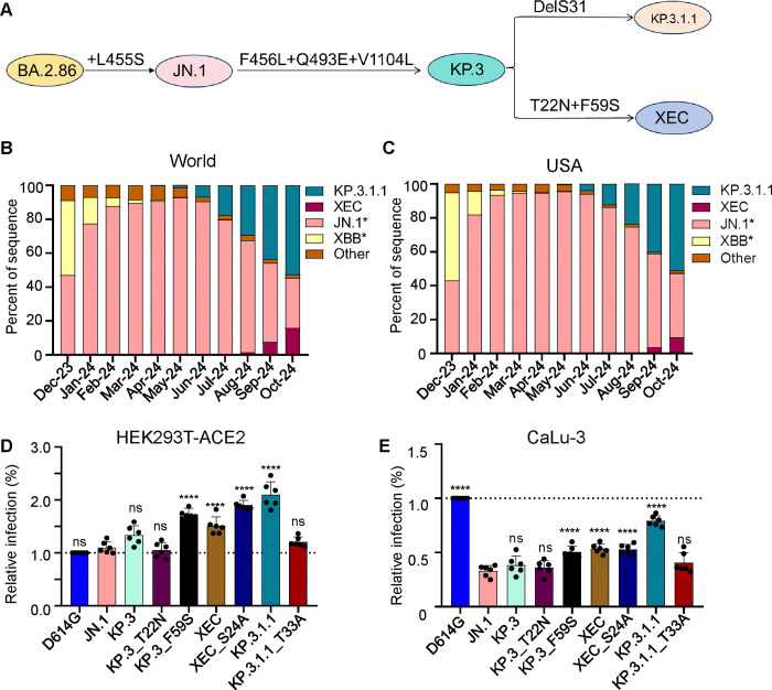
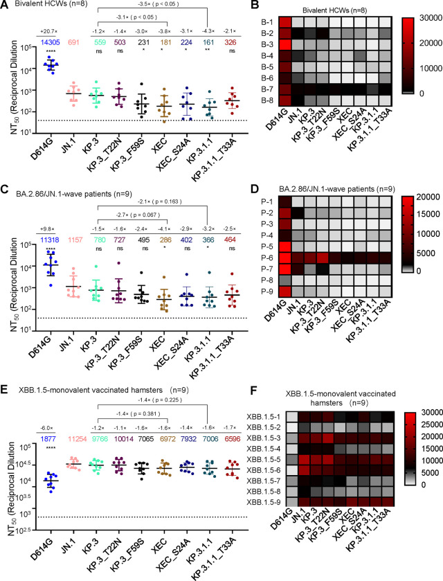
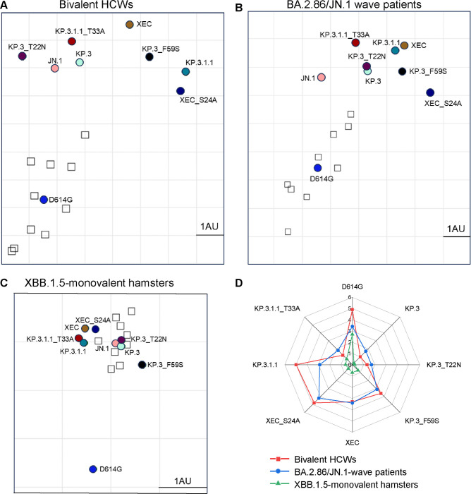
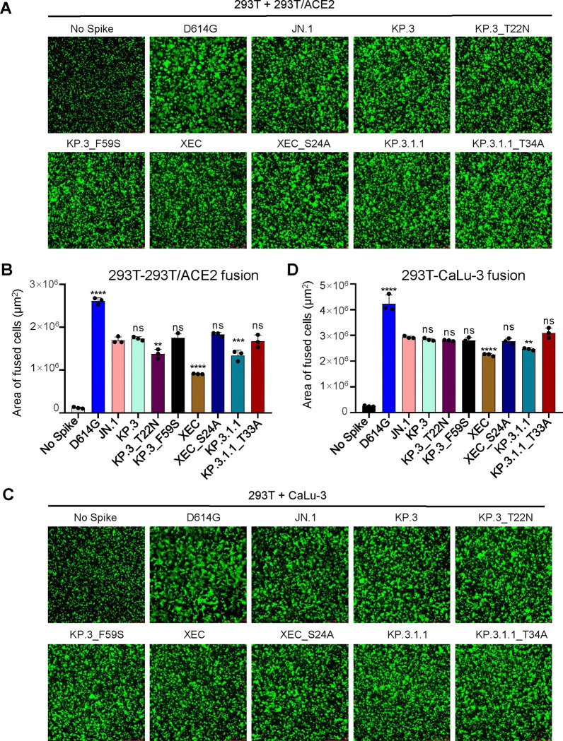
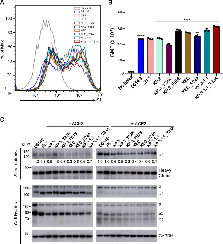
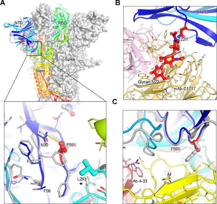
SARS-CoV-2 continues to evolve, producing new variants that drive global COVID-19 surges. XEC, a recombinant of KS.1.1 and KP.3.3, contains T22N and F59S mutations in the spike protein's N-terminal domain (NTD). The T22N mutation, similar to the DelS31 mutation in KP.3.1.1, introduces a potential N-linked glycosylation site in XEC. In this study, we examined the neutralizing antibody (nAb) response and mutation effects in sera from bivalent-vaccinated healthcare workers, BA.2.86/JN.1 wave-infected patients, and XBB.1.5 monovalent-vaccinated hamsters, assessing responses to XEC alongside D614G, JN.1, KP.3, and KP.3.1.1. XEC demonstrated significantly reduced neutralization titers across all cohorts, largely due to the F59S mutation. Notably, removal of glycosylation sites in XEC and KP.3.1.1 substantially restored nAb titers. Antigenic cartography analysis revealed XEC to be more antigenically distinct from its common ancestral BA.2.86/JN.1 compared to KP.3.1.1, with the F59S mutation as a determining factor. Similar to KP.3.1.1, XEC showed reduced cell-cell fusion relative to its parental KP.3, a change attributed to the T22N glycosylation. We also observed reduced S1 shedding for XEC and KP.3.1.1, which was reversed by ablation of T22N and DelS31 glycosylation mutations, respectively. Molecular modeling suggests that T22N and F59S mutations of XEC alters hydrophobic interactions with adjacent spike protein residues, impacting both conformational stability and neutralization. Overall, our findings underscore the pivotal role of NTD mutations in shaping SARS-CoV-2 spike biology and immune escape mechanisms.


Conflict of interest statement

DECLARATION OF INTERESTS The authors have no competing interests to disclose.

Figures

Figure 1:
Figure 1:. Mutations, circulation, and infectivity for JN.1-lineage variants XEC, KP.3.1.1 and KP.3.
(A) Schematic depiction of spike-defining mutations and relationships of JN.1, KP.3, KP.3.1.1, and XEC. (B-C) Frequency of sequences of KP.3.1.1, XEC, JN.1, and XBB worldwide (B) and the United States (C) represented by percentage. (D-E) Relative infectivity of lentivirus pseudotypes bearing spikes of interest as determined by secreted Gaussia luciferase, with D614G set to 1.0 for comparison, in HEK293T cells expressing human ACE2 (D) and CaLu-3 cells (E). Bars represent means with standard deviation of 3 biological replicate and 6 separate luciferase readings. Significance was determined by repeated measures one-way ANOVA in comparison to JN.1 and represented as ns p > 0.05 and ****p < 0.0001.
Figure 2:
Figure 2:. XEC exhibits strong nAb escape.
NAb titers were determined using a HIV-1 pseudotyped vector neutralization assay for three cohorts of sera. (A-B) OSU Wexner Medical Center healthcare workers that received 3 doses of monovalent (WT) mRNA vaccine and 1 dose of bivalent (WT + BA.4/5) mRNA vaccine (n=8). (C-D) COVID-19 patients at the OSU Wexner Medical Center that were admitted during the BA.2.86/JN.1 wave of infection in Columbus, OH (n=9). (E-F) Golden Syrian hamsters vaccinated twice with a monovalent, recombinant Mumps XBB.1.5 mRNA vaccine (n=9). (A, C, and E) Plots represent geometric mean nAb titers at 50% (NT50) with standard error. Geometric mean values are listed at the top of the plots and significance was determined in comparison to JN.1 unless otherwise noted. Fold changes relative to JN.1 are listed above the geometric mean values. (B, D, and F) Heatmaps depicting the nAb titers for each individual in each cohort. Significance was determined in (A, C, and E) using log10 transformed NT50 values using repeated measures one-way ANOVA and represented as ns p > 0.05, *p < 0.05, and ****p < 0.0001.
Figure 3:
Figure 3:. Analysis of antigenicity of XEC and related variants.
(A-C) Antigenic cartography analysis was conducted for the nAb titers results for the bivalent vaccinated HCWs (A), the BA.2.86/JN.1 wave infected patients (B), and the XBB.1.5-monovalent vaccinated hamsters (C). One antigenic distance unit (AU) (AU = 1) represents an approximate two-fold change in NT50. Circles represent the different spike antigens while boxes represent individual sera samples. (D) The antigenic distances of each variant relative to JN.1 from three groups of cohorts (n=3) were averaged and plotted. The scale bar represents 1 antigenic distance unit (AU).
Figure 4:
Figure 4:. XEC exhibits decreased fusogenicity in HEK293T-ACE2 and CaLu-3 cells.
Fusion of spikes was determined in HEK293T-ACE2 cells (A-B) and CaLu-3 cells (C-D). Representative images of fusion are depicted for 293T-ACE2 (A) and CaLu-3 (C), and quantification of total areas of fusion across 3 images are represented for in (B) 293T-ACE2 and (D) CaLu-3 cells. Areas of fused cells were determined using microscope software (see Methods). Plots represent means with standard deviation with significance determined in comparison to JN.1. Significance was calculated using repeated measures one-way ANOVA and represented as ns p > 0.05, **p < 0.01, ****p < 0.0001.
Figure 5:
Figure 5:. The surface expression, processing, and S1 shedding of XEC spike and related variants.
(A-B) Surface expression of spikes on the membrane of HEK293T cells was determined using flow cytometry using anti-S1 antibody. (A) Representative histograms and (B) plots of averaged geometric mean fluorescence intensity (GMFI) are depicted. Plots represented geometric mean fluorescence intensities with standard deviation. Significance in (B) was determined using repeated measures one-way ANOVA and represented as ****p < 0.0001.(C) Spike expression in transfected cells and S1 shedding. HEK293T cells were transfected with spike constructs of interest and treated with or without sACE2 (10 μg/ml) for 4 h. Cell culture media and lysates were collected, with shed S1 proteins being immunoprecipitated with an anti-S1 antibody. Cell lysates with or without sACE2 were blotted with anti-S2, anti-S1 and anti-GAPDH antibodies, and relative signals were quantified by NIH ImageJ (30) by setting the value of JN.1 to 1.0. Images from one representative experiment are shown.
Figure 6.
Figure 6.. Structural modeling of key mutations in XEC spike.
(A) Structural representation of the spike protein domains, with the location of NTD mutations T22N and F59S highlighted. The spike is shown with two protomers displayed as a grey surface and one as a ribbon in rainbow colors. Inset: The F59S mutation alters its side-chain interaction with several nearby hydrophobic residues, including F32, F58, and L293, while introducing a hydrogen bond with residue N30. (B) The glycosylation at N22 (shown as sticks) interferes with the recognition of certain NTD-targeting antibodies, such as C1717, potentially reducing antibody binding efficiency. (C) The F59S mutation disrupts the epitopes of NTD-targeting antibodies, such as 4–33, by abolishing the interaction with a hydrophobic cluster, thereby impairing antibody recognition and contributing to immune evasion.

References

    1. Nesteruk I. 2023. Endemic characteristics of SARS-CoV-2 infection. Sci Rep 13:14841. - PMC - PubMed
    1. Contreras S, Iftekhar EN, Priesemann V. 2023. From emergency response to long-term management: the many faces of the endemic state of COVID-19. Lancet Reg Health Eur 30:100664. - PMC - PubMed
    1. Lavine JS, Bjornstad ON, Antia R. 2021. Immunological characteristics govern the transition of COVID-19 to endemicity. Science 371:741–745. - PMC - PubMed
    1. Gangavarapu K, Latif AA, Mullen JL, Alkuzweny M, Hufbauer E, Tsueng G, Haag E, Zeller M, Aceves CM, Zaiets K, Cano M, Zhou X, Qian Z, Sattler R, Matteson NL, Levy JI, Lee RTC, Freitas L, Maurer-Stroh S, Core G, Curation T, Suchard MA, Wu C, Su AI, Andersen KG, Hughes LD. 2023. Outbreak.info genomic reports: scalable and dynamic surveillance of SARS-CoV-2 variants and mutations. Nat Methods 20:512–522. - PMC - PubMed
    1. Callaway E. 2023. Why a highly mutated coronavirus variant has scientists on alert. Nature 620:934. - PubMed

Publication types

LinkOut - more resources