[Preprint]. 2024 Nov 17:2024.11.16.623943.
doi: 10.1101/2024.11.16.623943.

IL-33 protects from recurrent C. difficile infection by restoration of humoral immunity

Affiliations

IL-33 protects from recurrent C. difficile infection by restoration of humoral immunity

Farha Naz et al. bioRxiv. .

Update in

Abstract

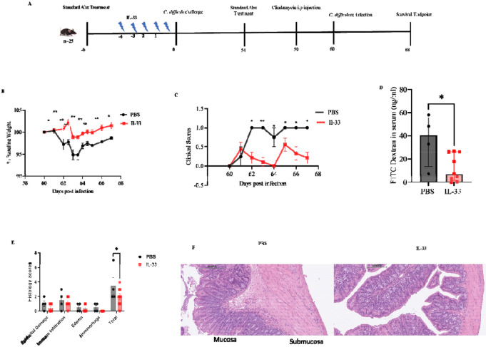
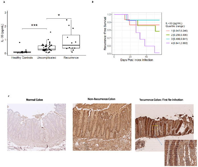
Clostridioides difficile infection (CDI) recurs in one of five patients. Monoclonal antibodies targeting the virulence factor TcdB reduce disease recurrence, suggesting that an inadequate anti-TcdB response to CDI leads to recurrence. In patients with CDI, we discovered that IL-33 measured at diagnosis predicts future recurrence, leading us to test the role of IL-33 signaling in the induction of humoral immunity during CDI. Using a mouse recurrence model, IL-33 was demonstrated to be integral for anti-TcdB antibody production. IL-33 acted via ST2+ ILC2 cells, facilitating germinal center T follicular helper (GC-Tfh) cell generation of antibodies. IL-33 protection from reinfection was antibody-dependent, as μMT KO mice and mice treated with anti-CD20 mAb were not protected. These findings demonstrate the critical role of IL-33 in generating humoral immunity to prevent recurrent CDI.

Keywords: Clostridioides difficile; GC-TFH; IL-33 signaling; ILC2s; TH17 cells; dysbiosis; recurrent C. difficile infection; toxin-specific antibody.


Figures

Fig 1:
Fig 1:. IL-33 increases toxin-specific antibody in mice after first infection with C. difficile
IL-33 (0.75 μg) was administered i.p. on days −4 to 0 to C57BL/6J mice (A–H) and/or ST2−/− mice (I-M). Mice were infected with C. difficile strain R20291 (A-E and G-M) or VPI 10463 (F). On post-infection day 15 antibodies were measured in plasma and cecal content. (A) Schematic diagram showing infection and treatment timeline; (B) survival curves; (C) weight loss; (D) clinical scores; (E) plasma toxin B specific IgG from mice infected with C. difficile strain R20291 ; (F) plasma toxin B specific IgG from mice infected with C. difficile strain VPI 10463; (G) plasma toxin B specific IgM from mice infected with C. difficile strain R20291 ; (H) cecal content toxin B specific IgA from mice infected with C. difficile strain R20291. (I-M) WT vs ST2−/− mice infected with C. difficile strain R20291 : (I) Schematic diagram showing infection and treatment timeline; (J) plasma IgG; (K) cecal IgG; (L) cecal IgA; and (M) plasma IgM. B, Comparison made by log-rank test ( n = 30 in both groups). C, D, Comparison made by two-tailed Student’s t-test (C, D n = 30). E, F, G, H, J, K, L, M, A two-tailed t-test for normally distributed data and a Mann-Whitney test for non-normally distributed data were used. *P < 0.05, **P < 0.01, and ***P < 0.001. The error bar indicates SEM.
Fig 2:
Fig 2:. IL33 protects from a 2nd C. difficile infection
IL-33 (0.75 μg) was administered i.p. on days −4 to 0 and wild-type mice were infected on day 0 and again on day 60 with C. difficile strain R20291. (A) Experimental design for 2nd infection; (B) 2nd infection weight loss; (C) clinical scores; (D) FITC-dextran gut permeability test; (E) epithelial damage scoring; (F) representative H&E stain of the colon. C, D, Comparison made by two-tailed Student’s t-test (C, D n = 13). D, Mann-Whitney test for non-normally distributed data was used E, Šídák's multiple comparisons test was used to determine the statistical significance between groups. Statistical significance is demarked as *P < 0.05, **P < 0.01, and ***P < 0.001. The error bar indicates SEM in B, D, and E but D indicates the median with interquartile range.
Fig 3:
Fig 3:. IL33 protection from a 2nd C. difficile infection is antibody-dependent
WT and μMT KO mice were administered IL-33 (0.75 μg) i.p. on days −4 to 0 and mice infected on day 0 and reinfected on day 60 with C. difficile strain R20291. (A) Experimental design; (B) 1st infection weight loss; (C) clinical scores. WT and μMT KO mice were reinfected with C. difficile R20291 60 days after the first infection. Reinfection (D) weight loss; (E) stool C. difficile toxin A and B; (F) FITC-dextran gut permeability assay; (G) H&E stain; (I) epithelial damage scoring. B, C, D, Comparison made by two-tailed Student’s t-test (B, C n = 30, D n=18). E, a two-tailed t-test was used and the error bar indicates SEM. F, a Mann-Whitney test was used and the error bar indicates the median with interquartile range. H, Šídák's multiple comparisons test was used. Statistical significance is demarked as *P < 0.05, **P < 0.01, and ***P < 0.001.
Fig 4:
Fig 4:. Antibody deficient mice (αCD20 treated) lost IL-33 mediated protection from a 2nd C. difficile infection
αCD20 was administered to deplete B cells on days −7, 3, 18, 33, 48, and 63, and mice infected on day 0 and 2nd time infected on day 60 with C. difficile strain R20291. (A) Experimental design; (B) survival curve; (C) 1st infection weight loss; (D) 1st infection clinical scores; (E) reinfection weight loss ; (F) reinfection clinical scores (G) epithelial damage scoring; (H) H&E stain. (I) toxin B specific plasma IgG; (J) IgA; (K) cecal IgG; and (L) cecal IgA measured on day 10 post 2nd infection. MLN and colon were harvested on day 10 post-reinfection. Colonic (M, N) B cells ( CD45+ CD3− CD19+); (O, P) TH2 cells (CD45+CD3+ CD4+ GATA3+); (Q, R) neutrophils; (S, T, U) Treg (CD45+CD3+ CD4+ FOXP3+)and TH17(CD45+CD3+ CD4+ RORγt+) cells; and (V, W) MLN B cells. B, Comparison made by log-rank test ( n = 26 in both groups). C, D, E, F Comparison made by two-tailed Student’s t-test (C, D n = 26, E, F n=15). G, Šídák's multiple comparisons test was used. I, J, K, L, M, P, Q, T, U, V A two-tailed t-test for normally distributed data and a Mann-Whitney test for non-normally distributed data were used. Statistical significance is demarked as *P < 0.05, **P < 0.01, ***P < 0.001, and ****P < <0.0001. The error bar indicates SEM.
Fig 5:
Fig 5:. IL-33 increased mesenteric lymph node ILC2 and decreased TH1 cells during 1st C. difficile infection
IL-33 (0.75 μg) was administered i.p. on days −4 to 0 and mice infected on day 0. (A, B) ILC populations on day 0, prior to infection, and (C, D) at 2 days post-infection. TH1, TH2 populations (E-H) before infection, day 0; (I-L) day 2; and (M-P) day 6 post-infection. A two-tailed t-test for normally distributed data and a Mann-Whitney test for non-normally distributed data were used. Statistical significance is demarked as *P < 0.05, **P < 0.01, ***P < 0.001, and ****P < <0.0001. The error bar indicates SEM.
Fig 6:
Fig 6:. IL-33 increased mesenteric lymph node GC-TFH, Tregs, and decreased TH17 cells during a primary C. difficile infection
IL-33 (0.75 μg) was administered i.p. on days −4 to 0 and mice infected on day 0. Mesenteric lymph nodes were harvested to analyze T helper cells before infection, day 2, and day 6 post-first infection. (A-C) TH17 cells (CD45+, CD3+, CD4+, RORγt+) and Treg cells (CD45+, CD3+, CD4+, FOXP3+) on day 0 prior before infection; (D-F) on day 2; (G-I) on day 6 post-infection. (J-M) TFH cells were defined as germinal center (GC) TFH (CD45+ CD3+ CD4+ CD44+ PD1 high CXCR5+) and non-GC TFH cells (CD45+ CD3+ CD4+ CD44+ PD1 low CXCR5+) by flow cytometry. (J-K) TFH subsets on day 0 prior to infection; (L-M) TFH subsets on day 6 post-infection. A two-tailed t-test for normally distributed data and a Mann-Whitney test for non-normally distributed data were used. Statistical significance is demarked as *P < 0.05, **P < 0.01, and ***P < 0.001. The error bar indicates SEM.
Fig 7:
Fig 7:. Adoptive transfer of ST2+ ILC2s into ST2 KO mice induces TFH cells and increased Toxin B specific antibody to protect from reinfection
ST2+ ILC2s (from uninfected IL-33 treated mice) were ex-vivo expanded, purified by flow-sorting and adoptively transferred into ST2 KO mice. (A-C) Mice were pretreated with antibiotics and injected with 0.75μg per dose per mouse of IL-33 in the gut one day after the adoptive transfer and one day before the R20291 infection. At 16 days post-primary infection, plasma toxin B-specific (B) IgG and (C) IgA antibodies were measured in plasma. (D-N) Mice were rechallenged with C. difficile on day 60. On day 70 (10 days post-2nd infection), (D-F), ILC2 were measured in the colon and mesenteric lymph nodes. (G-H) GC-TFH measured in the MLN (I) Day 70 representative epithelial damage (H&E) of treatment groups and (J) assessed by blinded scoring of infected tissue. A two-tailed t-test for normally distributed data and a Mann-Whitney test for non-normally distributed data were used. J , Šídák's multiple comparisons test was used. Statistical significance is demarked as *P < 0.05, **P < 0.01, and ***P < 0.001. The error bar indicates SEM.
Fig 8:
Fig 8:. IL-33 is a biomarker for recurrent C. difficile infection in humans
(A) Plasma IL-33 was measured in healthy controls (n = 17), patients with uncomplicated CDI (n=41), and recurrent CDI (n=12) (excluding 5 patients who died) within 8 weeks of diagnosis. (B) Recurrence-free survival among the patients with C. difficile infection (1A), grouped by serum IL-33 quartile (Wilcoxon P=0.002). (C) Immunohistochemical staining of IL-33 from colon tissue biopsies of patients without or with recurrence. The Kaplan–Meier method was used to measure recurrence-free survival curves and to evaluate the effects of IL-33 on risk for recurrent C. difficile infection. To account for competing risk against recurrent infection, death within the 8-week follow-up period was treated as a censoring event.

References

    1. Guh AY, Mu Y, Winston LG, Johnston H, Olson D, Farley MM, et al. Trends in US Burden of Clostridioides difficile Infection and Outcomes. New Engl J Med. 2020;382(14):1320–30. - PMC - PubMed
    1. Naz F, and Petri WA. Host Immunity and Immunization Strategies for Clostridioides difficile Infection. Clin Microbiol Rev. 2023;36(2):e0015722. - PMC - PubMed
    1. van Opstal E, Kolling GL, Moore JH 2nd, Coquery CM, Wade NS, Loo WM, et al. Vancomycin Treatment Alters Humoral Immunity and Intestinal Microbiota in an Aged Mouse Model of Clostridium difficile Infection. J Infect Dis. 2016;214(1):130–9. - PMC - PubMed
    1. Merrick B, Allen L, Masirah MZN, Forbes B, Shawcross DL, and Goldenberg SD. Regulation, risk and safety of Faecal Microbiota Transplant. Infect Prev Pract. 2020;2(3):100069. - PMC - PubMed
    1. Moreau GB, Naz F, and Petri WA Jr., Fecal microbiota transplantation stimulates type 2 and tolerogenic immune responses in a mouse model. Anaerobe. 2024;86:102841. - PMC - PubMed

Publication types