[Preprint]. 2024 Nov 18:2024.11.18.622547.
doi: 10.1101/2024.11.18.622547.

Accurate de novo design of high-affinity protein binding macrocycles using deep learning

Affiliations

Accurate de novo design of high-affinity protein binding macrocycles using deep learning

Stephen A Rettie et al. bioRxiv. .

Update in

  • Accurate de novo design of high-affinity protein-binding macrocycles using deep learning.
    Rettie SA, Juergens D, Adebomi V, Bueso YF, Zhao Q, Leveille AN, Liu A, Bera AK, Wilms JA, Üffing A, Kang A, Brackenbrough E, Lamb M, Gerben SR, Murray A, Levine PM, Schneider M, Vasireddy V, Ovchinnikov S, Weiergräber OH, Willbold D, Kritzer JA, Mougous JD, Baker D, DiMaio F, Bhardwaj G. Rettie SA, et al. Nat Chem Biol. 2025 Jun 20. doi: 10.1038/s41589-025-01929-w. Online ahead of print. Nat Chem Biol. 2025. PMID: 40542165

Abstract

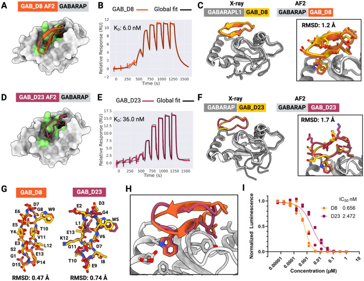
The development of macrocyclic binders to therapeutic proteins typically relies on large-scale screening methods that are resource-intensive and provide little control over binding mode. Despite considerable progress in physics-based methods for peptide design and deep-learning methods for protein design, there are currently no robust approaches for de novo design of protein-binding macrocycles. Here, we introduce RFpeptides, a denoising diffusion-based pipeline for designing macrocyclic peptide binders against protein targets of interest. We test 20 or fewer designed macrocycles against each of four diverse proteins and obtain medium to high-affinity binders against all selected targets. Designs against MCL1 and MDM2 demonstrate KD between 1-10 μM, and the best anti-GABARAP macrocycle binds with a KD of 6 nM and a sub-nanomolar IC50 in vitro. For one of the targets, RbtA, we obtain a high-affinity binder with KD < 10 nM despite starting from the target sequence alone due to the lack of an experimentally determined target structure. X-ray structures determined for macrocycle-bound MCL1, GABARAP, and RbtA complexes match very closely with the computational design models, with three out of the four structures demonstrating Ca RMSD of less than 1.5 Å to the design models. In contrast to library screening approaches for which determining binding mode can be a major bottleneck, the binding modes of RFpeptides-generated macrocycles are known by design, which should greatly facilitate downstream optimization. RFpeptides thus provides a powerful framework for rapid and custom design of macrocyclic peptides for diagnostic and therapeutic applications.


Figures

Figure 1:
Figure 1:. RFpeptides is a diffusion-based pipeline for the de novo design of protein-binding macrocycles.
(A) Introduction of a cyclically symmetric relative position encoding to RFdiffusion enables the generation of macrocyclic peptide backbones with N- and C-termini covalently linked via a peptide bond. The relative position encodings are cyclized by switching from positive relative position encodings (i.e., “to the right”) to negative encodings (i.e., “to the left”) when index j is more than halfway around the peptide relative to index i. (B) RFpeptides produces diverse and designable cyclic peptides traversing α-helical, β-strand, and loop/coil secondary structures. Left, Structural clusters calculated using t-SNE to reduce the dimensionality of an all-by-all TMscore matrix computed with TMalign on an unfiltered set of 1200 macrocycles generated using RFpeptides. Clustering of the 1200 (now 2D) points was then performed using a 40-component Gaussian mixture model in Scikit-learn. Right, Six RFpeptides outputs from differing structural clusters, all with < 1 Å backbone RMSD between the design model (blue) and the structure predicted by AfCycDesign (gold). (C) Self-consistency benchmark results for RFpeptides cyclic peptide design at various lengths. For each peptide length, the fraction (with N=200 per length) of backbones with at least one out of the of eight LigandMPNN sequences predicted by AfCycDesign to refold with pLDDT > 0.8 and within 2.0 Å backbone RMSD of the designed structure. Success rates for all sampled backbones are in blue, and success rates only counting unique structural clusters (as calculated using MaxCluster, at a TMscore threshold of 0.5) are in orange. (D) For multi-chain diffusion trajectories (e.g., macrocycle binder design), the relative position encodings for the macrocycle chain are cyclized, whereas inter-chain and target chain relative positional encoding are kept as standard. (E) Pipeline for the design of protein-binding macrocycles using RFpeptides. Macrocycle backbones are generated from randomly initialized atoms by a stepwise RFdiffusion-based denoising process, followed by fixed-backbone amino acid sequence design using ProteinMPNN. Design models are downselected based on the computational metrics from structure prediction using AfCycDesign and physics-based interface quality metrics using Rosetta. (F) RFpeptides pipeline generates diverse macrocycles against selected targets. Four diverse cyclic peptide binders against the same target were generated using RFpeptides, with AfCycDesign iPAE < 0.3 and < 1.5 Å Cα RMSD between the design model (blue) and AfCycDesign prediction (gold).
Figure 2:
Figure 2:. De novo design and characterization of macrocyclic binders to MCL1 and MDM2
(A) AfCycDesign prediction of MCB_D2 (purple) bound to MCL1 (gray surface). MCB_D2 side chains are shown as sticks. (B) Affinity determination of MCB_D2 using SPR. SPR sensorgram from a 9-point single-cycle kinetics experiment (2-fold dilution, highest concentration: 20 μM). Experimental data are shown in purple and global fits are shown with black lines. The dissociation constant (KD) is also shown on the plot. (C) Experimentally determined complex structures closely match the design model. Overlap of the X-ray crystal structure (gold and gray) with the design model for MCB_D2 (purple); Cα RMSD for macrocycle is 0.7 Å when experimental structure and design models are aligned by MCL1 residues. Close-up views demonstrate strong agreement between the side chain rotamers of the design model and the X-ray structure. (D) Overlay of the macrocycle model to the crystal structure shows a Cα RMSD of 0.4 Å with nearly identical backbones and side chain rotamers. (E) Close-up view of the macrocycle-bound MCL1 structure showing the cation-π interaction at the interface. (F) Close-up view of the macrocycle-bound MCL1 structure showing the hydrophobic contacts at the interface. (G) AfCycDesign prediction of MDB_D8 design (blue) in complex with MDM2 (gray) shown as cartoons with interacting side chains shown as sticks. (H) Affinity determination of MDB_D8 using SPR. SPR sensorgram from a 9-point single cycle kinetics experiment (5-fold dilution, highest concentration: 50 μM). Experimental data are shown in blue and global fits are shown with black lines. The dissociation constant (KD) is also shown on the plots. (I) Overall and close-up views of the AfCycDesign prediction of the MDB_D8 design model highlighting key interactions with the MDM2.
Figure 3:
Figure 3:. De novo design of high-affinity macrocycle binders to GABARAP
(A) AfCycDesign predicted model for design GAB_D8 bound to GABARAP shown as surface, with hotspot residues highlighted in green. (B) Affinity determination of GAB_D8 using SPR. SPR sensorgram from 9-point single-cycle kinetics experiments (5-fold dilution, highest concentration 20 μM). Experimental data are shown in orange and global fits are shown with black lines. The dissociation constant (KD) is also shown on the plot. (C) Superposition of chains E and F from the X-ray crystal structure of GAB_D8 bound to GABARAPL1 and the AfCycDesign model. (D) AfCycDesign predicted model for design GAB_D23 bound to GABARAP shown as surface, with hotspot residues highlighted in green. (E) Affinity determination of GAB_D23 using SPR. SPR sensorgram from 9-point single-cycle kinetics experiments (5-fold dilution, highest concentration 20 μM). Experimental data are shown in pink and global fits are shown with black lines. The dissociation constant (KD) is also shown on the plot. (F) Alignment of chains A and B from the X-ray crystal structure of GAB_D23 bound to GABARAP and the AfCycDesign model. (G) Alignments of GAB_D8 and GAB_D23 macrocycle models to X-ray crystal structures show close matches. (H) Comparison of GAB_D8 and GAB_D23 binding modes in the design models. (I) Competitive AlphaScreen response vs. concentration plot, IC50 from the average of three experiments. Donor and acceptor beads in the assay are bound to GABARAP and GABARAP-binding peptide K1, respectively.
Figure 4:
Figure 4:. Accurate de novo design of a high-affinity cyclic peptide binder against the predicted structure of RbtA from A. baumannii
(A) AfCycDesign prediction of design RBB_D10 (violet cartoon) bound to the AF2 predicted β-helix domain of RbtA shown in gray surface. Hotspot residues from RbtA used during the backbone design step are shown in green. (B) SPR sensorgram from 9-point single-cycle kinetics experiment (5-fold dilution, highest concentration 20 μM). KD determined from the SPR experiment also denoted on the plot. (C) Close agreement of the RF2-predicted structure of RbtA (gray) with the X-ray structure (gold) of the RbtA N-terminal domain determined here confirms the predicted structure of the target used for the macrocycle design calculations. (D) Alignment of the design model of RbtA-bound RBB_D10 (violet/gray) to the X-ray structure (in gold) shows a close match between the design model and the experimentally determined structure (Cɑ RMSD for macrocycle: 1.4 Å). Close-up view of the RbtA-bound RBB_D10 with side chains shown as sticks. (E) Overlay of RBB_D10 design model (after the AfCycDesign prediction step) aligned to the X-ray structure without RbtA demonstrates a nearly identical match for backbone coordinates and side chain rotamers (Cɑ RMSD: 0.4 Å). The design model and X-ray structure are shown in violet and gold, respectively. (F-H) Close-up views of the macrocycle-bound RbtA structure and the design model showing the accurate design of electrostatic and hydrophobic interactions at the binding interface.

References

    1. Vinogradov A. A., Yin Y. & Suga H. Macrocyclic Peptides as Drug Candidates: Recent Progress and Remaining Challenges. J. Am. Chem. Soc. 141, 4167–4181 (2019). - PubMed
    1. Muttenthaler M., King G. F., Adams D. J. & Alewood P. F. Trends in peptide drug discovery. Nat. Rev. Drug Discov. (2021) doi: 10.1038/s41573-020-00135-8. - DOI - PubMed
    1. Tsomaia N. Peptide therapeutics: targeting the undruggable space. Eur. J. Med. Chem. 94, 459–470 (2015). - PubMed
    1. Atanasov A. G., Zotchev S. B., Dirsch V. M., International Natural Product Sciences Taskforce & Supuran, C. T. Natural products in drug discovery: advances and opportunities. Nat. Rev. Drug Discov. 20, 200–216 (2021). - PMC - PubMed
    1. Bhardwaj G. et al. Accurate de novo design of membrane-traversing macrocycles. Cell (2022) doi: 10.1016/j.cell.2022.07.019. - DOI - PMC - PubMed

Publication types