. 2024 Dec 11;146(49):33675-33711.
doi: 10.1021/jacs.4c11556. Epub 2024 Nov 28.

Leveraging Dual-Ligase Recruitment to Enhance Protein Degradation via a Heterotrivalent Proteolysis Targeting Chimera

Affiliations

Leveraging Dual-Ligase Recruitment to Enhance Protein Degradation via a Heterotrivalent Proteolysis Targeting Chimera

Adam G Bond et al. J Am Chem Soc. .

Abstract

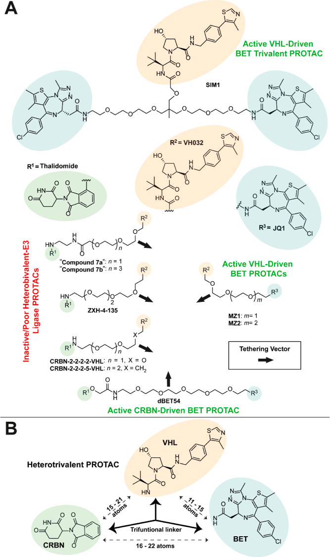
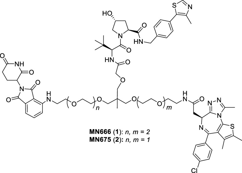
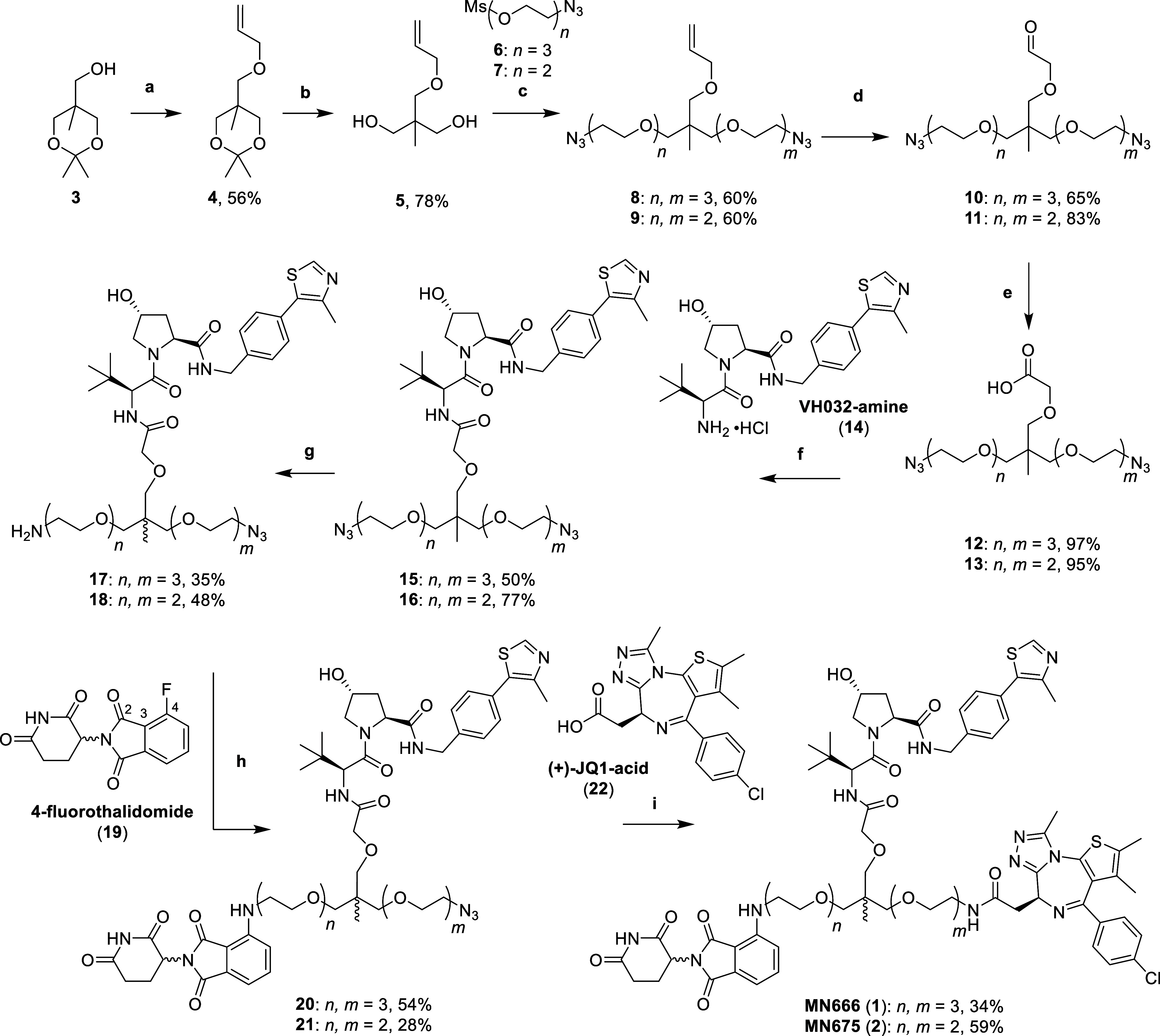
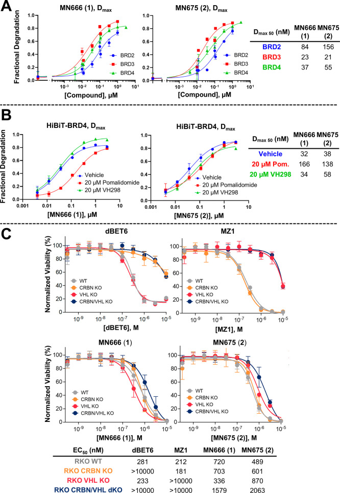
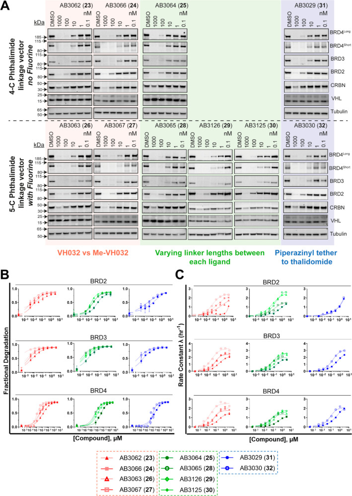
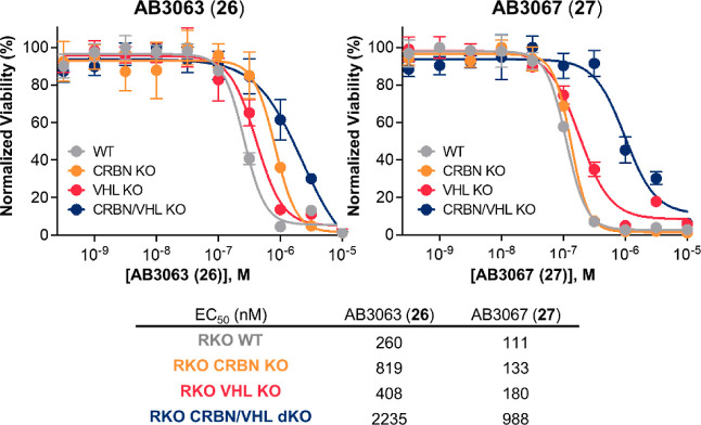
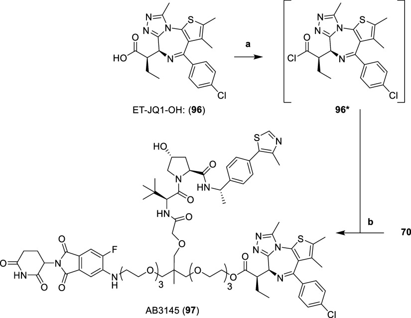
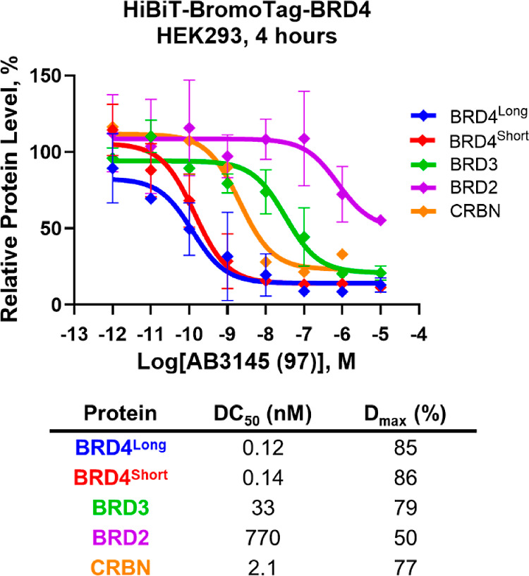
Proteolysis targeting chimera (PROTAC) degraders are typically bifunctional with one E3 ligase ligand connected to one target protein ligand via a linker. While augmented valency has been shown with trivalent PROTACs targeting two binding sites within a given target protein, or used to recruit two different targets, the possibility of recruiting two different E3 ligases within the same compound has not been demonstrated. Here we present dual-ligase recruitment as a strategy to enhance targeted protein degradation. We designed heterotrivalent PROTACs composed of CRBN, VHL and BET targeting ligands, separately tethered via a branched trifunctional linker. Structure-activity relationships of 12 analogues qualifies AB3067 as the most potent and fastest degrader of BET proteins, with minimal E3 ligase cross-degradation. Comparative kinetic analyses in wild-type and ligase single and double knockout cell lines revealed that protein ubiquitination and degradation induced by AB3067 was contributed to by both CRBN and VHL in an additive fashion. We further expand the scope of the dual-ligase approach by developing a heterotrivalent CRBN/VHL-based BromoTag degrader and a tetravalent PROTAC comprising of two BET ligand moieties. In summary, we provide proof-of-concept for dual-E3 ligase recruitment as a strategy to boost degradation fitness by recruiting two E3 ligases with a single degrader molecule. This approach could potentially delay the outset of resistance mechanisms involving loss of E3 ligase functionality.


Conflict of interest statement

The authors declare the following competing financial interest(s): The University of Dundee has filed a PCT patent application PCT/GB2024/051314 on May 24, 2024 covering the chemical structures and their use. A.C., A.G.B., and N. M. are inventors of this patent. A.C. is a scientific founder and shareholder of Amphista Therapeutics, a company that is developing targeted protein degradation therapeutic platforms. The Ciulli laboratory receives or has received sponsored research support from Almirall, Amgen, Amphista Therapeutics, Boehringer Ingelheim, Eisai, Merck KaaG, Nurix Therapeutics, Ono Pharmaceutical and Tocris-Biotechne. G.E.W. is scientific founder and shareholder of Proxygen and Solgate. The Winter lab received research funding from Pfizer. C.M.B., E.A.C., M.U. and K.M.R. are or were employees of Promega Corporation. Promega Corporation is the commercial owner by assignment of patents of the HaloTag, NanoLuc, NanoBRET target engagement, and HiBiT technologies and their applications. Other authors declare no competing interests.

Figures

Figure 1
Figure 1
Heterotrivalent PROTAC design rationale. (A) Active VHL-driven BET trivalent PROTAC, SIM1 (top), and bivalent PROTACs, MZ1 and MZ2 (middle right). Active CRBN-driven BET PROTAC, dBET54 (bottom). Inactive CRBN-VHL Heterobifunctional-E3 Ligase PROTACs, “Compounds 7a & 7b”, ZXH-4–135, CRBN-2–2–2–2-VHL and CRBN-2–2–2–5-VHL (middle left). VHL ligand, VH032 (orange), BET ligand, JQ1 (blue), and CRBN ligand, thalidomide (green) are highlighted. Black arrows indicate potential vectors for linker tethering. (B) Simplified structure of a heterotrivalent PROTAC labeled with optimal linker lengths required between each ligand to have active VHL/CRBN driven BET degradation and to avoid cross-ligase degradation of VHL and/or CRBN.
Figure 2
Figure 2
Chemical structures of first generation heterotrivalent PROTACs. Chemical structures of MN666 (1) and MN675 (2).
Scheme 1
Scheme 1. Synthesis of Initial Heterotrivalent PROTACs MN666 (1) and MN675 (2)
Reaction conditions: (a) allyl bromide, KOH, TBAB, toluene, H2O, r.t., 16 h; (b) TFA, MeOH, H2O, r.t., 3 h; (c) (i) NaH, DMF, 0 °C, 30 min, (ii) 6 or 7, DMF, 60 °C, 16 h; (d) OsO4, NaIO4, 2,6-lutidine, dioxane, H2O, r.t., 16 h; (e) 2-methyl-2-butene, NaH2PO4, NaClO2, t-BuOH, H2O, r.t., 16 h; (f) VH032-amine (14), HATU, DIPEA, DMF, r.t., 2 h; (g) PPh3, EtOAc, 1.0 M HCl (aq); (h) 19, DIPEA, NMP, 100–120 °C, 4 h; (i) (i) H2, 10% Pd/C, MeOH, r.t., 16 h, (ii) (+)-JQ1-acid (22), HATU, DIPEA, DMF, r.t., 2 h.
Figure 3
Figure 3
Cellular evaluation of MN666 (1) and MN675 (2). (A) (B) Degradation potency of 1 and 2 from live cell kinetic profiles in HiBiT-BRD CRISPR knock-in HEK293 cells plotted as fractional degradation at Dmax versus concentration of 1 (left) and 2 (right). Cells were treated with DMSO and a threefold serial dilution of 1 or 2 over a concentration range of 4 nM to 3 μM without (A) or with (B) 20 μM of either CRBN inhibitor pomalidomide, or VHL inhibitor VH298. Dmax 50 is tabulated. Mean ± S.D.; n = 3 biological replicates (A) or n = 1 biological replicates (B). (C) Cell viability assay in BET sensitive wild-type and CRBN/VHL knockout RKO cell lines. Cell antiproliferation of MZ1 and dBET6 (top) compared to 1 and 2 (bottom) after 316 pM to 10 μM treatment in WT, CRBN KO, VHL KO or CRBN/VHL dKO RKO cell lines. Mean ± S.D.; n = 6 biological replicates. EC50 values are tabulated below and in Table S1 with 95% CI.
Scheme 2
Scheme 2. Synthesis of Trifunctional Linkers 53–57
Reaction conditions: (a) MEMCl or MOMBr, DIPEA, DCM, r.t., 16 h; (b) MsCl, DIPEA, DCM, r.t., 3 h; (c) (i) NaH, DMF, 0 °C, 30 min, (ii) 6 or 7, DMF, 60 °C, 16 h; (d) (i) NaH, DMF, 0 °C, 30 min, (ii) 39 or 40, DMF, 60 °C, 16 h; (e) (i) NaH, DMF, 0 °C, 30 min, (ii) 38, DMF, 60 °C, 16 h; (f) OsO4, NaIO4, 2,6-lutidine, dioxane, H2O, r.t., 16 h; (h) 2-methyl-2-butene, NaH2PO4, NaClO2, t-BuOH, H2O, r.t., 16 h. Products 4347 formed through step (f) then (g). Product 47 formed directly from step (e).
Scheme 3
Scheme 3. Synthesis of Aniline Tethered Heterotrivalent PROTACs 23–30
Reaction conditions: (a) HATU, DIPEA, DMF, r.t., 2 h; (b) (i) 5964, H2, 10% Pd/C, MeOH, r.t., 16 h, (ii) 19 or 65, DIPEA, DMSO, 90 °C, 16 h; (c) SOCl2, DCM, r.t., 3 h; (d) (i) 4 N HCl in dioxane, MeOH, r.t., 3 h, (ii) 22*, DIPEA, DCM, r.t., 16 h. Product used further in Scheme 4.
Scheme 4
Scheme 4. Synthesis of Piperazinyl Tethered Heterotrivalent PROTACs 31 & 32
Reaction conditions: (a) (i) 4 N HCl in dioxane, MeOH, r.t., 1 h, (ii) 22*, DIPEA, DCM, r.t., 16 h; (b) (i) MsCl, DIPEA, DCM, 0 °C, 20 min, (ii) r.t., 1 h; (c) 1-Boc-piperazine, DIPEA, DMSO, 90 °C, 16 h; (d) (i) 76 or 77, 4 N HCl in dioxane, DCM, r.t., 16 h, (ii) 75, DIPEA, DMF, 80 °C, 16 h.
Figure 4
Figure 4
Evaluation of cellular BET degradation for heterotrivalent PROTACs 23–32 in HEK293 cells. (A) Western blot data for BET, CRBN and VHL protein levels monitored from 1 μM to 100 pM compound treatments over 6 h in HEK293 cells. Blots arranged with nonfluorinated compounds 2325 and 31 on top, and fluorinated compounds 2630 and 32 on bottom. Bands are normalized to tubulin and vehicle control (DMSO) to derive DC50 values that enable rank ordering of each PROTAC. (B) Degradation potency and (C) rate constants extracted from kinetic degradation profiles of HEK293 HiBiT-BRD2, HiBiT-BRD3, or HiBiT-BRD4 cells treated with 3 μM to 4 nM compound. Compounds with fluorine represented by open symbols, compounds with no fluorine represented by closed symbols. Mean ± S.D.; n = 2 biological replicates (six technical replicates) (BRD4) or n = 1 biological replicate (three technical replicates) (BRD2 and BRD3). Dmax 50 and λmax values are tabulated in Tables S2 and S3, respectively, with 95% CI.
Figure 5
Figure 5
Cell viability assay with 26 and 27 in BET sensitive WT and CRBN/VHL KO RKO cell lines. Effect on cellular proliferation of 26 (left) and 27 (right) after 316 pM to 10 μM treatment in WT, CRBN KO, VHL KO or CRBN/VHL dKO RKO cell lines. Mean ± S.D.; n = 3 biological replicates. EC50 values are tabulated below and in Table S5 with 95% CI.
Figure 6
Figure 6
Live cell ternary complex formation between VHL or CRBN with 26 or 27 and BRD4. NanoBRET kinetic ternary complex formation in endogenous HiBiT-BRD4 HEK293 cells stably expressing LgBiT and transiently expressing (A) HaloTag-VHL or (B) HaloTag-CRBN. Cells were pretreated with 1 μM of proteasome inhibitor MG132, and subsequently treated with 0.01, 0.1, and 1 μM of 26, 27 or DMSO control. Donor and acceptor signal was continuously monitored for 3.5 h after compound addition. N = 1 biological replicate, data is presented as mean values with error bars representing the S.D. of technical triplicates.
Figure 7
Figure 7
NanoBRET lytic and live cell target engagement assay of 26 and 27. (A) & (B) Competitive displacement profiles of HEK293 cells transiently transfected with NanoLuc-VHL, which are incubated with a VHL fluorescent tracer in the presence of serial dilutions of 26, 27 or VH298 in cells lysed with digitonin (A) or in live cells for 2 h (B). (C) & (D) Competitive displacement profiles of HEK293 cells transiently transfected with NanoLuc-CRBN which are incubated with a CRBN fluorescent tracer in the presence of serial dilutions of 26, 27 or lenalidomide in cells lysed with digitonin (C) or in live cells for 2 h (D). Data are represented as NanoBRET ratios normalized to 0 μM compound. Error bars are expressed as S.D. of the mean of n = 2 biological replicates (each consisting of 3 technical replicates) (A) & (B) or n = 3 biological replicates (each consisting of 3 technical replicates) (C) & (D). IC50 values are tabulated below for indicated target, compound, and assay format.
Scheme 5
Scheme 5. Synthesis of Heterotetravalent PROTAC AB3124 (86)
Reaction conditions: (a) (i) NaH, DMF, 0 °C, 15 min, (ii) allyl bromide, r.t., 16 h; (b) (i) NaH, DMF, r.t., 30 min, (ii) 7, DMF, 60 °C, 16 h; (c) (i) NaH, DMF, r.t., 30 min, (ii) 40, DMF, 60 °C, 16 h; (d) OsO4, NaIO4, 2,6-lutidine, dioxane, H2O, r.t., 16 h; (e) 2-methyl-2-butene, NaH2PO4, NaClO2, t-BuOH, H2O, r.t., 16 h; (f) 14, HATU, DIPEA, DMF, r.t., 2 h; (g) (i) H2, 10% Pd/C, MeOH, r.t., 16 h, (ii) 65, DIPEA, DMSO, 90 °C, 16 h; (h) (i) 4 N HCl in dioxane, MeOH, r.t., 3 h, (ii) 22*, DIPEA, DCM, r.t., 16 h.
Figure 8
Figure 8
Cellular evaluation of heterotetravalent PROTAC AB3124 (86). (A) Western blot data for BET, CRBN and VHL protein levels monitored after 1 μM to 100 pM treatments of 86 over 6 h in HEK293 cells. Bands are normalized to tubulin and vehicle control (DMSO) to derive DC50 values that enable rank order of each PROTAC. (B) Plots of Dmax expressed as fractional degradation versus concentration of 86 and 27 from live cell degradation kinetics in HiBiT-BRD4 CRISPR knock in HEK293 cells. Cells were treated with DMSO and a threefold serial dilution of 86 and 27 over a concentration range of 5 pM to 3 μM in HiBiT-BRD4 knock in cells. Data points ≥333 μM for 86 were excluded from the data fitting due to appeared onset of hook-effect. Mean ± S.D.; n = 2 biological replicates (each consisting of 3 technical replicates). (C) Cell viability assay with 86 and 27 in BET sensitive WT and CRBN/VHL KO RKO cell lines. Cell antiproliferation of 86 (top) and 27 (bottom) after 316 pM to 10 μM treatment in WT, CRBN KO, VHL KO or CRBN/VHL dKO RKO cell lines. Mean ± S.D.; n = 3 biological replicates. EC50 values are tabulated below and in Table S5 with 95% CI.
Figure 9
Figure 9
Degradation and ubiquitination profiles for AB3067 (27) in HiBiT-BRD4 CRISPR knock-in HEK293 cells with/without CRBN or VHL knocked out. Plots of (A) Dmax expressed as fractional degradation and (B) rate constant λ (h–1) versus concentration of 27 from live cell degradation kinetics in HiBiT-BRD4 CRISPR knock in HEK293 cells with normal E3 ligase expression or with a CRBN or VHL KO. Cells were treated with DMSO and a threefold serial dilution of 27 over a concentration range of 5 pM to 3 μM. N = 2 biological replicates, a single representative experiment is shown. Error bars in A represent S.D. of technical triplicates. (C) Ubiquitination plots of HiBiT-BRD4 parental (left), with CRBN KO (middle), and with VHL KO (right) CRISPR knock-in HEK293 cells. Cells were first transiently transfected with HaloTag-Ubiquitin and were then treated with DMSO and a threefold serial dilution of 27 over a concentration range of 12 nM to 3 μM. The BRET signal was then measured at regular time points over 4 h. N = 3 biological replicates, a single representative experiment is shown. Error bars in (C) represent S.D. of technical triplicates.
Scheme 6
Scheme 6. Synthesis of AB3067 (27) Control Compounds, 93–95
Reaction conditions: (a) MeI, K2CO3, DMF, 0 °C – r.t., 5.5 h; (b) HATU, DIPEA, DMF, r.t., 2 h; (b) (i) H2, 10% Pd/C, MeOH, r.t., 16 h, (ii) 87 or 65, DIPEA, DMSO, 90 °C, 16 h; (c) (i) 4 N HCl in dioxane, MeOH, r.t., 3 h, (ii) 22*, DIPEA, DCM, r.t., 16 h.
Figure 10
Figure 10
Cell viability assay with control compound 93–95 in BET sensitive wild-type and CRBN/VHL knockout RKO cell lines compared with, MZ1, dBET6 and 27. Cell antiproliferation of heterobivalent (MZ1 and dBET6) and heterotrivalent (27) BET degraders, and control compounds 9395 after 500 pM to 10 μM treatment in wild-type, CRBN knockout, VHL knockout or CRBN/VHL double knockout RKO cell lines. EC50 values are tabulated below and in Table S6 with 95% CI.
Figure 11
Figure 11
Proteomics of AB3067 (27) and neg-cis-AB3067 (95) treated HEK293 cells. Volcano plot showing impact on the proteome of HEK293 cells after 4 h following a 250 nM treatment of either 27 (blue) or 95 (red) relative to a vehicle control (DMSO). The data plotted is log2 of the normalized fold change in abundance against -log10 of the P value per protein identified from TMT (tandem mass tagging) mass spectrometry analysis produced from five independent repeats. A total of 7276 proteins were identified in this experiment. Dashed lines on the x-axis indicates boundary line for proteins to be considered differentially expressed at [Log22 = 1]. Dashed line on the y-axis indicates boundary line for proteins to be considered statistically significant; any proteins with a -log10(P value) ≥ 1.5 to have a P value ≤ 0.03.
Scheme 7
Scheme 7. Synthesis of Heterotrivalent BromoTag PROTAC AB3145 (97)
Reaction conditions: (a) SOCl2, DCM, r.t., 3 h; (b) (i) 4 N HCl in dioxane, MeOH, r.t., 3 h, (ii) 96*, DIPEA, DCM, r.t., 16 h.
Figure 12
Figure 12
Western blot evaluation of heterotrivalent BromoTag PROTAC AB3145 (97) in homozygous CRISPR knock-in HiBiT-BromoTag-BRD4 HEK293 cell line. Plot of Western blot data for BET and CRBN protein levels after 10 μM to 1 pM treatments of 97 over 4 h in a homozygous endogenous HiBiT-BromoTag-BRD4 HEK293 cell line. Protein levels are normalized to tubulin and vehicle controls (DMSO) to derive DC50 values. Data is mean ± S.D.; n = 2 biological replicates (BRD4, BRD3 and BRD2) or n = 1 biological replicate (CRBN). Calculated DC50 and Dmax values are tabulated below.

References

    1. Békés M.; Langley D. R.; Crews C. M. PROTAC targeted protein degraders: the past is prologue. Nat. Rev. Drug Discovery 2022, 21 (3), 181–200. 10.1038/s41573-021-00371-6. - DOI - PMC - PubMed
    1. Cowan A. D.; Ciulli A. Driving E3 ligase substrate specificity for targeted protein degradation: lessons from nature and the laboratory. Annu. Rev. Biochem. 2022, 91, 295–319. 10.1146/annurev-biochem-032620-104421. - DOI - PubMed
    1. Teng M.; Gray N. S. The rise of degrader drugs. Cell Chem. Biol. 2023, 30 (8), 864–878. 10.1016/j.chembiol.2023.06.020. - DOI - PubMed
    1. Dale B.; Cheng M.; Park K.-S.; Kaniskan H. U. ¨.; Xiong Y.; Jin J. Advancing targeted protein degradation for cancer therapy. Nat. Rev. Cancer 2021, 21 (10), 638–654. 10.1038/s41568-021-00365-x. - DOI - PMC - PubMed
    1. Samarasinghe K. T. G.; Crews C. M. Targeted protein degradation: A promise for undruggable proteins. Cell Chem. Biol. 2021, 28 (7), 934–951. 10.1016/j.chembiol.2021.04.011. - DOI - PMC - PubMed

MeSH terms

Substances

LinkOut - more resources