Ultrasound-Guided Puncture Pneumoperitoneum for Laparoscopy. Pilot Study of a New Technique in an Animal Model
- PMID: 39607186
- PMCID: PMC11548872
- DOI: 10.1590/0100-6991e-20243789-en
Ultrasound-Guided Puncture Pneumoperitoneum for Laparoscopy. Pilot Study of a New Technique in an Animal Model
Abstract
Introduction: All forms of access to the peritoneal cavity in laparoscopy could damage intra-abdominal structures. Currently, ultrasound (USG) is being used in several procedures to guide needles: breast biopsy, central venous access puncture, anesthetic nerve blocks, etc. Therefore, this research seeks to verify the feasibility and viability of performing pneumoperitoneum using USG-guided puncture in a pilot study using a porcine model.
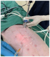
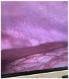
Methods: The cross-sectional study was carried out with a sample of 10 anesthetized sows in the IRCAD-América Latina Barretos Unit laboratory. The experiment consisted of an abdominal puncture guided by USG with a linear transducer to create the pneumoperitoneum. After the puncture, the drop test was performed, and CO2 was insufflated into the cavity. Subsequently, a 10mm trocar was introduced to insert the optic. The parameters from the USG were the thickness of the abdominal wall layers, intraperitoneal needle measurement, drop test, and the presence of complications.
Results: The average measurement of the layers was 0.45 centimeters of subcutaneous tissue, 0.67 centimeters of muscle, and 0.15 centimeters of peritoneum. The mean measurement of the intraperitoneal needle was 1.17cm. Furthermore, the drop test was positive in 100% of cases, and there was no bleeding or lesions on any attempt.
Conclusion: Ultrasound-guided pneumoperitoneum is feasible and safe in the porcine model. The subcutaneous, muscular, and peritoneum layers are identifiable and measurable in this model. Subsequent studies are necessary to verify the importance of this new procedure.
Introdução:: Todas as formas de acesso a cavidade peritoneal na laparoscopia possuem riscos de lesionar as estruturas intra-abdominais. Atualmente, a ultrassonografia (USG) está sendo utilizada em diversos procedimentos para direcionar algum tipo de punção: biópsia de mama, acesso venoso central, bloqueios anestésicos de nervos etc. Diante disso, esta pesquisa busca verificar a factibilidade e viabilidade da realização do pneumoperitônio por punção guiada por USG, em um estudo piloto em modelo porcino.
Métodos:: O estudo experimental foi feito com uma amostra de 10 porcas anestesiadas, no laboratório do IRCAD-América Latina Unidade de Barretos. O experimento consistiu na punção abdominal guiado por USG com transdutor linear para confecção do pneumoperitônio. Após a punção, foi realizado o teste da gota e insuflado CO2 na cavidade, posteriormente, um trocarte de 10mm foi introduzido para inserção da óptica. Os parâmetros a partir do USG foram: as espessuras das camadas da parede abdominal; medida da agulha intraperitoneal; teste da gota; e presença de complicações.
Resultados:: A mensuração da média das camadas foi de 0,45 centímetros (cm) de subcutâneo, 0,67cm de muscular e 0,15cm de peritônio. A média da medida da agulha intraperitoneal foi de 1,17cm. Ademais, o teste da gota foi positivo em 100% dos casos e não houve sangramento ou lesões em nenhuma tentativa.
Conclusão:: É factível e seguro a realização de pneumoperitônio guiado por ultrassonografia no modelo porcino. As camadas subcutâneas, muscular e peritônio são identificáveis e mensuráveis no modelo. Estudos subsequentes são necessários para verificar a importância deste novo procedimento.
Conflict of interest statement
Conflict of interest: no.
Figures
Similar articles
-
Preliminary pneumoperitoneum facilitates transgastric access into the peritoneal cavity for natural orifice transluminal endoscopic surgery: a pilot study in a live porcine model.Endoscopy. 2007 Oct;39(10):849-53. doi: 10.1055/s-2007-966844. Endoscopy. 2007. PMID: 17968798
-
Latif's point: A new point for Veress needle insertion for pneumoperitoneum in difficult laparoscopy.Asian J Endosc Surg. 2018 May;11(2):133-137. doi: 10.1111/ases.12418. Epub 2017 Aug 30. Asian J Endosc Surg. 2018. PMID: 28856845
-
Veress needle insertion in the left hypochondrium in creation of the pneumoperitoneum.Acta Cir Bras. 2006 Sep-Oct;21(5):296-303. Acta Cir Bras. 2006. PMID: 16981032 Clinical Trial.
-
Access techniques: Veress needle--initial blind trocar insertion versus open laparoscopy with the Hasson trocar.Endosc Surg Allied Technol. 1995 Feb;3(1):35-8. Endosc Surg Allied Technol. 1995. PMID: 7757437 Review.
-
Methods of creating pneumoperitoneum: a review of techniques and complications.Obstet Gynecol Surv. 1998 Mar;53(3):167-74. doi: 10.1097/00006254-199803000-00022. Obstet Gynecol Surv. 1998. PMID: 9513987 Review.
References
-
- Campos FGCM, Roll S. Abdominal Access and Pneumoperitonium Related Complications in Laparoscopic Surgery - Causes, Prevention & Treatment. Rev. bras. vídeo-cir. 2003;1(1):21–28.
-
- Pantoja Garrido M, Frías Sánchez Z, Zapardiel Gutiérrez I, Torrejón R, Jiménez Sánchez C, Polo Velasco A. Direct trocar insertion without previous pneumoperitoneum versus insertion after insufflation with Veress needle in laparoscopic gynecological surgery a prospective cohort study. J Obstet Gynaecol. 2019;39(7):1000–1005. doi: 10.1080/01443615.2019.1590804. - DOI - PubMed
MeSH terms
LinkOut - more resources
Full Text Sources


