Review
. 2024 Oct;121(10):e20240190.
doi: 10.36660/abc.20240190.

Cardiac Abnormalities in Hypereosinophilic Syndromes

[Article in Portuguese, English]
Affiliations
Review

Cardiac Abnormalities in Hypereosinophilic Syndromes

[Article in Portuguese, English]
Viviane Tiemi Hotta et al. Arq Bras Cardiol. 2024 Oct.

Erratum in

  • October 2024 Issue, vol. 121(10): e20240190.
    [No authors listed] [No authors listed] Arq Bras Cardiol. 2025 Feb 10;122(2):e20250038. doi: 10.36660/abc.20250038. Arq Bras Cardiol. 2025. PMID: 39936739 Free PMC article. English, Portuguese.

Abstract

Hypereosinophilia (HE) is defined as an eosinophil count exceeding 1500 cells/microL in peripheral blood in two tests, performed with an interval of at least one month and/or anatomopathological confirmation of HE, with eosinophils comprising more than 20% of all nucleated cells in the bone marrow. Hypereosinophilic syndrome (HES) indicates the presence of HE with organ involvement due to eosinophil action, which can be classified as primary (or neoplastic), secondary (or reactive), and idiopathic. Cardiac involvement occurs in up to 5% of cases in the acute phase and 20% of the chronic phase of the disease, ranging from oligosymptomatic cases to fulminant acute myocarditis or chronic restrictive cardiomyopathy (Loeffler endomyocarditis). However, the degree of cardiac dysfunction does not directly correlate with the degree of eosinophilia. The cardiac involvement of HES occurs in three phases: initial necrotic, thrombotic, and finally necrotic. It can manifest as heart failure, arrhythmias, and thromboembolic phenomena. The diagnosis of cardiopathy is based on multimodality imaging, with an emphasis on the importance of echocardiography (echo) as the primary examination. TTE with enhanced ultrasound agents can be used for better visualization, allowing greater accuracy in assessing ventricular apex, and myocardial deformation indices, such as longitudinal strain, may be reduced, especially in the ventricular apex (reverse apical sparing). Cardiac magnetic resonance imaging allows the characterization of subendocardial late gadolinium enhancement, and endomyocardial biopsy is considered the gold standard in diagnosing cardiopathy. Treatment is based on the etiology of HES.

Figura Central : Anormalidades Cardíacas nas Síndromes Hipereosinofílicas Abordagem sugerida em pacientes com Síndromes Hipereosinofílicas (SHE) e suspeita de acometimento cardíaco. BNP: peptídeo natriurético tipo B; ETT: ecocardiograma transtorácico; RMC: ressonância magnética cardíaca; TMO: tratamento medicamentoso otimizado.

Plain language summary

Central Illustration : Cardiac Abnormalities in Hypereosinophilic Syndromes Suggested approach in patients with HES and suspected cardiac involvement. Source: Adapted.29 BNP: brain natriuretic peptide; CMR: cardiac magnetic resonance imaging; TTE: transthoracic echocardiogram; GDMT: guideline-directed medical treatment; ECG: electrocardiogram; HES: hypereosinophilic syndromes.


Conflict of interest statement

Potencial conflito de interesse: Não há conflito com o presente artigo

Figures

Figura Central
Figura Central. : Anormalidades Cardíacas nas Síndromes Hipereosinofílicas
Figura 1
Figura 1. – Imagens de ecocardiograma transtorácico de um paciente diagnosticado com SHE e obliteração apical do ventrículo esquerdo no corte apical de 2 câmaras (A), corte apical de 4 câmaras (B) e do ventrículo direito no corte apical modificado para avaliação do VD (C). Observa-se insuficiência mitral de grau discreto ao Doppler colorido.
Figura 2
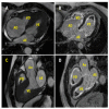
Figura 2. – Imagem de ressonância magnética cardíaca de um paciente com SHE e anormalidades cardíacas. Eixo longitudinal quatro câmaras com deposição de tecido fibroso endocárdico apical biventricular e microtrombos no átrio direito (A); Imagens para avaliação de fibrose pela técnica de realce tardio pelo gadolínio (RTG) (B). Eixo longitudinal da via de saída do ventrículo esquerdo com deposição de tecido fibroso endocárdico apical biventricular (C) ce avaliado pela técnica de RTG (D). Ao: aorta; AE: átrio esquerdo; VE: ventrículo esquerdo; AD: átrio direito; VD: ventrículo direito.
Central Illustration
Central Illustration. : Cardiac Abnormalities in Hypereosinophilic Syndromes
Figure 1
Figure 1. – Transthoracic echocardiogram images of a patient diagnosed with HES and apical obliteration of the left ventricle in the apical 2-chamber view (A), apical 4-chamber view (B) and from the right ventricle to the apical 4-chamber view directed towards the RV (C). A mild degree of mitral regurgitation is observed on color Doppler echocardiography.
Figure 2
Figure 2. – Cardiac magnetic resonance imaging of a patient with HES and cardiac abnormalities. Four chamber view with biventricular apical endocardial fibrous tissue deposition and microthrombi in the right atrium (A) with late gadolinium enhancement (LGE) CMRI (B). Left ventricular outflow tract view with biventricular apical endocardial fibrous tissue deposition (C) with LGE (D). Ao,:aorta; LA: left atrium; LV: left ventricle; RA: right atrium; RV: right ventricle.

References

    1. Valent P, Klion AD, Horny HP, Roufosse F, Gotlib J, Weller PF, et al. Contemporary Consensus Proposal on Criteria and Classification of Eosinophilic Disorders and Related Syndromes. J Allergy Clin Immunol. 2012;130(3):607–12.e9. doi: 10.1016/j.jaci.2012.02.019. - DOI - PMC - PubMed
    1. Nair P, Ochkur SI, Protheroe C, Simms E, Lee NA, Lee JJ. The Identification of Eosinophilic Gastroenteritis in Prednisone-dependent Eosinophilic Bronchitis and Asthma. 4Allergy Asthma Clin Immunol. 2011;7(1) doi: 10.1186/1710-1492-7-4. - DOI - PMC - PubMed
    1. Protheroe C, Woodruff SA, Petris G, Mukkada V, Ochkur SI, Janarthanan S, et al. A Novel Histologic Scoring System to Evaluate Mucosal Biopsies from Patients with Eosinophilic Esophagitis. Clin Gastroenterol Hepatol. 2009;7(7):749–55.e11. doi: 10.1016/j.cgh.2009.03.022. - DOI - PMC - PubMed
    1. Chusid MJ, Dale DC, West BC, Wolff SM. The Hypereosinophilic Syndrome: Analysis of Fourteen Cases with Review of the Literature. Medicine. 1975;54(1):1–27. - PubMed
    1. Butt NM, Lambert J, Ali S, Beer PA, Cross NC, Duncombe A, et al. Guideline for the Investigation and Management of Eosinophilia. Br J Haematol. 2017;176(4):553–572. doi: 10.1111/bjh.14488. - DOI - PubMed

MeSH terms

LinkOut - more resources