Randomized Controlled Trial
. 2025 Jan;32(1):e16559.
doi: 10.1111/ene.16559. Epub 2024 Nov 28.

Enhancing multisensory rehabilitation of visual field defects with transcranial direct current stimulation: A randomized clinical trial

Affiliations
Randomized Controlled Trial

Enhancing multisensory rehabilitation of visual field defects with transcranial direct current stimulation: A randomized clinical trial

Lorenzo Diana et al. Eur J Neurol. 2025 Jan.

Abstract

Background and purpose: Visual rehabilitation is necessary for improving the quality of life of patients with acquired homonymous visual field defects (HVFDs). By modulating brain excitability and plasticity, transcranial direct current stimulation (tDCS) may accelerate and increase the effects of compensatory trainings, which are usually long and intensive. In the present proof-of-principle, double-blind, randomized, sham-controlled study, we assess whether anodal tDCS applied over ipsilesional occipital or parietal cortices can increase the effects of a compensatory audiovisual training for HVFDs.

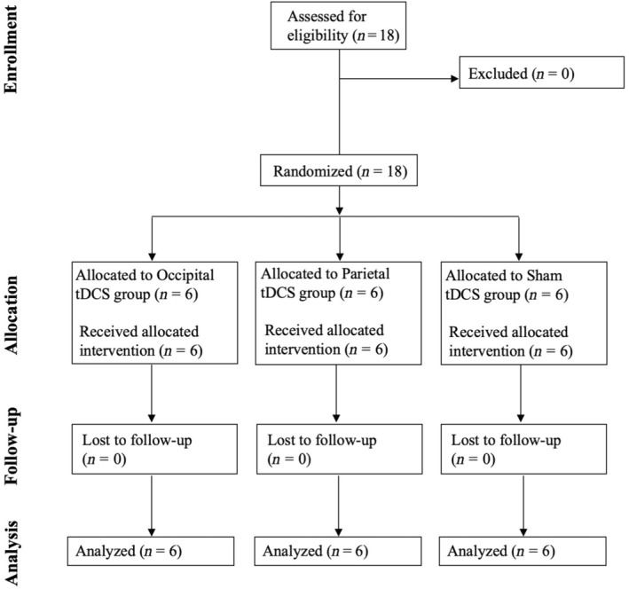
Methods: Eighteen participants with chronic HVFDs were randomized to receive anodal or sham tDCS over the ipsilesional parietal or occipital cortex during a 2-week (10 days, 2 h/day) audiovisual treatment aimed at improving oculomotor visual field exploration. Improvements were assessed by administering visual detection with eye movements and visual search tests, and a questionnaire for activities of daily living (ADLs) before the treatment, at its end, and at 1-month and 4-month follow-ups; lesion analyses were performed to look for predictors of treatment effects.

Results: Anodal ipsilesional tDCS, regardless of the target area (occipital vs. parietal), speeds up and increases daily improvements during the training. Whereas long-lasting (up to 4 months) post-treatment improvements in visual search and ADLs were observed in all groups, a greater and stable increase of visual detections in the blind hemifield was brought about only by the adjuvant use of occipital tDCS.

Conclusions: Compensatory audiovisual rehabilitation of HFVDs is effective and benefits from the adjuvant application of occipital and parietal tDCS, which speeds up and increases training-induced improvement.

Registry number: NCT06116760.

Keywords: hemianopia; rehabilitation; stroke; transcranial direct current stimulation.


Conflict of interest statement

The authors declare no conflict of interest.

Figures

FIGURE 1
FIGURE 1
CONSORT (Consolidated Standards of Reporting Trials) flow diagram. tDCS, transcranial direct current stimulation.
FIGURE 2
FIGURE 2
Lesion localization of brain‐damaged patients. Overlay lesion plots (frequencies of overlapping lesions, from dark blue, i.e., minimum overlap, to red, i.e., maximum overlap) for (a) the occipital transcranial direct current stimulation (tDCS) group, (b) the parietal tDCS group, and (c) the sham tDCS group. The mean lesion volume was 36.5 ± 31.5 cm3 (range = 3.95–127 cm3). The most affected areas, irrespective of the lesion side, were the calcarine sulcus (n = 15), the lingual gyrus (n = 15), the superior (n = 12), the middle (n = 12), and the inferior (n = 11) occipital lobes, as well as the cuneus (n = 9), and the fusiform gyrus (n = 12). Moreover, a number of intra‐ and interhemispheric white matter bundles were affected: corpus callosum (n = 15), inferior fronto‐occipital fasciculus (n = 15), inferior longitudinal fasciculus (n = 15), optic radiation (n = 14), posterior cingulum bundle (n = 12), and the first and second branches of the superior longitudinal fasciculus (n = 9). Left‐hemispheric lesions were mirrored onto the right hemisphere (neurological convention).
FIGURE 3
FIGURE 3
Improvements during the audiovisual training (AVT) and in the visual detection task with eye movements. (a) Performance during the AVT, showing mean percentage of audiovisual detection accuracy (ACC) in the whole visual field on each day of the 2‐week AVT (1st week = day 1–5, 2nd week = day 6–10), representing the significant 'Day × tDCS Group' interaction. Black lines = performance of patients who received occipital tDCS, gray lines = patients who received parietal tDCS, dotted black lines = sham tDCS. Asterisks indicate a significant improvement with respect to the 1st day of training (all p < 0.001). (b) Performance in the visual detection task with eye movements, showing mean percentage of visual detection AC in the sighted hemifield (white bars) and in the blind hemifield (black bars) pre‐training, post‐training, and at 1‐month and 4‐month follow‐ups (FUs) for each experimental group (occipital, parietal, and sham tDCS). Asterisks indicate a significant improvement (all p < 0.03) with respect to pre‐training performance. Error bars = standard error.
FIGURE 4
FIGURE 4
Visual search tests and questionnaire about vision‐related activities of daily living (ADLs; e). (a–d) Mean accuracy as a percentage (a, b) and response times (RTs; c, d) for the EF and the Triangles tests. White bars = pre‐training, black bars = post‐training, gray bars = follow‐up at 1 month (FU1), striped black/gray bars = follow‐up at 4 months (FU4). Error bars = standard error. Asterisks indicate a significant improvement with respect to pre‐training. tDCS, transcranial direct current stimulation.

Similar articles

Cited by

References

    1. Zihl J. Rehabilitation of Visual Disorders after Brain Injury. 1st ed. Psychology Press; 2021. http://digital.casalini.it/9781000443271. doi:10.4324/9781003210535 - DOI
    1. de Haan GA, Heutink J, Melis‐Dankers BJM, Brouwer WH, Tucha O. Difficulties in daily life reported by patients with homonymous visual field defects. J Neuroophthalmol. 2015;35(3):259‐264. doi:10.1097/WNO.0000000000000244 - DOI - PubMed
    1. Gall C, Lucklum J, Sabel BA, Franke GH. Vision‐ and health‐related quality of life in patients with visual field loss after postchiasmatic lesions. Invest Ophthalmol Vis Sci. 2009;50(6):2765‐2776. doi:10.1167/iovs.08-2519 - DOI - PubMed
    1. Pollock A, Hazelton C, Henderson CA, et al. Interventions for visual field defects in patients with stroke. Stroke. 2012;43(4):e37‐e38. doi:10.1161/strokeaha.111.639815 - DOI - PubMed
    1. Hanna KL, Hepworth LR, Rowe FJ. The treatment methods for post‐stroke visual impairment: a systematic review. Brain Behav. 2017;7(5):e00682. doi:10.1002/brb3.682 - DOI - PMC - PubMed

Publication types

Associated data

LinkOut - more resources