. 2024 Dec;29(1):2430882.
doi: 10.1080/13510002.2024.2430882. Epub 2024 Nov 28.

Palmitic acid and eicosapentaenoic acid supplementation in 3T3 adipocytes: impact on lipid storage and oxidative stress

Affiliations

Palmitic acid and eicosapentaenoic acid supplementation in 3T3 adipocytes: impact on lipid storage and oxidative stress

Edina Bakondi et al. Redox Rep. 2024 Dec.

Abstract

Objectives: Obesity is a worldwide public health problem, predisposing individuals to serious cardiovascular and metabolic complications such as type 2 diabetes mellitus. White adipose tissue serves as an important regulator of energy balance, and its expansion in obesity can trigger inflammatory reactions and oxidative stress, which can also lead to insulin resistance. Adipocytes, with a key role in regulating metabolic homeostasis, respond to increased calorie intake and altered fatty acid composition with hypertrophy or hyperplasia. Of particular interest are saturated fatty acids such as palmitic acid and omega-3 polyunsaturated fatty acids such as eicosapentaenoic acid (EPA), which have differential effects on adipocyte function and inflammation.

Methods: Using 3T3-L1 cells as a model for adipocytes, we evaluated the effects of PA and EPA on lipid accumulation, droplet size, and oxidative stress markers.

Results: We were able to show that EPA supplementation in 3T3 adipocytes does not lead to excessive lipid accumulation, but rather reduces the size of lipid droplets and also induces redox changes due to the unsaturated nature of EPA.

Discussion: These results emphasize the contrasting roles of PA and EPA and the importance of fatty acid composition in the regulation of adipocyte function.

Keywords: Obesity; adipocytes; eicosapentaenoic acid; fatty acids; lipid droplets; lipid storage; oxidative stress; palmitic acid.


Conflict of interest statement

No potential conflict of interest was reported by the author(s).

Figures
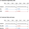
Figure 1.
Figure 1.
Scheme of the treatment of 3T3-L1 adipocytes with different fatty acids. 3T3-L1 adipocytes were treated with palmitate (PA) or eicosapentaenoic acid (EPA) in differentiation medium (DM) and maintaining medium (MM), either with the same fatty acid for 6 days (A) or with swapping the type of fatty acids at the third day (B). Control cells (Con. in all further figures) were treated according to the same time schedule (3 days of differentiation medium followed by 3 days of maintaining medium) but without fatty acid treatment.
Figure 2.
Figure 2.
Accumulation of lipids in 3T3-L1 adipocytes treated with Palmitate (PA). Panel (A) demonstrates a representative Oil Red O staining after incubation with different concentrations of PA. The viability of adipocytes after 6 days incubation with different concentrations of PA is shown in panel (B). PA concentration-dependent quantification of lipid accumulation (Oil Red O staining) is depicted in panel (C). An untreated control (Con.) was included in all experiments. All data are represented as mean ± SEM. Statistical significance was assessed by one-way ANOVA followed by Dunnet’s post-hoc test where each group of treatments was compared to the control group (**P < 0.01).
Figure 3.
Figure 3.
Comparison of effects of palmitate (PA) and eicosapentaenoic acid (EPA) on lipid accumulation in 3T3-L1 adipocytes. The viability of adipocytes after 6 days incubation with 500 µM PA and 250 µM EPA is shown in panel (A). Oil Red O based quantification of lipid accumulation in adipocytes after 6 days incubation with 500 µM PA and 250 µM EPA is demonstrated in panel (B), whereas a representative Oil Red O staining after incubation with 500 µM PA and 250 µM EPA is shown in (C). An untreated control (Con.) was included in all experiments. Results of the measurement of lipid droplet size are presented in panel (D). Data are represented as mean ± SEM. Statistical significance was assessed by a one-way ANOVA followed by Dunnet’s post-hoc test where each group of treatments was compared to the control group (****P < 0.0001).
Figure 4.
Figure 4.
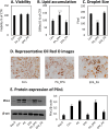
Effects of ‘swapping’ fatty acid exposure on lipid accumulation. The viability of 3T3-L1 adipocytes after 3 days of incubation with 500 µM PA followed by 3 days with 250 µM EPA (PA_EPA) or 250 µM EPA followed by 3 days with 500 µM PA (EPA_PA) is shown in panel (A). Lipid content (B), lipid droplet size (C) and a representative Oil Red O staining (D) are shown for this treatment scheme. An untreated control (Con.) was included in all experiments. Panel (E) shows a representative immunoblot and the quantification of PLIN1 after a 6-day treatment of adipocytes according to the treatment regimen PA_EPA and EPA_PA (250 µM PA/250 µM EPA) and additionally after treatment with the individual fatty acids (PA and EPA) as well as an untreated control (Con.), and non-differentiated cells (Day 0). Data are represented as mean ± SEM. Statistical significance was assessed by a one-way ANOVA followed by Dunnet’s post-hoc test where each group of treatments was compared to the control group (*P < 0.05; ****P < 0.0001).
Figure 5.
Figure 5.
Fatty acid composition in neutral lipids and phospholipids in adipocytes after fatty acid treatment. The fatty acid composition in neutral lipids (mainly triglycerides, lipid storage) and membrane lipids (mainly phospholipids) is shown after 6 days of incubation with 250 µM PA or 250 µM EPA (A,B) or after 3 days of incubation with 250 µM PA followed by 3 days with 250 µM EPA (PA_EPA) or 250 µM EPA followed by 3 days with 250 µM PA (EPA_PA) (C,D). An untreated control (Con.) was included. Data are represented as mean ± SEM. Statistical significance was assessed by a one-way ANOVA followed by Dunnet’s post-hoc test where each group of treatments was compared to the control group. (***P < 0.001; ****P < 0.0001).
Figure 6.
Figure 6.
Accumulating EPA induces pro-oxidative changes and contributes to lipid peroxidation. DCF staining (A,D), MitoSOX™ Red staining (B,E) and Malondialdehyde (MDA) concentration (C,F) are shown after incubation with 500 µM PA and 250 µM EPA. Redox-associated effects of fatty acid swapping (250 µM PA for 3 days followed by another 3 days with 250 µM EPA (PA_EPA) or vice versa (EPA_PA)) is demonstrated by DCF (D), MitoSOX™ (E) and Malondialdehyde (F). An untreated control (Con.) was included in all experiments. Data are represented as mean ± SEM. Statistical significance was assessed by a one-way ANOVA followed by Dunnet’s post-hoc test where each group of treatments was compared to the control group (*P < 0.05; ****P < 0.0001).

References

    1. James WP. WHO recognition of the global obesity epidemic. Int J Obes (Lond). 2008;32(Suppl 7):S120–S126. doi:10.1038/ijo.2008.247 - DOI - PubMed
    1. Arroyo-Johnson C, Mincey KD.. Obesity epidemiology worldwide. Gastroenterol Clin North Am. 2016;45(4):571–579. doi:10.1016/j.gtc.2016.07.012 - DOI - PMC - PubMed
    1. Kahn SE, Hull RL, Utzschneider KM.. Mechanisms linking obesity to insulin resistance and type 2 diabetes. Nature. 2006;444(7121):840–846. doi:10.1038/nature05482 - DOI - PubMed
    1. Himanshu D, Ali W, Wamique M.. Type 2 diabetes mellitus: pathogenesis and genetic diagnosis. J Diabetes Metab Disord. 2020;19(2):1959–1966. doi:10.1007/s40200-020-00641-x - DOI - PMC - PubMed
    1. de Heredia FP, Gomez-Martinez S, Marcos A.. Obesity, inflammation and the immune system. Proc Nutr Soc. 2012;71(2):332–338. doi:10.1017/S0029665112000092 - DOI - PubMed

LinkOut - more resources