. 2024 Nov 27;12(11):e010157.
doi: 10.1136/jitc-2024-010157.

Degradation of PARP1 by MARCHF3 in tumor cells triggers cCAS-STING activation in dendritic cells to regulate antitumor immunity in hepatocellular carcinoma

Affiliations

Degradation of PARP1 by MARCHF3 in tumor cells triggers cCAS-STING activation in dendritic cells to regulate antitumor immunity in hepatocellular carcinoma

Jun Cao et al. J Immunother Cancer. .

Abstract

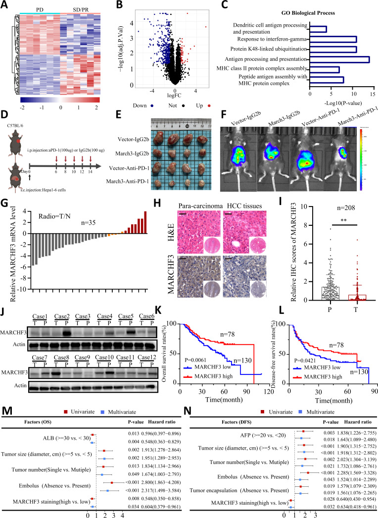
Background: Resistance to immune checkpoint inhibitors (ICIs) significantly limits the efficacy of immunotherapy in patients with hepatocellular carcinoma (HCC). However, the mechanisms underlying immunotherapy resistance remain poorly understood. Our aim was to clarify the role of membrane-associated ring-CH-type finger 3 (MARCHF3) in HCC within the framework of anti-programmed cell death protein-1 (PD-1) therapy.

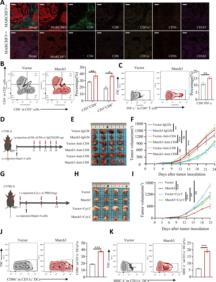
Methods: MARCHF3 was identified in the transcriptomic profiles of HCC tumors exhibiting different responses to ICIs. In humans, the correlation between MARCHF3 expression and the tumor microenvironment (TME) was assessed via multiplex immunohistochemistry. In addition, MARCHF3 expression in tumor cells and immune cell infiltration were assessed by flow cytometry.

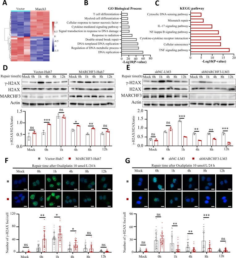
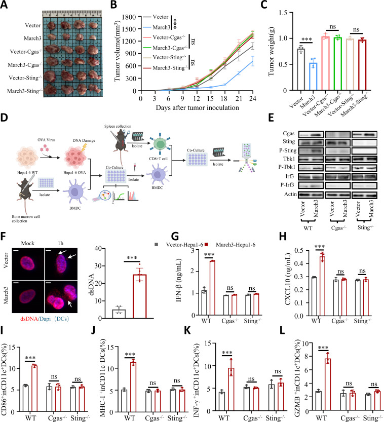
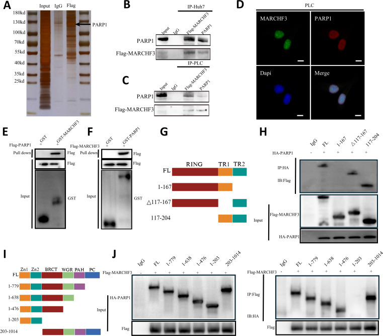
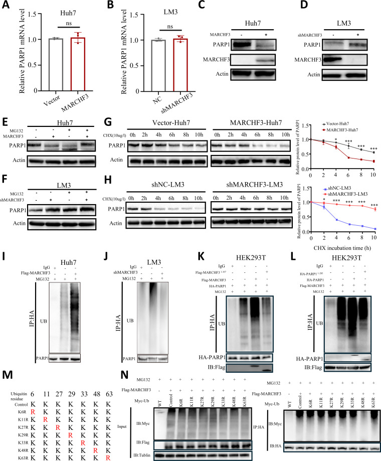
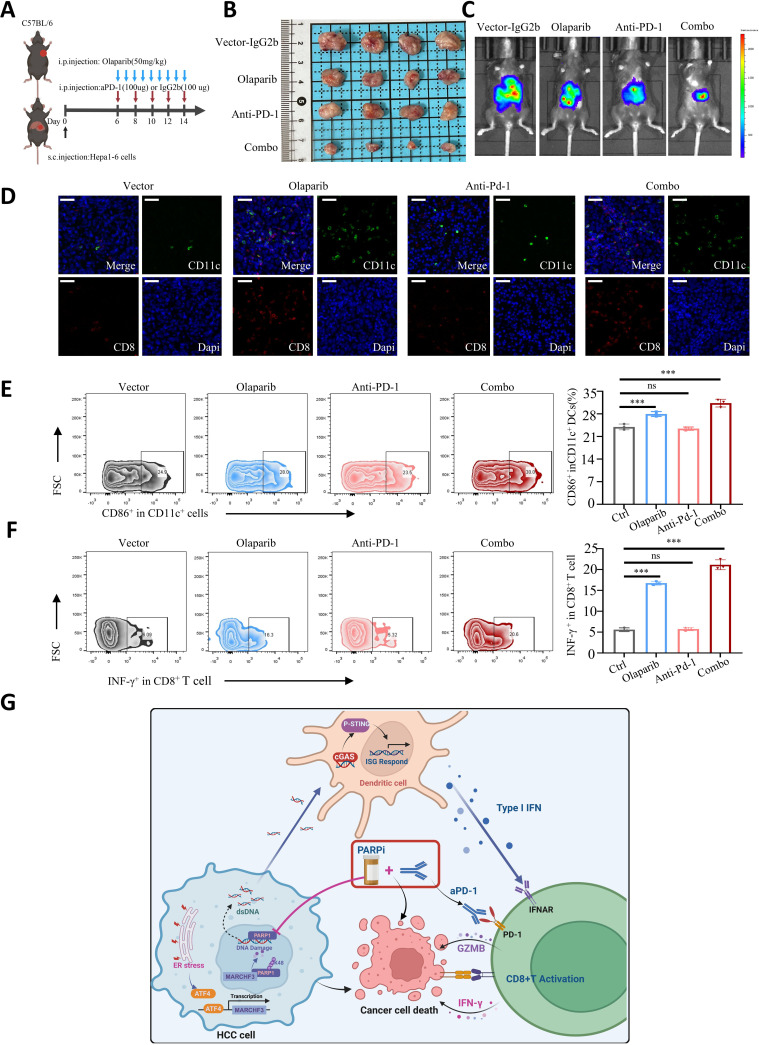
Results: MARCHF3 was significantly upregulated in tumors from patients who responded to ICIs. Increased MARCHF3 expression in HCC cells promoted dendritic cell (DC) maturation and stimulated CD8+ T-cell activation, thereby augmenting tumor control. Mechanistically, we identified MARCHF3 as a pivotal regulator of the DNA damage response. It directly interacted with Poly(ADP-Ribose) Polymerase 1 (PARP1) via K48-linked ubiquitination, leading to PARP1 degradation. This process promoted the release of double-strand DNA and activated cCAS-STING in DCs, thereby initiating DC-mediated antigen cross-presentation and CD8+ T-cell activation. Additionally, ATF4 transcriptionally regulated MARCHF3 expression. Notably, the PARP1 inhibitor olaparib augmented the efficacy of anti-PD-1 immunotherapy in both subcutaneous and orthotopic HCC mouse models.

Conclusions: MARCHF3 has emerged as a pivotal regulator of the immune landscape in the HCC TME and is a potent predictive biomarker for HCC. Combining interventions targeting the DNA damage response with ICIs is a promising treatment strategy for HCC.

Keywords: Hepatocellular Carcinoma; Immune Checkpoint Inhibitor; Immunotherapy; Tumor microenvironment - TME.


Conflict of interest statement

Competing interests: No, there are no competing interests.

Figures

Figure 1
Figure 1. MARCHF3 affects the patient response to ICIs and is correlated with better outcomes. (A) Heatmap showing the differentially expressed genes between responders and non-responders to ICIs. (B) Volcano plot of the differentially expressed genes between ICI responders and non-responders. (C) GO analyses of the DEGs. (D) Scheme of the experimental procedure. (E) Tumor burdens in C57BL/6 mice subcutaneously injected with control vector or March3-overexpressing Hepa1-6 cells and treated with anti-PD-1 antibodies. (F) In vivo imaging systems were used to measure the fluorescence intensity in HCC tumors. (G) The mRNA expression of MARCHF3 in HCC tissues, presented as log(T/N) values. (H–I) Representative image of IHC staining showing MARCHF3 expression in HCC tumors and matched adjacent tissues. Scale bar, 40µm. (J) Protein imprinting analysis revealed the expression of the MARCHF3 protein in HCC tumors and matched adjacent tissues. (K) Kaplan-Meier OS and (L) DFS curves for patients with HCC in the high and low MARCHF3 expression tissue microarray cohorts. (M) Univariate and (N) multivariate analyses of factors associated with OS and DFS. Scale bar: 100 mm (top panel). **p<0.01, Student’s t-test. DFS, disease-free survival; GO, Gene Ontology; HCC, hepatocellular carcinoma; ICI, immune checkpoint inhibitor; IHC, immunohistochemistry; MARCHF3, membrane-associated ring-CH-type finger 3; MHC, major histocompatibility complex; mRNA, messenger RNA; OS, overall survival; PD-1, programmed cell death protein-1.
Figure 2
Figure 2. MARCHF3 reshaped the tumor microenvironment by inducing DC maturation and CD8+ T-cell activation. (A) Representative multiplex immunohistochemistry staining results for CD4, CD8, CD11c, CD56, and CD163 in the MARCHF3 high-expression or low-expression groups (scale bar, 50 µm). (B) Flow cytometry analysis of the profiles of infiltrating immune cells, including CD8+ T cells and CD4+ T cells, in tumor tissue. (C) Detection of the cytotoxic activities of CD8+ IFN-γ+ T cells in the MARCHF3 and WT groups by flow cytometry. (D) Schematic representation of the experimental procedure. (E–F) Tumor burdens in C57BL/6 mice subcutaneously injected with tumor cells and administered anti-CD4 or anti-CD8 antibodies. (G) Scheme of the experimental procedure. C57BL6 mice bearing control vector-overexpressing or March3-overexpressing Hepa1-6 tumors were treated with phosphate-buffered saline or Cyt c (depleted DCs). (H) Macroscopic images and (I) average volumes are shown as the means±SDs. n=4 mice per group. (J–K) Flow cytometry analysis of CD86+ CD11c+ DCs (G) and MHC-I+ CD11c+ DCs (H) in the MARCHF3 and WT groups. Cyt c, cytochrome c; DC, dendritic cell; IFN, interferon; MARCHF3, membrane-associated ring-CH-type finger 3; MHC, major histocompatibility complex; WT, wild-type.
Figure 3
Figure 3. MARCHF3 inhibited DNA repair after DNA damage. (A) Heatmap displaying the differentially expressed genes between vector-treated and MARCHF3-overexpressing tumor tissues. (B) GO and (C) KEGG pathway enrichment analyses of the differentially expressed genes. (D–E) Western blot analysis of rH2AX expression in (D) Huh7 cells and (E) HCCLM3 cells. (F–G) The number of rH2AX foci in (F) Huh7 and (G) HCCLM3 cells. Scale bars, 50 µm. GO, Gene Ontology; IL, interleukin; KEGG, Kyoto Encyclopedia of Genes and Genomes; MARCHF3, membrane-associated ring-CH-type finger 3; TNF, Tumor necrosis factor.
Figure 4
Figure 4. MARCHF3 triggered DC cCAS-STING activation to regulate antitumor immunity in HCC. (AC) Macroscopic images (A), average tumor volumes (B) and average tumor weights (C) in WT, Cgas−/−, and Sting−/− mice in the MARCHF3 and vector groups. (D) Scheme of the experimental procedure. (E) Western blot analysis was conducted to assess the expression of proteins in the cGAS-STING pathway in DCs from WT, Cgas−/−, and Sting−/− mice. (F) DCs from the WT and MARCHF3 groups were subjected to dsDNA (red) and nuclear (DAPI) immunofluorescence staining. Scale bar, 20µm. (GH) ELISAs were used to determine the (G) IFN-β and (H) CXCL10 levels in the supernatants of WT, Cgas−/−, and Sting−/− mouse DCs. (I–J) FC analysis of (I) CD86+ CD11c+ DCs and (J) MHC-I+ CD11c+ DCs from WT, Cgas−/−, and Sting−/− mice. (K) IFN-γ+ CD8+ T cells and (L) GZMB+ CD8+ T cells among CD8+T cells from WT, Cgas−/−, and Sting−/− mice were detected using flow cytometry. BMDC, bone marrow-derived dendritic cell; DAPI, 4',6-diamidino-2-phenylindole; DC, dendritic cell; dsDNA, double-strand DNA; GZMB, Granzyme B; HCC, hepatocellular carcinoma; IFN, interferon; MARCHF3, membrane-associated ring-CH-type finger 3; MHC, major histocompatibility complex; WT, wild-type.
Figure 5
Figure 5. MARCHF3 interacted with PARP1. (A) Silver staining of eluates resolved by SDS-PAGE. (B–C) Co-immunoprecipitation assays showing the interaction of MARCHF3 with PARP1 in (B) Huh7 and (C) PLC cells. (D) Immunofluorescence assays showing the interaction and nuclear colocalization of MARCHF3 with PARP1 in hepatocellular carcinoma cells. Scale bar, 20 µm. (E–F) The interaction between MARCHF3 and PARP1 was assayed via GST precipitation, and purified GST was used as the control. (G) Domain structures of MARCHF3 and the MARCHF3 deletion mutants used in the study. (H) The domain of MARCHF3 that interacts with PARP1. (I) Domain structures of the PARP1 and deletion mutants used in the study. (J) The domain of PARP1 that interacts with MARCHF3.GST, Glutathione-S-transferase; IP, immunoprecipitation; MARCHF3, membrane-associated ring-CH-type finger 3; PARP1, Poly [ADP-ribose] polymerase 1; SDS-PAGE, Sodium dodecyl sulfate polyacrylamide gel electrophoresis.
Figure 6
Figure 6. MARCHF3 ubiquitinated PARP1. (A–B) MARCHF3 expression did not affect PARP1 mRNA expression in (A) Huh7-MARCHF3 and (B) HCCLM3-shMARCHF3 cells. (C–D) Western blot analysis of the effect of MARCHF3 on PARP1 protein levels in (C) Huh7 and (D) PLC cells. (E–F) Western blot analysis of PARP1 levels in (E) Huh7-MARCHF3 and (F) HCCLM3-shMARCHF3 cells cultured in the presence of MG132. (G–H) Western blot analysis of the effect of MARCHF3 on the half-life of PARP1 in (G) Huh7-MARCHF3 and (H) HCCLM3-shMARCHF3 cells treated with cycloheximide (10 mg/mL) for the indicated periods. (I–J) Ubiquitination of PARP1 in Huh7-MARCHF3 cells (I) or HCCLM3-shMARCHF3 cells (J) treated for 6 hours with 10 µM MG132. (K–L) Ubiquitination of PARP1 in HEK293T cells transfected with Flag1-176 (K) or HA1-203 (L) and treated for 6 hours with 10 µM MG132. (M) A schematic diagram of MARCHF3 and its mutants. (N) Immunoblotting was used to detect the ubiquitination of PARP1 mutants in HEK293T cells cotransfected with Myc-UB mutants, Flag-MARCHF3, and HA-PARP1. IP, immunoprecipitation; MARCHF3, membrane-associated ring-CH-type finger 3; mRNA, messenger RNA; PARP1, Poly [ADP-ribose] polymerase 1; WT, wild-type.
Figure 7
Figure 7. Blockade of PARP1 potentiated the efficacy of anti-PD-1 therapy in HCC. (A) Schematic showing the schedule of treatment with the anti-PD-1 antibody and the PARP1 inhibitor olaparib in the HCC model C57BL/6 mice. (B) Subcutaneous tumor xenografts in mice. (C) In vivo imaging systems were used to measure the fluorescence intensity in HCC tumors. (D) Representative images of multiplex immunohistochemistry staining for CD11c and CD8 in orthotopic HCC tissues. Scale bars, 30 µm. (E–F) Flow cytometry analysis of the proportions of infiltrating (E) CD86+ CD11c+ DCs and (F) INF-γ+CD8+ T cells in xenograft HCC tissues from each group. (G) ATF4 induced MARCHF3 expression on ER stress. Increased MARCHF3 expression inhibits PARP1-mediated DNA repair that induces DNA damage and cytoplasmic dsDNA release, leading to DC cGAS/STING-dependent activation of type I IFN signaling and thereby reprogramming of the tumor microenvironment. Targeting the DNA damage pathway, such as with the PARP1 inhibitor olaparib. Immune checkpoint inhibitor efficacy against HCC is potentiated. DC, dendritic cell; dsDNA, double-strand DNA; ER, endoplasmic reticulum; GZMB, Granzyme B; HCC, hepatocellular carcinoma; IFN, interferon; MARCHF3, membrane-associated ring-CH-type finger 3; PARP1, Poly [ADP-ribose] polymerase 1; PARPi, PARP inhibitors; PD-1, programmed cell death protein-1.

Similar articles

Cited by

References

    1. Siegel RL, Miller KD, Wagle NS, et al. Cancer statistics, 2023. CA Cancer J Clin. 2023;73:17–48. doi: 10.3322/caac.21763. - DOI - PubMed
    1. El-Serag HB. Hepatocellular carcinoma. N Engl J Med. 2011;365:1118–27. doi: 10.1056/NEJMra1001683. - DOI - PubMed
    1. Kudo M, Finn RS, Qin S, et al. Lenvatinib versus sorafenib in first-line treatment of patients with unresectable hepatocellular carcinoma: a randomised phase 3 non-inferiority trial. The Lancet. 2018;391:1163–73. doi: 10.1016/S0140-6736(18)30207-1. - DOI - PubMed
    1. Rimini M, Persano M, Tada T, et al. Real-World Data for Atezolizumab Plus Bevacizumab in Unresectable Hepatocellular Carcinoma: How Does Adherence to the IMbrave150 Trial Inclusion Criteria Impact Prognosis? Target Oncol. 2023;18:221–33. doi: 10.1007/s11523-023-00953-x. - DOI - PubMed
    1. Pinter M, Scheiner B, Pinato DJ. Immune checkpoint inhibitors in hepatocellular carcinoma: emerging challenges in clinical practice. Lancet Gastroenterol Hepatol. 2023;8:760–70. doi: 10.1016/S2468-1253(23)00147-4. - DOI - PubMed

MeSH terms