Clinical Trial
. 2024 Nov 28;15(1):10332.
doi: 10.1038/s41467-024-54580-9.

Enhanced and sustained biodistribution of HIV-1 neutralizing antibody VRC01LS in human genital and rectal mucosa

Affiliations
Clinical Trial

Enhanced and sustained biodistribution of HIV-1 neutralizing antibody VRC01LS in human genital and rectal mucosa

Maria P Lemos et al. Nat Commun. .

Abstract

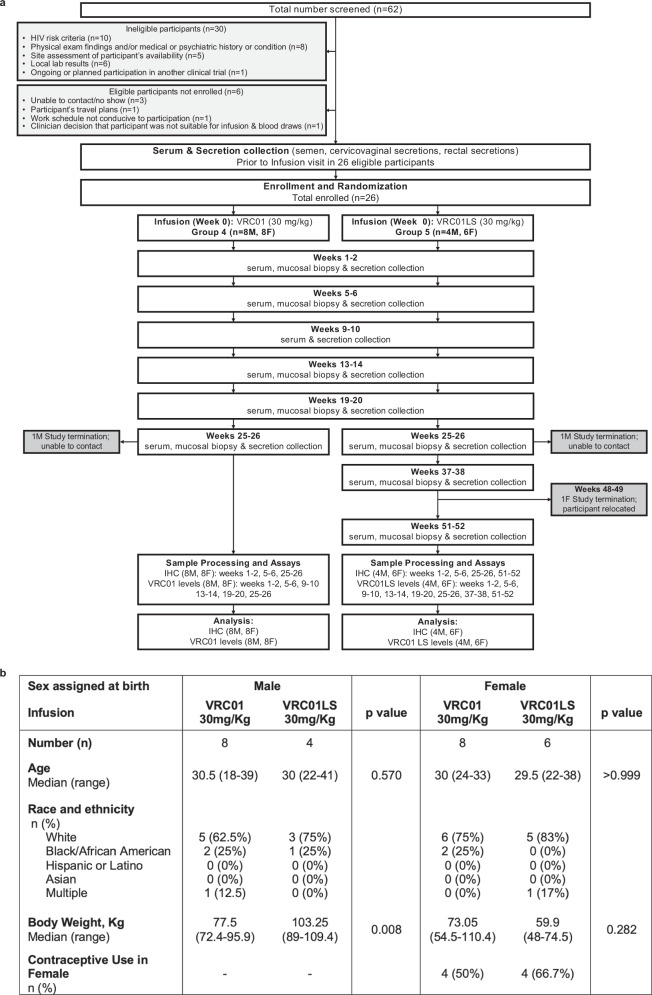
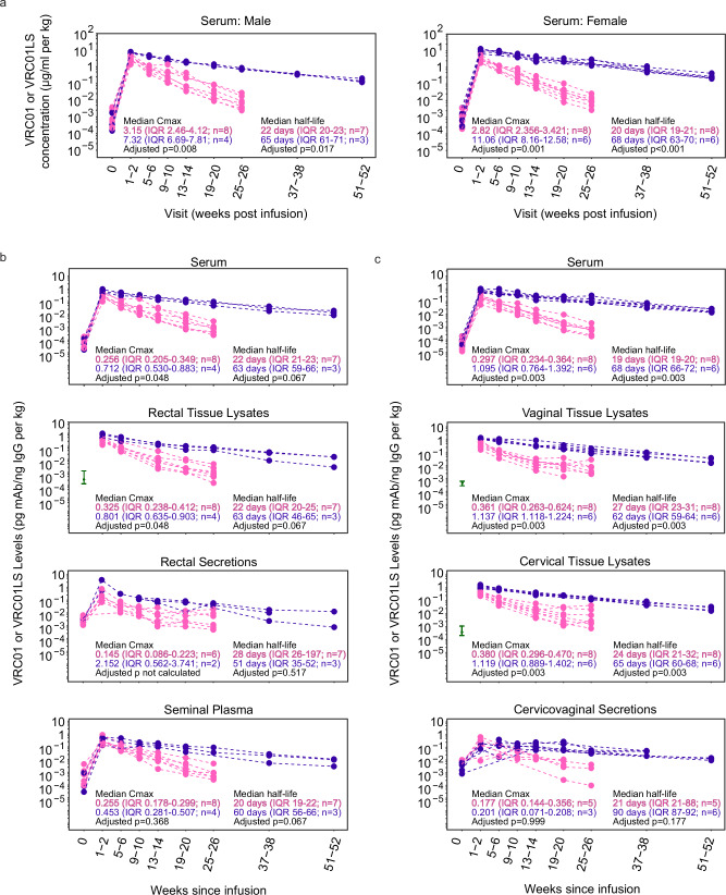
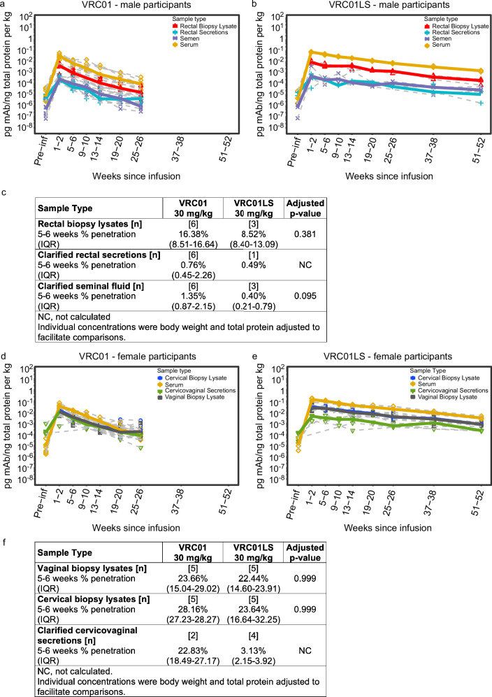
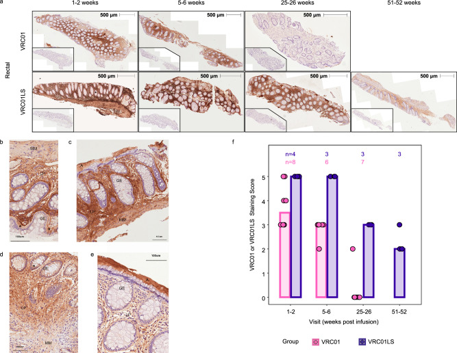
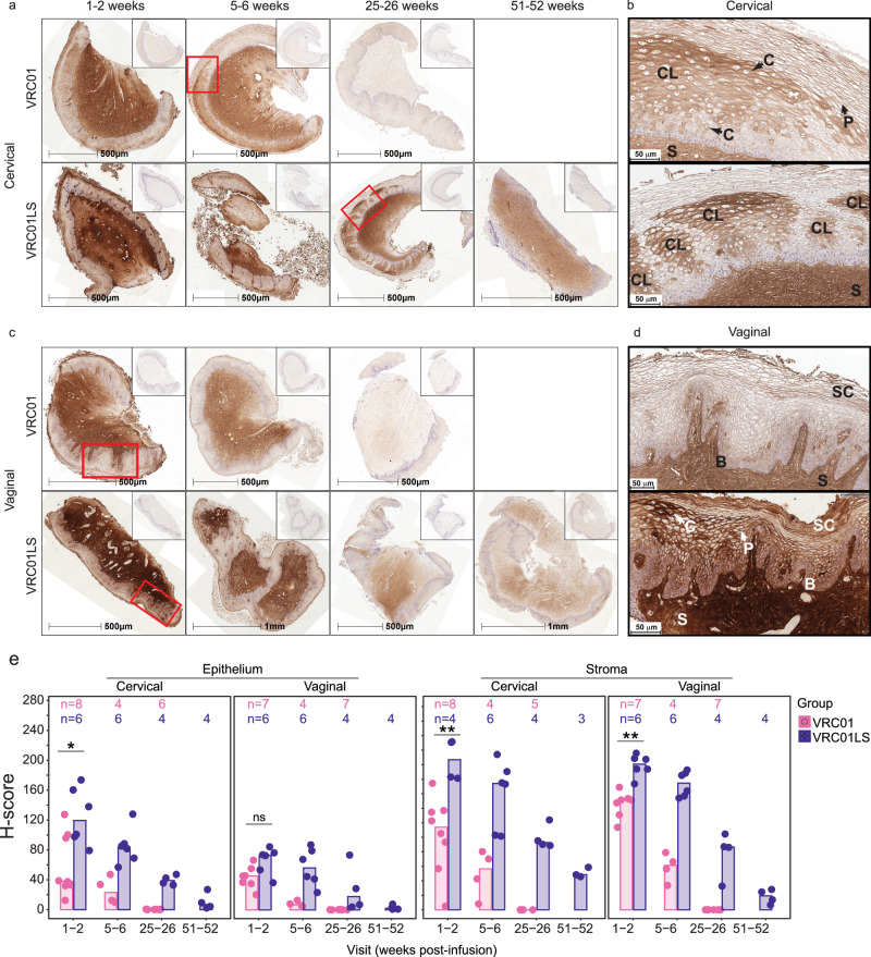
To prevent sexually-acquired HIV-1 infection by immunoprophylaxis, effective concentrations of broadly neutralizing antibodies are likely needed at mucosal sites of exposure. Here, we examine the biodistribution of monoclonal antibody VRC01 and its extended half-life variant, VRC01LS, in colorectal and genitourinary tracts of healthy adults 1-52 weeks after intravenous infusion. At 1-2 weeks, VRC01LS levels are ~3-4 times higher than VRC01 in serum (p = 0.048), rectal (p = 0.067), vaginal (p = 0.003) and cervical tissues (p = 0.003); these differences increase over time. Both antibodies primarily localize within rectal lamina propria and cervicovaginal stroma, with limited and variable epithelial distribution. Although 8-28% of serum mAb levels reach mucosal tissues, <3% are in seminal and rectal secretions. Elimination half-lives in mucosal tissues are 20-28 days for VRC01 and 51-68 days for VRC01LS. Thus, VRC01LS infusion achieves higher, sustained concentrations in human mucosal tissues than VRC01, supporting the future investigation of potent, long-acting LS-modified antibodies to prevent HIV-1.

Trial registration: ClinicalTrials.gov NCT02797171.


Conflict of interest statement

Competing interests: J.R.M. is listed on patent 9738703 for the VRC01 antibody (patent held by the US National Institutes of Health). M.P., J.H., and S.N. are current employees of the US NIH. All other authors have no competing interests to declare. The study funders had no role in the study design, data collection and analysis, decision to publish, or preparation of the manuscript.

Figures

Fig. 1
Fig. 1. Study conduct and participant demographics of HVTN 116 single infusion groups receiving intravenous infusions of 30 mg/kg VRC01 or VRC01LS.
a CONSORT diagram depicting study enrollment and time points of sample collection and analyses. Abbreviations: immuno-histochemistry (IHC); male (M); female (F). b Demographic characteristics of enrolled participants. P-values are two-sided Wilcoxon rank tests.
Fig. 2
Fig. 2. VRC01LS infusion leads to a higher mucosal Cmax and longer mucosal half-life compared to VRC01 infusion.
a Serum mAb concentrations were measured using Singulex Erenna single-molecule counting technology in participants who received a single IV infusion of 30 mg/kg VRC01 (pink) or VRC01LS (violet). Serum samples were diluted at 1:1000 to 1:50,000 for quantitation using 5C9 in the bead-based assay. Pre-infusion (timepoint 0) serum concentrations are included for reference and were comparable among both mAb groups (p = 0.852 for females, p = 0.562 for males). b IgG-normalized concentrations of VRC01 (pink) and VRC01LS (violet) in males receiving VRC01 or VRC01LS. Serum, rectal biopsy lysates, rectal secretions, and semen are depicted in individual graphs. Rectal biopsies were not collected pre-infusion (timepoint 0), so the baseline comparator (green interval line) is the 25–75 percentile range of pre-infusion samples collected from the other HVTN 116 study arms. Fourteen of the rectal secretions (n = 4 males; 21.5% of collections) were excluded due to evidence of high hemoglobin, an evidence of blood contamination. Three additional rectal secretion samples did not have sufficient IgG for quantitation at 1:5 dilution, so their denominator was replaced by ½ the LLOQ of their IgG ELISA runs. c IgG-normalized concentrations of VRC01 (pink) and VRC01LS (violet) in females who received a single 30 mg/kg IV infusion of VRC01 or VRC01LS. Serum and cervicovaginal secretions are depicted in individual graphs. Cervical and vaginal biopsies were not collected pre-infusion (timepoint 0); the baseline comparators (green interval line) are the 25–75 percentile ranges from pre-infusion samples from the other HVTN 116 study arms. Seven cervicovaginal secretions (n = 6 females; 8.9% of collections) did not contain sufficient fluid to run any assay, and 7 (n = 5 females; 8.9% of collections) had evidence of hemoglobin contamination; these samples were excluded. All mAb measurements in (b and c) are divided by the local concentration of IgG in the sample, and body weight-normalized. The p-values in (ac) are two-sided Wilcoxon ranked sum tests adjusted for multiple comparisons using the Holm-Bonferroni method. Abbreviations mAb: monoclonal antibody; IgG immunoglobulin G; IQR: inter-quartile range; Cmax: Maximal concentration at 1-2 weeks post-infusion.
Fig. 3
Fig. 3. Penetration of infused VRC01 and VRC01LS into the male intestinal compartment and the lower FGT.
Protein-normalized mAb levels of (a) VRC01 and (b) VRC01LS in male participants who received a single IV infusion at 30 mg/kg. Blood serum (yellow diamonds), rectal tissue lysates (red triangles), clarified Seminal Fluid (purple asterisks), and clarified rectal secretions (blue crosses) are depicted. Gray dashed lines denote each individual participant, and colored bold lines represent the median of each group. Fourteen of the rectal secretions (n = 4; 21.5% of collections) were excluded due to high hemoglobin, implying blood contamination. Eight rectal secretions had insufficient quantity for protein quantitation and were excluded. Three rectal secretions and two rectal biopsy lysates were below the Quant-iT LLOQ, so their denominator was replaced by ½ the LLOQ of their QuantiT runs. c Percent penetration in tissues and secretions from male participants. Protein-normalized concentrations of VRC01 and VRC01LS in female participants who received (d) VRC01 or (e) VRC01LS. Median serum (yellow diamonds), cervical biopsy lysates (blue circles), vaginal biopsy lysates (black squares), and cervicovaginal secretions (green triangles) are depicted. Gray dashed lines denote each individual participant, and colored bold lines represent the median of each group. Seven cervicovaginal secretions (n = 6 females; 8.9% of collections) did not contain sufficient fluid to assay, and 7 cervicovaginal secretions (n = 5; 8.9% collections) had evidence of hemoglobin contamination; these were excluded. Two cervical biopsy lysates and four vaginal biopsy lysates did not have sufficient protein for quantitation, so their denominator was replaced by ½ the LLOQ of their QuantiT runs. f Percent penetration in tissues and secretions from female participants. The p-values in (c and f) are two-sided Wilcoxon ranked sum tests adjusted for multiple comparisons using the Holm-Bonferroni method. Abbreviations: mAb monoclonal antibody, IQR inter-quartile range, NC not calculated.
Fig. 4
Fig. 4. Infused VRC01 and VRC01LS localizes into the rectal lamina propria and muscularis layers.
PAXgene-fixed, paraffin-embedded rectal tissue biopsies were sectioned (4 μm) and stained with 5C9 (VRC01 and VRC01LS detection) or mouse IgG2a (isotype control), followed by anti-mouse IgG-DAB (brown staining), and counterstained with hematoxylin (blue). All images were acquired on the TissueFAXS microscope in a bright field using a 10 × objective and identical exposure times. a Whole tissue section images of two representative individuals from VRC01-infused (n = 8) and VRC01LS-infused recipients (n = 4) are shown at multiple time points. Whole tissue section images indicate 5C9 staining of VRC01 or VRC01LS with 500 μm measurements to indicate the size of the tissue section analyzed; inset images are consecutive sections to the 5C9 stains, stained with mouse IgG isotype control. be Sections from 4 different participants at high magnification, with adjacent isotype control stained images. b, c Sections of rectal biopsies from 2 different VRC01-infused participants at 5, 6 weeks post infusion. d, e Sections of rectal biopsies from 2 different VRC01LS-infused participants at 5, 6 weeks post infusion. Abbreviations: A: adherent mucus layer, GE: glandular epithelium, LP: lamina propria, MM: muscularis mucosa, *: selected epithelium depicting mAb staining. A 100 μm ruler indicates size. f Manual scoring of 5C9 staining at different time points post-infusion. The number of participant samples analyzed is indicated above each bar. Scores were subtracted for any mouse IgG2a background to remove any artifacts. No data was excluded.
Fig. 5
Fig. 5. Localization of VRC01 and VRC01LS in cervical and vaginal tissues after infusion.
a, c Whole tissue section images showing persistence of VRC01LS compared to VRC01 in (a) cervical and (c) vaginal tissues; inset images show isotype control staining on consecutive sections. All time points for each tissue type are from the same representative VRC01 (n = 8) or VRC01LS (n = 6) recipient. b, d Higher magnification views of regions from (a) and (c), outlined in red, showing specific localization patterns of VRC01 or VRC01LS within (b) cervical and (d) vaginal epithelium. Arrows labeled (c) and P indicate examples of cytoplasmic and pericellular localization, respectively. Examples of localization in the stratum corneum and basal (and parabasal) layers are labeled SC and (b), respectively, and stromal staining is labeled S. In panel (b), there is a combination of pericellular, cytoplasmic, and clustered localization (labeled CL) in the intermediate layer in both cervical images; upper and lower images show a single large area and five smaller areas, respectively, of clustered localization in the intermediate layers. In panel (d), the upper vaginal image shows pericellular localization with some concentration in the basal, parabasal, and stratum corneum. The lower image shows pericellular localization and regions of cytoplasmic localization with some concentrated deposition in the intermediate layer as well as the basal, parabasal, and most superficial layers of the stratum corneum. e Area quantitation of VRC01 or VRC01LS staining over time post-infusion in cervical and vaginal epithelium and stroma. The number of participant samples analyzed is indicated above each bar. Individual data points correspond to the H-scores for each participant sample; bar heights represent the medians. Three cervical and 2 vaginal samples had insufficient tissue, and 1 cervical sample had insufficient stroma and were therefore excluded from analysis. Two-sided Wilcoxon rank sum test comparisons were done at 1-2 weeks post-infusion. The exact p-values are as follows: cervical (*) p = 0.020 and vaginal (ns, not significant) p = 0.051 epithelium; cervical (**) p = 0.004 and vaginal (**) p = 0.001 stroma. Of note, VRC01LS recipients donated week 1-2 samples slightly earlier (median 7.5 days, range 4–14) than VRC01 recipients (median 11 days, range 2–15).

References

    1. Wu, X. et al. Rational design of envelope identifies broadly neutralizing human monoclonal antibodies to HIV-1. Science329, 856–861 (2010). - PMC - PubMed
    1. Zhou, T. et al. Structural basis for broad and potent neutralization of HIV-1 by antibody VRC01. Science329, 811–817 (2010). - PMC - PubMed
    1. Corey, L. et al. Two randomized trials of neutralizing antibodies to prevent HIV-1 acquisition. N. Engl. J. Med.384, 1003–1014 (2021). - PMC - PubMed
    1. Gilbert, P. B. et al. Neutralization titer biomarker for antibody-mediated prevention of HIV-1 acquisition. Nat. Med.28, 1924–1932 (2022). - PMC - PubMed
    1. Gaudinski, M. R. et al. Safety and pharmacokinetics of the Fc-modified HIV-1 human monoclonal antibody VRC01LS: A Phase 1 open-label clinical trial in healthy adults. PLoS Med.15, e1002493 (2018). - PMC - PubMed

Publication types

MeSH terms