. 2024 Nov 28;15(1):10354.
doi: 10.1038/s41467-024-54529-y.

SPLICER: a highly efficient base editing toolbox that enables in vivo therapeutic exon skipping

Affiliations

SPLICER: a highly efficient base editing toolbox that enables in vivo therapeutic exon skipping

Angelo Miskalis et al. Nat Commun. .

Abstract

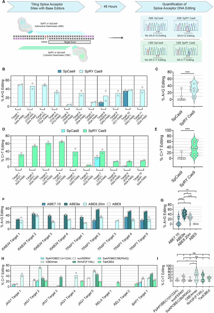
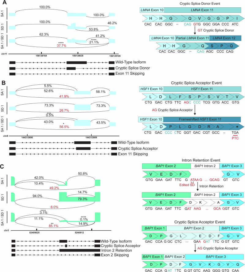
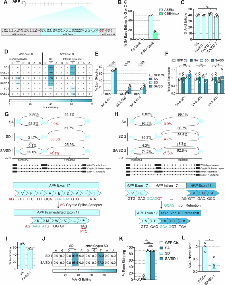
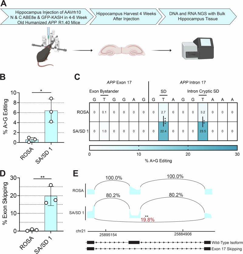
Exon skipping technologies enable exclusion of targeted exons from mature mRNA transcripts, which have broad applications in medicine and biotechnology. Existing techniques including antisense oligonucleotides, targetable nucleases, and base editors, while effective for specific applications, remain hindered by transient effects, genotoxicity, and inconsistent exon skipping. To overcome these limitations, here we develop SPLICER, a toolbox of next-generation base editors containing near-PAMless Cas9 nickase variants fused to adenosine or cytosine deaminases for the simultaneous editing of splice acceptor (SA) and splice donor (SD) sequences. Synchronized SA and SD editing improves exon skipping, reduces aberrant splicing, and enables skipping of exons refractory to single splice site editing. To demonstrate the therapeutic potential of SPLICER, we target APP exon 17, which encodes amino acids that are cleaved to form Aβ plaques in Alzheimer's disease. SPLICER reduces the formation of Aβ42 peptides in vitro and enables efficient exon skipping in a mouse model of Alzheimer's disease. Overall, SPLICER is a widely applicable and efficient exon skipping toolbox.


Conflict of interest statement

Competing interests: P.P.P., M.G., and J.W. are named inventors in pending patent application US17/260,828 filed by the University of Illinois. Methods for exon skipping and AAV-mediated delivery of base editors are covered in the patent application. The remaining authors declare no competing interests.

Figures

Fig. 1
Fig. 1. SpRY Cas9 in combination with different deaminases enables efficient disruption of targeted splice acceptors.
A Schematic representation of the approach to disrupting splicing elements with near-PAMless ABEs (blue) or CBEs (green) by tiling eight sgRNAs per exon (Created in BioRender. Miskalis, A. (2024) https://BioRender.com/x92p416). Comparison of SpCas9 and SpRY Cas9 editing efficiency at the splice acceptors (SAs) of target exons using ABEs (B, C) or CBEs (D, E). Summary of editing efficiency at individual targets accomplished with SpCas9 or SpRY Cas9 ABEs (C) or CBEs (E). Comparison of DNA editing efficiency at multiple SAs using SpRY Cas9 fused with one of four different adenosine deaminases (F, G) or one of six different cytosine deaminases (H, I). Summary of editing efficiency at individual targets accomplished with different SpRY ABEs (G) or different SpRY CBEs (I). For (F−I), DNA editing rates were normalized to the protein expression of each deaminase from Fig. S1 by dividing each DNA editing rate by the normalized expression of each base editor. Values represent means and error bars indicate SD. For (B, D, F, and H), n = 2; n = 10 for (C); n = 9 for (E, G); n = 8 for (I). All replicates are biological replicates; ns, not significant; *p < 0.05, **p < 0.01; ***p < 0.001; two-tailed, unpaired t-test except for (G, I), which were analyzed via one-way ANOVA with Tukey’s post hoc. Source data are provided as a Source Data file.
Fig. 2
Fig. 2. Simultaneous editing of splice sites with SPLICER enhances exon skipping.
A Illustration of SPLICER strategy for simultaneously targeting SAs and SDs with ABEs or CBEs followed by screening to identify effective BE systems and characterization next-generation sequencing (Created in BioRender. Miskalis, A. (2024) https://BioRender.com/x43x713). BD Editing of AG dinucleotides in the SA (top panel) and GT dinucleotides in the SD (medium panel) and exon skipping (bottom panel) with ABEs (highlighted blue) or CBEs (highlighted green). Improvements in exon skipping were synergistic when targeting LMNA exon 11, EGFR exon 23 and JAG1 exon 12 (B). Improvements in exon skipping were additive when targeting HSF1 exon 11 and RELA exon 7 (C). Editing of SA and SD of AHCY exon 9 with ABEs did not improve exon skipping rates compared with editing of SA or SD alone (D). All measurements of full-length exon skipping were performed by NGS except for LMNA exon 11, which were measured via Sanger Sequencing. Values represent means and error bars indicate SD. All replicates are biological replicates originating from three independent transfections of each sgRNA set; n = 3 for all experiments; ns, not significant; *p < 0.05; **p < 0.01; ****p < 0.0001; one-way ANOVA, Tukey’s post hoc comparing SA/SD to SA and SD except for the comparison showing the DNA editing rates for JAG1 exon 12, which was performed via a two-tailed, unpaired t test. Source data are provided as a Source Data file.
Fig. 3
Fig. 3. SPLICER improves exon skipping through reduction of Both cryptic splicing and intron retention.
A Sashimi plot describing exon splicing following ABE editing of LMNA exon 11 with SPLICER, in which a cryptic SD site is recognized in the middle of the exon when editing the SD (left), leading to no exon skipping and partial retention of exon 11 (right). This aberrant splicing event is reduced 3-fold by ABE editing of both SA and SD sites. B Sashimi plot representing splicing of HSF1 exon 11 before and after ABE editing of splicing elements demonstrating a cryptic SA recognized in HSF1 exon 11 (left). This cryptic splicing leads to a frameshift mutation in a protein, which results in a premature termination codon in exon 11 (right). Cryptic splicing is reduced 10-fold when editing both sites (left). C Sashimi plot describing splicing of BAP1 exon 2 following editing with CBEs (left). Cryptic splicing and intron retention occur with SA and SD editing alone, leaving an in-frame, but mutated protein (right). Both events are minimized with SPLICER (left). All measurements of full and cryptic exon skipping were performed by NGS except for LMNA exon 11, in which full exon skipping was measured by RT-PCR densitometry. For sashimi plots, values represent the mean. All replicates are biological replicates originating from three independent transfections of each sgRNA set; n = 3 for all experiments; *p < 0.05; **p < 0.01; ***p < 0.001; ****p < 0.0001; one-way ANOVA, Tukey’s post hoc comparing cryptic splicing events in SA or SD to the rate of that cryptic splicing event in simultaneous SA/SD editing. Source data are provided as a Source Data file.
Fig. 4
Fig. 4. SPLICER Skips APP Exon 17 in vitro and Reduces Production of Aβ42.
A Schematic representation of APP and sequence of APP exon 17 and introns 16 and 17. B Genomic DNA editing rates at the SA of APP exon 17 with ABE8e and CBE4max fused with SpCas9 or SpRY Cas9 in HEK293T cells. C SA DNA editing rates with SpRY Cas9 ABE8e when editing the SA alone or in combination with a panel of sgRNAs targeting the SD. D SD DNA editing rates with sgRNAs targeting the SD alone or in combination with a sgRNA targeting the SA. Editing at the canonical and cryptic SDs is highlighted. E APP exon 17 exon skipping rates with an sgRNA targeting the SA in combination with one of 3 different sgRNAs targeting the SD measured by NGS. F qPCR quantification of total APP mRNA. G, H Quantification of APP exon 17 exon splicing (top) and schematic representation of the corresponding splicing event (bottom) following targeting of the SA with a sgRNA and SD with two different sgRNAs. (I) DNA editing at the SA and (J) SD of APP exon 17 in BE(2)-M17 neuroblastoma cells following enrichment with puromycin. K Exon skipping rates for APP exon 17 in puromycin selected BE(2)-M17 neuroblastoma cells. L Aβ42 levels in BE(2)-M17 cells following skipping of APP exon 17 in comparison with control cells using ELISA. All measurements of full-length and cryptic exon skipping were performed by NGS while all DNA editing rates were measured via Sanger sequencing. For all bar graphs, values represent means and error bars indicate SD. For heatmaps, values represent means. For sashimi plots, values represent means. In experiments performed in HEK293 cells, each replicate is a biological replicate originating from an independent transfection of each sgRNA. In experiments with BE(2)-M17 cells, a single transfection was performed with each sgRNA set to generate a puromycin selected cell line. Following cell line generation, each sgRNA set was maintained amongst three separate plates with each replicate deriving from one plate; n = 3 for all experiments; ns, not significant; *p < 0.05; **p < 0.01; ***p < 0.001; one-way ANOVA, Tukey’s post hoc comparing SA/SD to SA and SD except for (B, L), which were analyzed via a two-tailed, unpaired t-test. Source data are provided as a Source Data file.
Fig. 5
Fig. 5. SPLICER enables efficient DNA editing and full exon skipping in a humanized mouse model of Alzheimer’s Disease.
A Experimental workflow of in vivo editing following AAVrh10 injection of BEs targeting the SA and SD of APP exon 17 in the hippocampus of R1.40 mice (Created in BioRender. Miskalis, A. (2024) https://BioRender.com/f96o812). B In vivo genomic DNA editing of APP exon 17 SA and (C) SD in bulk hippocampus tissue measured using NGS. D In vivo exon skipping of APP exon 17 in bulk hippocampus tissue following treatment with BEs targeting the SA and SD. E Sashimi plot of splicing events in control mice and mice injected with BEs targeting the SA and SD of APP exon 17. For all bar graphs, values represent means and error bars indicate SD. For heatmaps, values represent means. For sashimi plots, values represent means. Each replicate value is from the hippocampus of an individual mouse. All measurements of exon skipping were performed by NGS and all DNA editing rates were measured by NGS. n = 3 for all groups; *p < 0.05; **p < 0.01; two-tailed, unpaired t-test. Source data are provided as a Source Data file.

Update of

References

    1. Anzalone, A. V., Koblan, L. W. & Liu, D. R. Genome editing with CRISPR–Cas nucleases, base editors, transposases and prime editors. Nat. Biotechnol.38, 824–844 (2020). - PubMed
    1. Adikusuma, F. et al. Large deletions induced by Cas9 cleavage. Nature560, E8–E9 (2018). - PubMed
    1. Leibowitz, M. L. et al. Chromothripsis as an on-target consequence of CRISPR–Cas9 genome editing. Nat. Genet.53, 895–905 (2021). - PMC - PubMed
    1. Haapaniemi, E., Botla, S., Persson, J., Schmierer, B. & Taipale, J. CRISPR–Cas9 genome editing induces a p53-mediated DNA damage response. Nat. Med.24, 927–930 (2018). - PubMed
    1. Tuladhar, R. et al. CRISPR-Cas9-based mutagenesis frequently provokes on-target mRNA misregulation. Nat. Commun.10, 4056 (2019). - PMC - PubMed

Publication types

Associated data