Review
. 2024 Nov 28;43(1):312.
doi: 10.1186/s13046-024-03218-1.

Understanding the role of TNFR2 signaling in the tumor microenvironment of breast cancer

Affiliations
Review

Understanding the role of TNFR2 signaling in the tumor microenvironment of breast cancer

Ali Mussa et al. J Exp Clin Cancer Res. .

Abstract

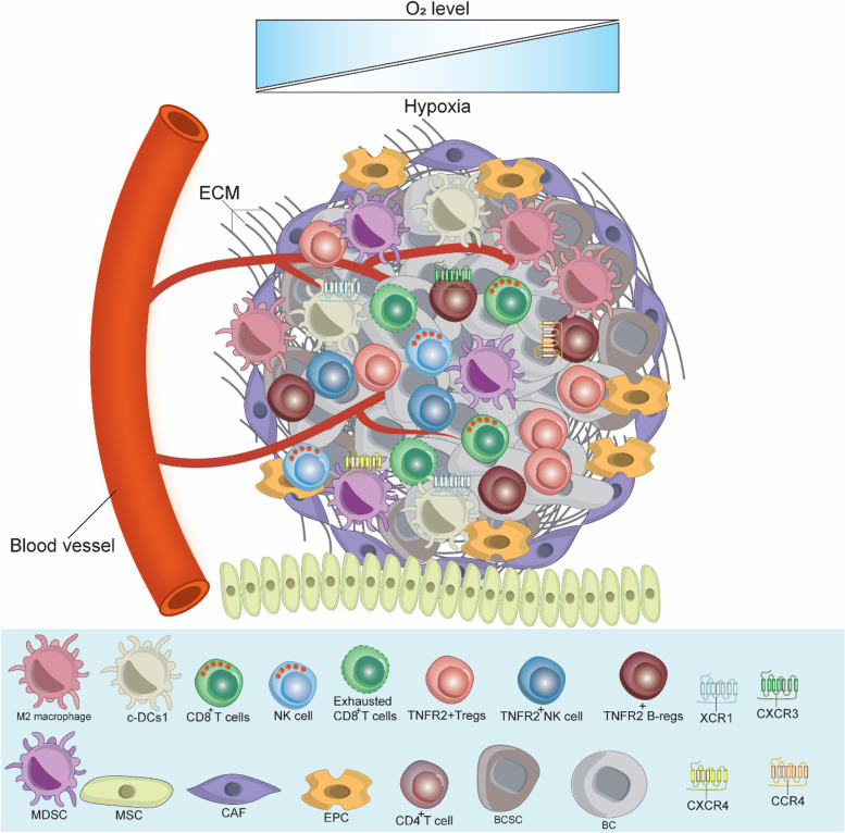
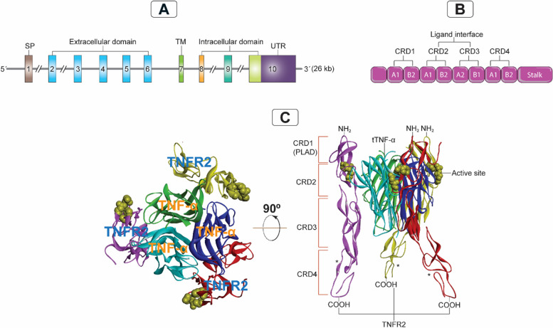
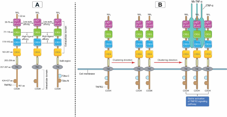
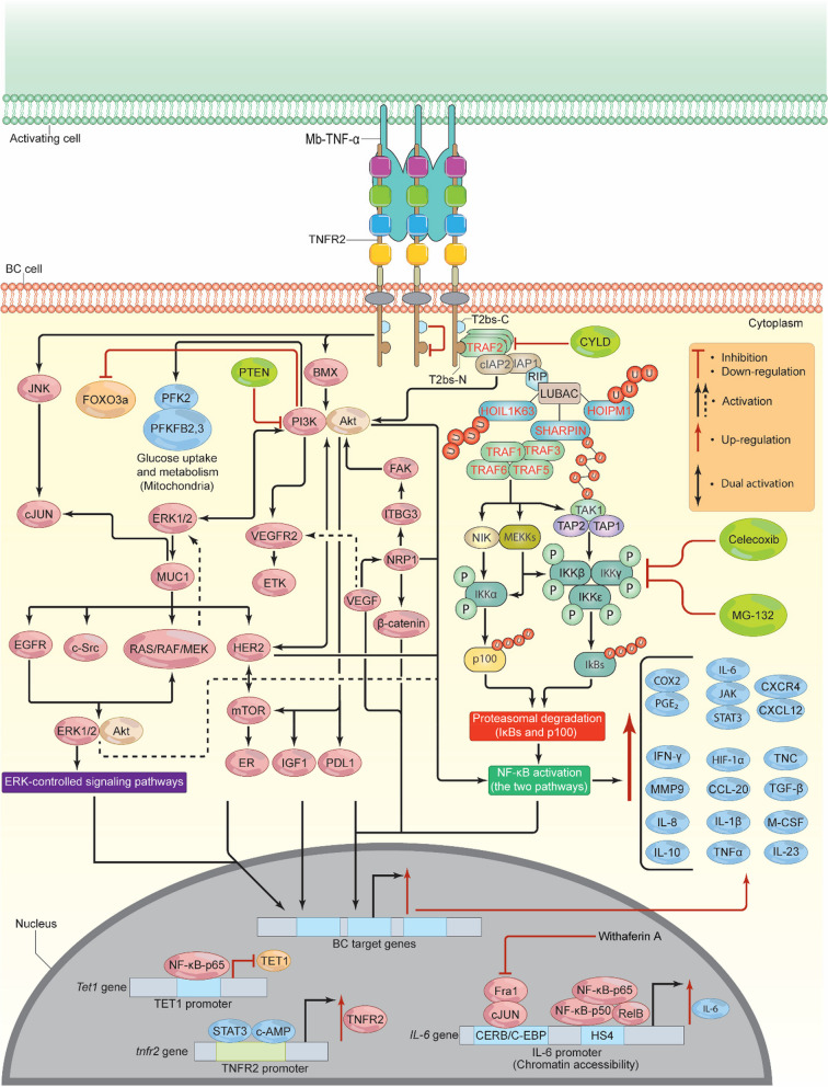
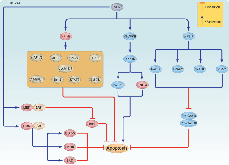
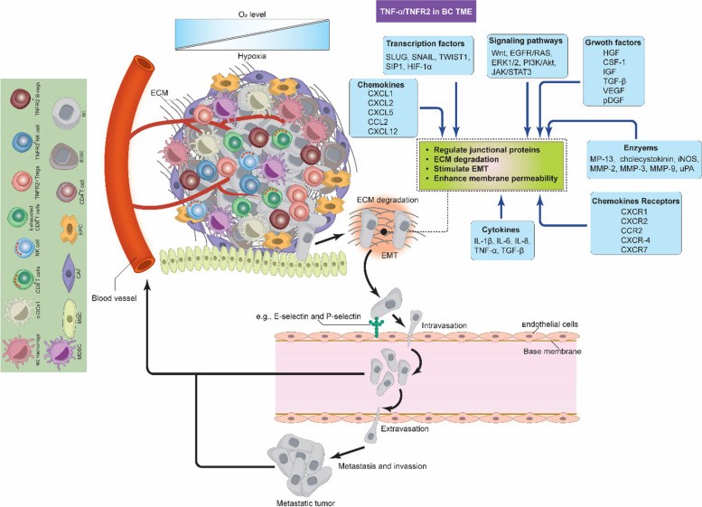
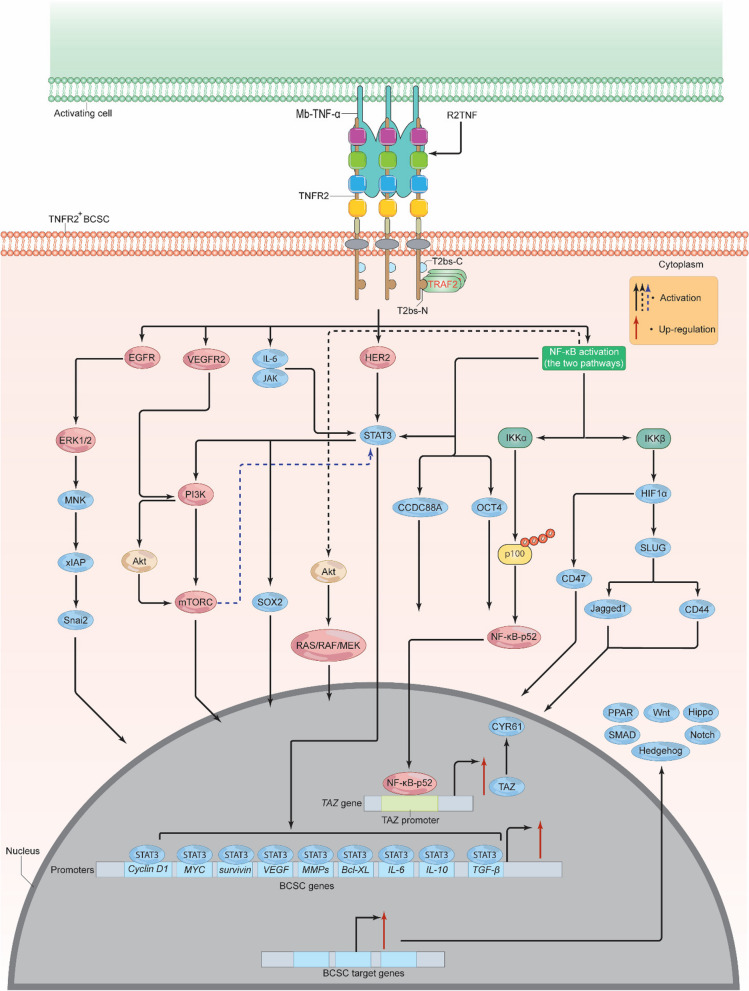
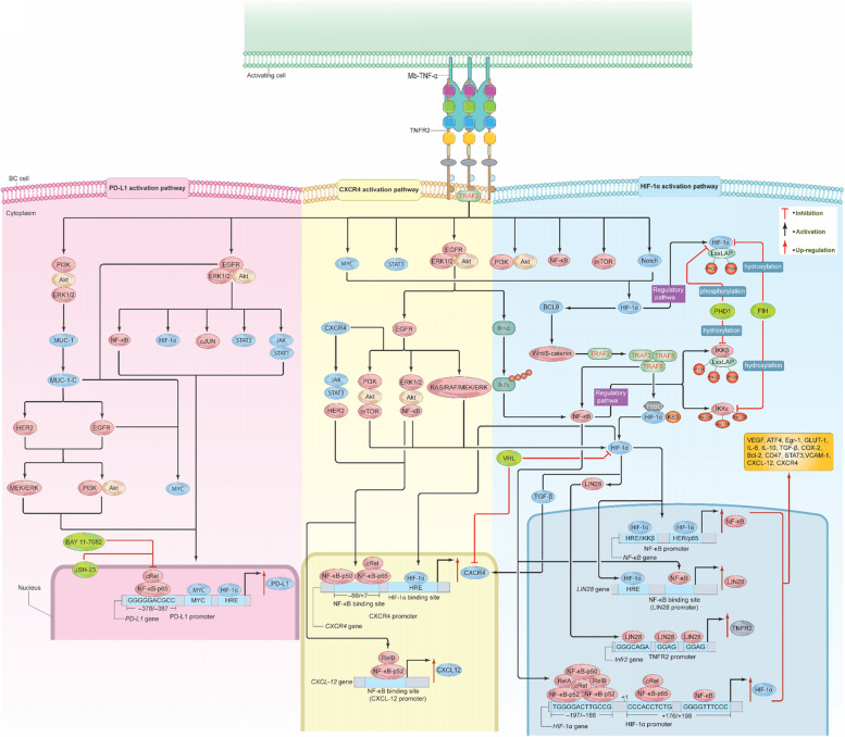
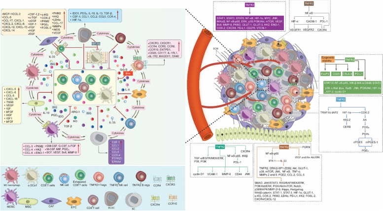
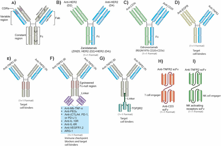
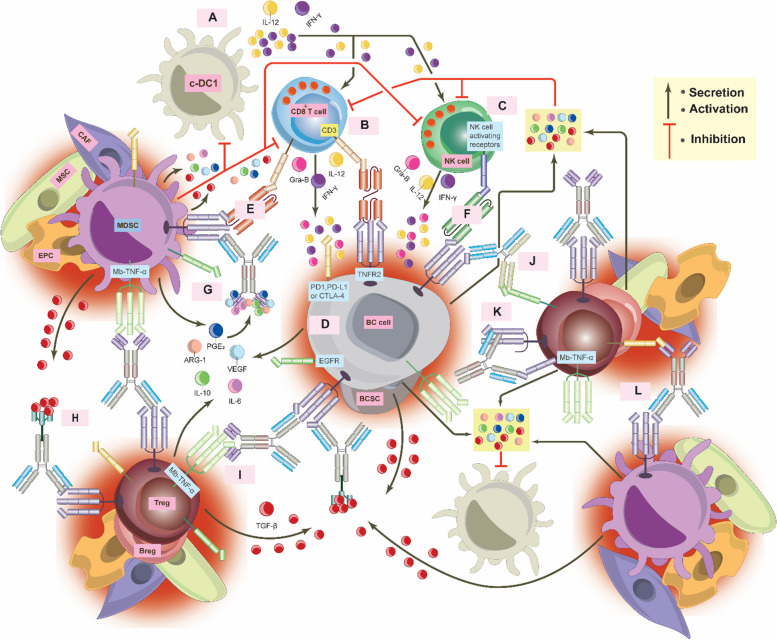
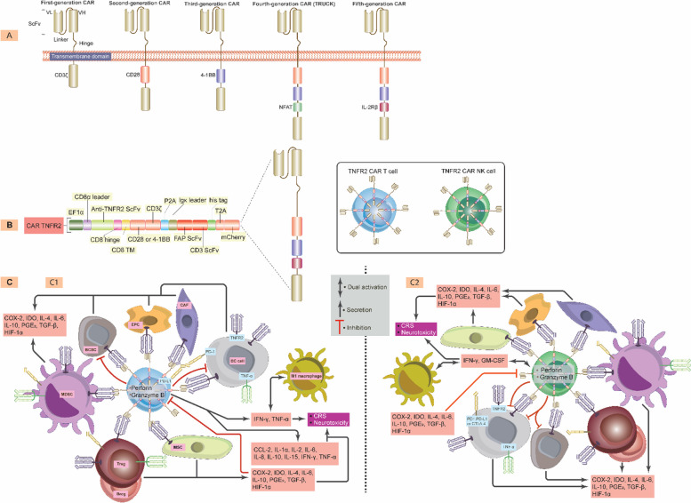
Breast cancer (BC) is the most frequently diagnosed malignancy among women. It is characterized by a high level of heterogeneity that emerges from the interaction of several cellular and soluble components in the tumor microenvironment (TME), such as cytokines, tumor cells and tumor-associated immune cells. Tumor necrosis factor (TNF) receptor 2 (TNFR2) appears to play a significant role in microenvironmental regulation, tumor progression, immune evasion, drug resistance, and metastasis of many types of cancer, including BC. However, the significance of TNFR2 in BC biology is not fully understood. This review provides an overview of TNFR2 biology, detailing its activation and its interactions with important signaling pathways in the TME (e.g., NF-κB, MAPK, and PI3K/Akt pathways). We discuss potential therapeutic strategies targeting TNFR2, with the aim of enhancing the antitumor immune response to BC. This review provides insights into role of TNFR2 as a major immune checkpoint for the future treatment of patients with BC.

Keywords: CD120b; Immune checkpoint; Immunosuppressive TME; TNF; TNFRSF1B.


Conflict of interest statement

Declarations. Ethics approval and consent to participate: Not applicable. Consent for publication: Not applicable. Competing interests: The authors declared that they have no competing interests.

Figures

Fig. 1
Fig. 1
Breast Cancer Tumor Microenvironment. As a solid tumor, the BC TME is highly complex and contains the ECM and various immune cells in addition to BC cells and BCSCs. These cells interact with each other, express, and release several immunomodulators to stimulate BC therapy resistance and escape from the anti-tumor immune response, which in turn promotes BC cell growth, invasion, and metastasis. c-DC1, type 1 conventional DCs; BC, breast cancer; BCSCs, breast cancer stem cells; MDSCs, myeloid-derived suppressor cells; T-regs, T regulatory cells; B-regs, B regulatory cells, CAFs, cancer associated fibroblast; EPCs, endothelial; MSCs, mesenchymal stem cells; TME, tumor microenvironment; ECM, extracellular matrix; CXCR3, C-X-C chemokine receptor 3; CXCR4, C-X-C chemokine receptor 4; XCR1, X-C motif chemokine receptor 1
Fig. 2
Fig. 2
Genomic organization of TNFR2 and its interaction with TNF-α. A: The TNFRSF1B gene organization on chromosome 1p36.2, consisting of 10 exons ranging from 35 to 2489 bp and 9 introns ranging from 338 to 7500 bp, totals 26 kb. B: Schematic representation of CRD 1–4 of TNFR2 indicating the component of each CRD of the two modules (A1, A2, B1 or B2), CRD 1 (PLAD; A1 and B2), CRD 2 (A1 and B2), CRD 3 (A2 and B1), and CRD 4 (A1 and B2). C: Showing the crystal structure of the 3ALQ (Protein Data Bank ID) of the TNFR2/TNF-α complex, indicating the confirmational interaction between three units of TNFR2 and three units of TNF-α. TNFR2, tumor necrosis factor receptor type 2; tTNF-α, triple tumor necrosis factor; TM, transmembrane; UTR, untranslated region; CRD, cysteine-rich domain; PLAD, pre-ligand assembly domain; kb, kilobases; bp, base pairs; (*), three identical TNFR2 units
Fig. 3
Fig. 3
TNFR2 protein structure and assembly on the cell surface. A. The full amino acid protein structure of the TNFR2 extracellular and intracellular domains. In the extracellular amino N-terminus domain, the organization of each CRD (1–4) is indicated, with the CRD1 harboring the self-structure interacting domain PLAD that interacts with low affinity with TNF-α and the regions of the CRD2 and CRD3 interacting with strong affinity with TNF-α. In the intracellular C-terminus domain, the receptor contains the TRAF binding sites (T2bs-C and T2bs-N) that actively recruit TRAF2 to inhibit or initiate signaling. B. A single chain of TNFR2 will start clustering with a second TNFR2 chain via their PLADs, then the two chains will cluster with a third chain via PLAD-PLAD interactions; then a Mb-TNF-α trimer binds to these clustering chains of TNFR2 to form a fully stable and active signalosome. CRD, cysteine-rich domain; PLAD, pre-ligand assembly domain; TNFR2, tumor necrosis receptor type two; Mb-TNF-α, membrane-bound tumor necrosis factor; TRAF, TNF receptor associated factor; T2bs-C, TRAF2-binding site C; T2bs-N, TRAF2-binding site N
Fig. 4
Fig. 4
TNFR2/Mb-TNF-α signalosome pathway. Upon Mb-TNF-α binding to TNFR2, the T2bs-N region recruits TRAF2, which then recruits E3 ligases (cIAP1, cIAP2, RIP, and LUBAC). LUBAC attaches M1-linked ubiquitin chains, stabilizing the signaling complex and enhancing downstream signaling. This leads to the accumulation of TRAFs 1, 3, 5, and 6, which phosphorylate TRAF 3, releasing NIK. NIK stimulates MEKK1, 2, 3, and TAK1, which then phosphorylate, IKKα, IKKβ, IKKε, and KKγ, inducing NF-κB pathways. Inhibitors like Celecoxib, CYLD, and MG-132 can prevent NF-κB activation. In addition, cIAP1 and cIAP2 stimulate BMX and JNK/cJUN, which induce PI3K/Akt, resulting in the activation and phosphorylation of several pathways (ETK/VEGFR2, ERK1/2, IGF1, HER2, mTOR, NF-κB, FOXO3a, and PD-L1). HER2 and ERK1/2 induce positive feedback loop signals, which enhance BC cell metabolism and proliferation via NF-κB, c-MYC, STATs, SAP-1a, AP-1, Elk-1, Cyclin D1, and ER. TNFR2 also stimulates HIF-1α, which in turn activates VEGF and its associated signaling pathways, leading to NF-κB-p65 activation. TNFR2 in its promoter contains binding sites for both STAT3 and c-AMP, the binding of which induces TNFR2 overexpression. Epigenetically, TNFR2 enhances IL-6 overexpression and down-regulates TET1 expression. As a result of TNFR2 activation, multiple proteins will be stimulated (CCL-2, PDL-1, IL-1β, IL-6, IL-8, IL-10, IL-23, M-CSF, MMP-9, COX-2/PGE2, TGF-β, CXCR4/CXCL-12, TNF-α, TNC, IFN-γ, and HIF-1α), leading to drug resistance, altered metabolism, migration, invasion, and the development of EMT. TNFR2, tumor necrosis receptor type 2; Mb-TNF-α, membrane-bound tumor necrosis factor; TRAF, TNF receptor associated factor; T2bs-C, TRAF2-binding site C; T2bs-N, TRAF2-binding site N; cIAP, cellular inhibitor of apoptosis; SHARPIN, shank-associated RH domain-interacting protein; HOIL-1-K63-Ub, heme-oxidized IRP2 ubiquitin ligase 1; HOIP-M1-Ub, HOIL-1-interacting protein; LUBAC, linear ubiquitin chain assembly complex; NIK, activate NF-κB inducing kinase; MEKK, MAP kinase/ERK kinase; TAK1, transforming growth factor-activated kinase 1; TAP, TAK1-binding protein complex; PI3K, phosphoinositide 3-kinases; Akt, protein kinase B/serine-threonine kinase; CYLD, cylindromatosis; PFK-2, phosphofructokinase-2; PFKFB2, 3, 6-phosophofrcto-2-kinase/fructose-2, 6-biphosphatase 3; mTOR, mammalian (or mechanistic) target of rapamycin; EGFR, epidermal growth factor receptor; PTEN, phosphatase and tensin homolog; VEGF, vascular endothelial growth factor; ETK, endothelial/epithelial tyrosine kinase; BMX, marrow x-linked kinase; VEGFR2, vascular endothelial growth factor receptor 2; EMT, epithelial-mesenchymal transition; MMP-9, matrix metalloproteinase-9; FOXO3a, forkhead box O3a; JNK, c-jun N-terminal kinase; ERK, extracellular signal-regulated kinase; MUC-1 and MUC1-C, Mucin; c-MYC, cellular myelocytomatosis; STAT, signal transducer and activator of transcriptions; AP-1, activator protein-1; IL-6, interleukin 6; IL-8, interleukin 8; IL-10, interleukin 10; IL-23, interleukin 23, M-CSF, macrophage colony stimulating factor; TET, ten-eleven translocation; COX-2, cyclooxygenase-2; PGE2, prostaglandin E2; TGF-β, transforming-growth factor β; IFN-γ, interferon gamma; HIF-1α, hypoxia inducible factor 1 alpha; NRP-1, Neuropilin-1; TNC, Tenascin C; ITBG3, integrin 3; FAK, focal adhesion kinase pathway; IKKs, IκB kinases; IGF1, insulin-like growth factor 1; HER2, human epidermal growth factor receptor 2
Fig. 5
Fig. 5
TNFR2-mediated apoptosis resistance in BC cells. TNFR2 triggers the activation of various proteins to help BC escape apoptosis. TNFR2-mediated Akt, NF-κB, and c-FLIP activation stimulate c-FLIP to activate FADD, RIPK1, TRAF2, and TRADD, inhibiting pro-Cas 8 and pro-Cas 10. While Akt activation leads to apoptosis via IKK/DR activation, inducing TNF-α and TWEAK, NF-κB activation induces a series of anti-apoptotic proteins, including Bcl-2, Bcl-XL, Bcl-W, xIAP, cIAP1/2, A1/BFL-1, Cyclin D1, STAT3, and MCL-1 to inhibit apoptosis. TNFR2 may also activate MEK/ERK and PI3K/Akt. MEK/ERK deactivates the protein Bim, while PI3K/Akt deactivates GSK-3, BAD, and FKHR. Together, these proteins inhibit apoptosis, leading to BC cell survival and growth. TNFR2, tumor necrosis receptor type 2; TNF-α, tumor necrosis factor; TRAF, TNF receptor associated factor; c-FLIP, cellular FLICE (FADD-like IL-1β-converting enzyme)-inhibitory protein; FADD, fas-associated protein-death domain; RIPK1, receptor-interacting protein kinase 1; TRADD, TNFR1-associated death domain; TWEAK, tumor necrosis-like weak inducer of apoptosis; BAD, Bcl-2-associated agonist of cell death; pro-cas8, pro-caspase 8; pro-cas10, pro-caspase 10; Bcl-2, B cell lymphoma 2; Bcl-XL, B cell lymphoma extra-large, Bcl-W, B cell lymphoma W; xIAP, X-linked inhibitor of apoptosis protein; cIAP, cellular inhibitor of apoptosis; STAT3, signal transducer and activator of transcription 3; MCL-1, Myeloid cell leukemia 1; GSK-3, glycogen kinase synthase-3; FKHR, fork-head in rhabdomyosarcoma; PI3K, phosphoinositide 3-kinases; Akt, protein kinase B/serine-threonine kinase; ERK, extracellular signal-regulated kinase
Fig. 6
Fig. 6
TNF-α/TNFR2-mediated invasion and metastasis in BC TME. TNFR2 signals can stimulate various proteins and signaling pathways, supporting almost all aspects of BC cell invasion and metastasis. For tumor cells to move from the primary tumor site by undergoing intravasation first and then extravasation, several enzymes, such as MMPs, must degrade the ECM. Many signaling pathways, in addition to several cytokines, chemokines, chemokine receptors, and growth factors, control the endothelial junctional proteins to induce membrane permeability. These events facilitate the movement of tumor cells, proliferation, vascularization, migration, and metastasis of BC to generate new tumors at distant sites and organs. c-DC1, type 1 conventional DCs; BC, breast cancer; BCSCs, breast cancer stem cells; MDSCs, myeloid-derived suppressor cells; T-regs, T regulatory cells; B-regs, B regulatory cells, CAFs, cancer associated fibroblast; EPCs, endothelial; MSCs, mesenchymal stem cells; TME, tumor microenvironment; ECM, extracellular matrix; MMPs, matrix metalloproteinases; iNOS, inducible nitric oxide synthase; NO, nitric oxide; TNF-α, membrane-bound tumor necrosis factor; TNFR2, tumor necrosis factor receptor type two; IL-1β, interleukin 1 beta; IL-6, interleukin 6; IL-8, interleukin 8; HIF-1α, hypoxia inducible factor 1 alpha; VEGF, vascular endothelial growth factor; pDGF, platelet-derived growth factor; IGF, insulin-like growth factor; FGF, fibroblast growth factor; HGF; CSF-1, colony stimulating factor 1; TGF-β, transforming-growth factor β; uPA, urokinase-type plasminogen; TWIST1, twist related protein-1; CCL-2, chemokine (C–C) motif ligand 2; CXCR1, C-X-C chemokine receptor 1; CXCR2, C-X-C chemokine receptor 2; CXCR-4, C-X-C chemokine receptor 4; CXCR7, C-X-C chemokine receptor 7; CCR2, C–C chemokine receptor type 2; CXCL-1, C-X-C motif chemokine ligand 1; CXCL-2, C-X-C motif chemokine ligand 2; CXCL-5, C-X-C motif chemokine ligand 5; CXCL-12, C-X-C motif chemokine ligand 12
Fig. 7
Fig. 7
TNFR2 supports BCSC growth and development. Once TNFR2 is activated via Mb-TNF-α or R2TNF, multiple proteins including EGFR, VEGFR2, IL-6/JAK, HER2, and NF-κB are activated. EGFR activation leads to ERK1/2 phosphorylation, inducing MNK, xIAP, and Snai 2. VEGFR2 activation triggers PI3K/Akt activation, stimulating mTORC, which along with IL-6/JAK, HER2, and NF-κB, promotes STAT3 activation. STAT3 induces various oncogenic proteins, including Cycline D1, MYC, surviving, VEGF, MMPs, Bcl-XL, IL-6, IL-10, and TGF-β. IKKα and IKKβ activation promote HIF-1α, OCT4, CCDC88A, and Akt stimulation and the release of NF-κB-p52 to the nucleus and up-regulate TAZ-enhancing CYR61 production. HIF-1α will trigger CD47 and SLUG/Jagged1 or SLUG/CD44, while Akt stimulates the RAS/RAF/MEK/ERK pathway. These proteins lead to Notch, Hedgehog, Wnt, Hippo, SMAD, and PPAR stimulation, promoting BCSC self-renewal, EMT development, angiogenesis, and survival. TNFR2, tumor necrosis receptor type 2; Mb-TNF-α, membrane-bound tumor necrosis factor; TRAF, TNF receptor associated factor; T2bs-C, TRAF2-binding site C; T2bs-N, TRAF2-binding site N; Akt, protein kinase B/serine-threonine kinase; mTOR, mammalian (or mechanistic) target of rapamycin; EGFR, epidermal growth factor receptor; VEGF, vascular endothelial growth factor; VEGFR2, vascular endothelial growth factor receptor 2; EMT, epithelial-mesenchymal; MMPs, matrix metalloproteinases; c-MYC, cellular myelocytomatosis; STAT, signal transducer and activator of transcriptions; IL-6, interleukin 6; IL-10, interleukin 10; TGF-β, transforming-growth factor β; HIF-1α, hypoxia inducible factor 1 alpha; IKKs, IκB kinases; Bcl-2, B cell lymphoma 2; HER2, human epidermal growth factor receptor 2; PI3K, phosphoinositide 3-kinases; ERK, extracellular signal-regulated kinase; MNK, MAPK interacting kinase; PPAR, peroxisome proliferator-activated receptor
Fig. 8
Fig. 8
Signals from Mb-TNF-α/TNFR2 stimulate HIF-1α, CXCR4, and PD-L1. Binding of Mb-TNF-α to TNFR2 activates the HIF-1α, CXCR4, and PD-L1 pathways. In the HIF-1α pathway, TNFR2 signals activate STAT3, mTOR, MYC, Notch, NF-κB, EGFR/ERK/Akt, PI3K/Akt, and PI3K/Akt/mTOR. HIF-1α can also activate BCL9, stimulating Wnt/β-catenin, which interacts with TRAF2, leading to TRAF 3, 5, and 6 accumulations inducing NF-κB activation. NF-κB subunits bind to specific sequences in the HIF-1α promoter, enhancing its stimulation. TRAF 6 can directly mediate HIF-1α activation via ubiquitination of K63. EGFR/ERK/Akt-mediated IκBα phosphorylation can also activate HIF-1α, creating a positive feedback loop of several signaling pathways. LIN28, activated by HIF-1α, upregulates TNFR2 expression. IKKβ and HIF-1α possess shared residues targeted at PHD1. FIH inhibits IKKε, preventing its binding to TRAF3/TBK1 proteins. In the CXCR4 pathway, TNFR2 signals stimulate NF-κB, HER2, PI3K, Akt, mTOR, Wnt/βcatenin, the RAS/RAF/MEK/ERK pathway, and JAK/STAT. NF-κB subunits bind to CXCR4 and CXCL-12 promoters, inducing its overexpression. HIF-1α, NF-κB, TGF-β, and HER2 can also enhance CXCR4 expression. VHL, a negative regulator of HIF-1α, down-regulates CXCR4, but is inactive in most solid tumors, leading to CXCR4 overexpression. In the PD-L1 pathway, TNFR2 signals activate EGFR/ERK1/2/Akt and PI3K/Akt/ERK1/2, which in turn promote NF-κB, HIF-1α, cJUN, STAT3, and JAK/STAT3, up-regulating PD-L1. NF-κB-p65 binds to the PD-L1 promoter, enhancing its expression. The PI3K/Akt/ERK1/2 pathway also stimulates Mucin-1, activating HER2, EGFR, and MYC, which further up-regulate PD-L1. Inhibition of NF-κB-p65 via BAY 11–7082 and JSH-23 abolished PD-L1 overexpression. These pathways support BC growth, development, immune escape, drug resistance, angiogenesis, and metastasis. TNFR2, tumor necrosis receptor type 2; Mb-TNF-α, membrane-bound tumor necrosis factor; TRAF, TNF receptor associated factor; PI3K, phosphoinositide 3-kinases; Akt, protein kinase B/serine-threonine kinase; mTOR, mammalian (or mechanistic) target of rapamycin; EGFR, epidermal growth factor receptor; VEGF, vascular endothelial growth factor; ERK, extracellular signal-regulated kinase; MYC, cellular myelocytomatosis; STAT, signal transducer and activator of transcriptions; IL-6, interleukin 6; IL-10, interleukin 10; COX-2, cyclooxygenase-2; TGF-β, transforming-growth factor β; HIF-1α, hypoxia inducible factor 1 alpha; CXCR4, C-X-C chemokine receptor 4; HRE, hypoxia response element; VHL, Von Hppel Lindau; PD-L1, programmed cell death ligand 1; T6BD, TRAF 6 binding domain; PHD1, prolyl-hydroxylase 1; FIH, Factor-inhibiting HIF-1α; Bcl-2, B cell lymphoma 2; VCAM-1, vascular cell adhesion molecule-1
Fig. 9
Fig. 9
TNFR2 expression in BC TME. TNFR2+ cells secrete IL-10, IL-4, IL-6, IL-33, IL-35, TGF-β, HIF-1α, IDO, COX-2/PGE2, ARG-1, iNOS, and others, establishing the ISG. This ISG depletes essential amino acids, impairs MHC class I antigen presentation, leading to suppression of DC1s, CD8+ T cells and promoting CD8+ T cell exhaustion. COX-2/PGE2 inhibits NK cell effector receptors and induces M2 macrophage differentiation, further enhancing immunosuppression. Hypoxia-induced HIF-1α supports BC metabolism and immunosuppressive cells, inhibits NK cell cytotoxicity, and promotes MDSC differentiation, as well as induces the activation of COX-2/PGE2, IL-6, IL-10, and TGF-1β, leading to PD-L1 overexpression, which inhibits c-DC1s. TNFR2 also induces the expression of immunosuppressive receptors and stabilizes FoxP3. In CAFs, TNFR2 activation leads to stromal mass formation, hindering CD8+ T cell infiltration, and also augments AICD- and FAS-mediated cell death. TNFR2-mediates the expression of chemokines, and their receptors recruit c-DC1s to BC TME, initiating their suppression and affecting CD8+ T cell stimulation. Wnt/β-catenin downregulates CCL-4, inhibiting c-DCs1 migration, recruitment, and tumor infiltration. Moreover, increased BC cell mass and reduced blood vessel formation act as physical barriers, hindering the infiltration and recruitment of c-DC1 cells into the TME. CXCR-3+T-regs compete with CD8+ T cells for the CXCL-9 gradient, restricting their activation by c-DCs1. Immunosuppressive cells can also mediate cell-to-cell contact inhibition via PD-L1, PD-1, FAS, and CTLA-4 expression. Collectively, this can lead to dysfunctional c-DC1s, ultimately limiting their presence in the TME and supporting BC growth. TNFR2, tumor necrosis factor receptor type two; Mb-TNF-α, membrane-bound tumor necrosis factor; c-DC1, type 1 conventional dendritic cells; BC, breast cancer; BCSCs, breast cancer stem cells; IL-4, interleukin 4; IL-6, interleukin 6; IL-10, interleukin 10; IL-12, interleukin 12; IL-33, interleukin 33; IL-35, interleukin IL-35; TGF-β, transforming-growth factor β; COX-2, cyclooxygenase-2; HIF-1α, hypoxia inducible factor 1 alpha; PEG2, prorstaglandin-E2; ARG-1, arginase-1; MMP-9, matrix metalloproteinase-9; CCL-4, chemokine (C–C) motif ligand 4; CCL-19, chemokine (C–C) motif ligand 19; CCL-21, chemokine (C–C) motif ligand 21; CXCR3, C-X-C chemokine receptor 3; CXCR4, C-X-C chemokine receptor 4; XCR1, X-C motif chemokine receptor 1; CCR-5, C–C chemokine receptor type 5; CCR-7, C–C chemokine receptor type 7; IFN-γ, interferon gamma; EGFR, epidermal growth factor receptor; PD-1, programmed cell death 1; PD-L1, programmed cell death ligand 1; CTLA-4, cytotoxic T lymphocyte antigen 4; VEGF, vascular endothelial growth factor; MHC-1, major histocompatibility complex class I; ISG, immunosuppressive gradient; STAT3, signal transducer and activator of transcription 3; STAT5, signal transducer and activator of transcription 5; c-MYC; cellular myelocytomatosis; GLUT-1, glucose transporter 1; GLUT-3, glucose transporter 3; α-KG, alpha ketoglutarate; PKM2, pyruvate kinase M2; ENO-1, enolase-1; LDHα, lactate dehydrogenase-alpha; IDO, indoleamine 2,3-dioxygenase; CSF-1,2, macrophage colony-stimulating factor 1, 2; c-TGF, connective-tissue growth factor; pDGF, platelet-derived growth factor; IGF-1, insulin-like growth factor 1; FGF, fibroblast growth factor; HGF, hepatocyte growth factor; EGF, epidermal growth factor; bFGF, basic insulin-like growth factor; G-CSF, granulocyte-colony stimulating factor; M-CSF, macrophage colony stimulating factor; MDSCs, myeloid-derived suppressor cells; T-regs, T regulatory cells; B-regs, B regulatory cells, CAFs, cancer associated fibroblast; EPCs, endothelial progenitor cells; MSCs, mesenchymal stem cells; TME, tumor microenvironment; ECM, extracellular matrix; AICD, antigen-mediated activation-induced cell death
Fig. 10
Fig. 10
Bs-Abs for neutralizing TNFR2. a. The standard structure of an IgG antibody. b. Established bs-Abs, zanidatamab (ZW25; HER2 (D2) × HER2 (D4)) targeting both forms of HER2 (D2 and D4)) with format (1 + 1). c. Established bs-Abs, odronextamab (REGN1979; CD20 × CD3ε) targeting bs-Ab for both CD20 and CD3ε with format (1 + 1). While d, e, f, and g represent proposed different bs-Abs for targeting TNFR2 on both BC cells and immunosuppressive cells, in addition to some immunosuppressive receptors (EGFR, CTLA-4, PD-1, PD-L1, Mb-TNF-α, TGFβR2, VEGFR1,2, IL-6R, and IL-10R) and mediators (ARG-1 and PGE2) with format (1 + 1), h and i bs-Abs engagers for CD8+ T cells (h) and for NK cells (i) that target TNFR2 on both BC cells and the immunosuppressive cells as well as targeting and stimulating immune activating receptors on NK cells such as NCR1, NCR2/NKp44, NCR3/NKp30, NCR1/NKp46, NKG2D, MICA/B, MICA, as well as on CD8+ T cells such as CD3ε. TNFR2, tumor necrosis factor receptor type two; Mb-TNF-α, membrane-bound tumor necrosis factor; Bs-Ab, bi-specific antibody; CDR, combativity determine region; HER2, epidermal growth factor receptor 2; IL-6R, interleukin 6 receptor; IL-10R, interleukin 10 receptor; PD-1, programmed cell death 1; PD-L1, programmed cell death ligand 1; CTLA-4, cytotoxic T lymphocyte antigen 4; TGF-βR2, transforming-growth factor β receptor 2; VEGFR 1,2, vascular endothelial growth factor receptor 1 and 2; PEG2, prostaglandin-E2; ARG-1, arginase-1; NCR1, natural cytotoxicity triggering receptor 1; NCR3, natural cytotoxicity triggering receptor 3; MICA and B, MHC class I chain-related polypeptide A and B; NKG2D, natural killer group 2-member D; NKp30, natural killer protein 30; NKp44, natural killer protein 44; NKp46, natural killer protein 46
Fig. 11
Fig. 11
Anti-TNFR2 bs-Ab mechanism of action. A. DCs activate both CD8+ T cells and NK cells by TAA-MHC-1 presentation and secretion of IL-12 and IFN-γ. B, C, and D. Following the activation, CD8+ T cells and NK cells will attack and destroy BC cells by producing high quantities of IL-12, IFN-γ, and gra-B. BC cells and BCSCs, in turn, express EGFR, Mb-TNF-α, TNFR2, PD-1, PD-L1, and CTLA-4, and also produce IL-6, IL-10, PEG2, VEGF, TGF-β, and ARG-1, which suppress DCs, CD8+ T cells, and NK cells. Other immunosuppressive cells, including MDSCs, T-regs, B-regs, CAFs, EPCs, and MSCs in the BC TME also produce tremendous quantities of IL-6, IL-10, PEG2, VEGF, ARG-1, and TGF-β, as well as over-express TNFR2, PD-1, PD-L1, EGFR, Mb-TNF-α, and CTLA-4, leading to DC, CD8+ T cell, and NK cell suppression. E. T cell engager bs-Ab (TNFR2 × CD3ε) targeting CD3ε on CD8+ T cells and TNFR2 on BC cells and BCSCs, blocking the signals of TNFR2 and stimulating CD8+ T cells to kill tumors. F. NK cells engager bs-Ab (TNFR2 × NK cell activating receptors) targeting NK cell activating receptors (NCR1, NCR2/NKp44, NCR3/NKp30, NCR3/NKp46, NKG2D, and MICA/B) on NK cells and TNFR2 on BC cells and BCSCs blocking the signals of TNFR2 and stimulating NK cells to kill tumor. G, I, and L. bs-Ab targeting TNFR2 on TNFR2+ cells to hinder its signal as well as targeting CTLA-4, PD-1, PD-L1, Mb-TNF-α, ARG-1, PGE2 TGFβR2, VEGFR1,2, IL-6R, and IL-10R expressed by the same cells inhibit their signaling pathway. These bs-Ab capture TGF-β, VEGF, IL-6, IL-10, ARG-1, and PGE2, which in turn diminishes the immunosuppressive effect in BC TME. K. bs-Ab (TNFR2 × EGFR) targeting TNFR2 and EGFR on BC cells, BCSCs, and the immunosuppressive cells to suppress their signals and induce an effective anti-tumor immune response. L. Bs-Ab (TNFR2 × TNFR2) binding to one chain of the tri-TNFR2 complex to prevent clustering on the surface of BC cells, BCSCs, MDSCs, T-regs, B-regs, CAFs, EPCs, and MSCs. TNFR2, tumor necrosis factor receptor type two; Mb-TNF-α, membrane-bound tumor necrosis factor; Bs-Ab, bi-specific antibody; c-DC1, type 1 conventional DCs; BC, breast cancer; BCSCs, breast cancer stem cells; TAA, tumor associated antigen; MHC-1, major histocompatibility complex class one; IL-6, interleukin 6; IL-10, interleukin 10; IL-12, interleukin 12; IFN-γ, interferon gamma; gra-B, granzyme B; EGFR, epidermal growth factor receptor; PD-1, programmed cell death 1; PD-L1, programmed cell death ligand 1; CTLA-4, cytotoxic T lymphocyte antigen 4; TGF-β, transforming-growth factor β; VEGF, vascular endothelial growth factor; PEG2, prorstaglandin-E2; ARG-1, arginase-1; MDSCs, myeloid-derived suppressor cells; T-regs, T regulatory cells; B-regs, B regulatory cells, CAFs, cancer associated fibroblast; EPCs, endothelial progenitor cells; MSCs, mesenchymal stem cells; TME, tumor microenvironment; NCR1, natural cytotoxicity triggering receptor 1; NCR3, natural cytotoxicity triggering receptor 3; MICA and B, MHC class I chain-related polypeptide A and B; NKG2D, natural killer group 2-member D; NKp30, natural killer protein 30; NKp44, natural killer protein 44; NKp46, natural killer protein 46
Fig. 12
Fig. 12
TNRT2-specific CAR T and CAR NK cells. A. Schematic representing the structure of the five generations of CARs. B. The proposed structure of TNFR2-specific CAR, including TNFR2 CAR T cells (blue) and TNFR2 CAR NK cells (green). C. The proposed mechanism of action: C1. First, TNFR2 CAR T cells will target TNFR2 on BC cells, BCSCs, MDSCs, T-regs, B-regs, CAFs, EPCs, and MSCs to deactivate TNFR2 to hinder its signal. These can produce the immunosuppressive mediators COX-2/PGE2, IDO, IL-4, IL-6, TGF-β, and HIF-1α which can inhibit TNFR2-specific CAR T cells and the effector CD8+ T cells. When targeting TNFR2 on BC cells and BCSCs, it will be able to block the interaction between Mb-TNF-α on activating cells (which could be any cell that over-expresses Mb-TNF-α in the BC TME) and TNFR2 on cancer cells, and at the same time, TNFR2-specific CAR T cells will produce perforin and granzyme B to kill the BC cells. C2. Second, TNFR2-specific CAR NK cells will fulfill the same functions as TNFR2-specific CAR T cells. While TNFR2-specific CAR T cells secrete CCL-2, IL-1α, IL-2, IL-6, IL-8, IL-10, IL-15, IFN-γ, and TNF-α, TNFR2-specific CAR NK cells will secrete IFN-γ and GM-CSF. TNFR2, tumor necrosis factor receptor type two; Mb-TNF-α, membrane-bound tumor necrosis factor; BC, breast cancer; BCSCs, breast cancer stem cells; IL-1α, interleukin 1 alpha; IL-2, interleukin 2; IL-4, interleukin 4; IL-6, IL-8, interleukin 8; IL-10, interleukin 10; IFN-γ, interferon-gamma; GM-CSF, granulocyte–macrophage colony stimulating factor; HIF-1α, hypoxia inducible factor 1 alpha; COX-2, cyclooxygenase-2; TGF-β, transforming-growth factor β; IDO, indoleamine 2,3-dioxygenase; PEG2, prostaglandin-E2; ScFv, single-chain variable fragment; MDSCs, myeloid-derived suppressor cells; T-regs, T regulatory cells; B-regs, B regulatory cells, CAFs, cancer associated fibroblast; EPCs, endothelial progenitor cells; MSCs, mesenchymal stem cells; TME, tumor microenvironment; CRS, cytokine release syndrome

References

    1. Sung, H., J. Ferlay, R.L. Siegel, et al.1, Global Cancer Statistics 2020: GLOBOCAN Estimates of Incidence and Mortality Worldwide for 36 Cancers in 185 Countries. CA: A Cancer Journal for Clinicians, 2021. 71(3): p. 209–249. 10.3322/caac.21660. - PubMed
    1. Collignon, E., A. Canale, C. Al Wardi, et al.1, Immunity drives TET1 regulation in cancer through NF-κB. Science Advances. 4(6): p. eaap7309. 10.1126/sciadv.aap7309. - PMC - PubMed
    1. Čelešnik, H. and U. Potočnik1, Peripheral Blood Transcriptome in Breast Cancer Patients as a Source of Less Invasive Immune Biomarkers for Personalized Medicine, and Implications for Triple Negative Breast Cancer. Cancers (Basel), 2022. 14(3): p. 10.3390/cancers14030591. - PMC - PubMed
    1. Yersal, O. and S. Barutca1, Biological subtypes of breast cancer: Prognostic and therapeutic implications. World J Clin Oncol, 2014. 5(3): p. 412–24. 10.5306/wjco.v5.i3.412. - PMC - PubMed
    1. Anderson, N.M. and M.C. Simon1, The tumor microenvironment. Current Biology, 2020. 30(16): p. R921-R925. 10.1016/j.cub.2020.06.081. - PMC - PubMed

Substances

LinkOut - more resources