. 2024 Nov 28;57(1):93.
doi: 10.1186/s40659-024-00576-0.

Heterogeneity in the formation of primary and secondary visual fields during human prenatal development

Affiliations

Heterogeneity in the formation of primary and secondary visual fields during human prenatal development

Olga Godovalova et al. Biol Res. .

Abstract

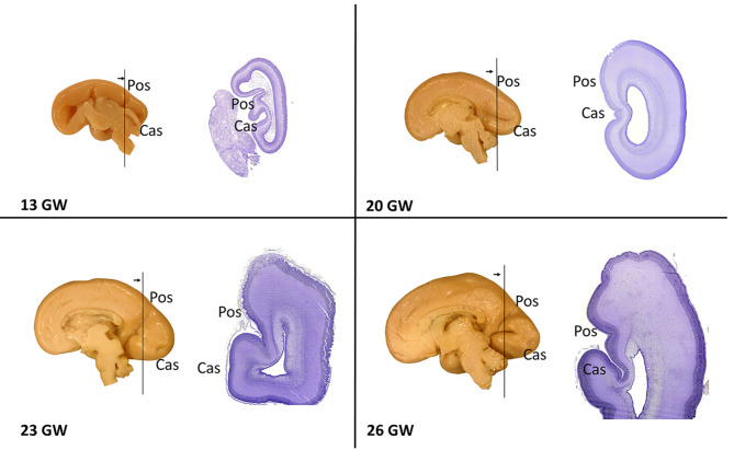
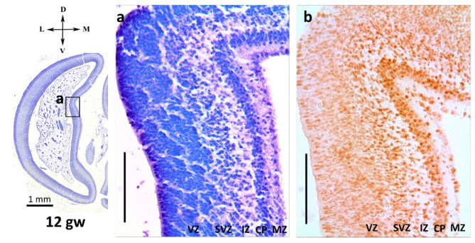
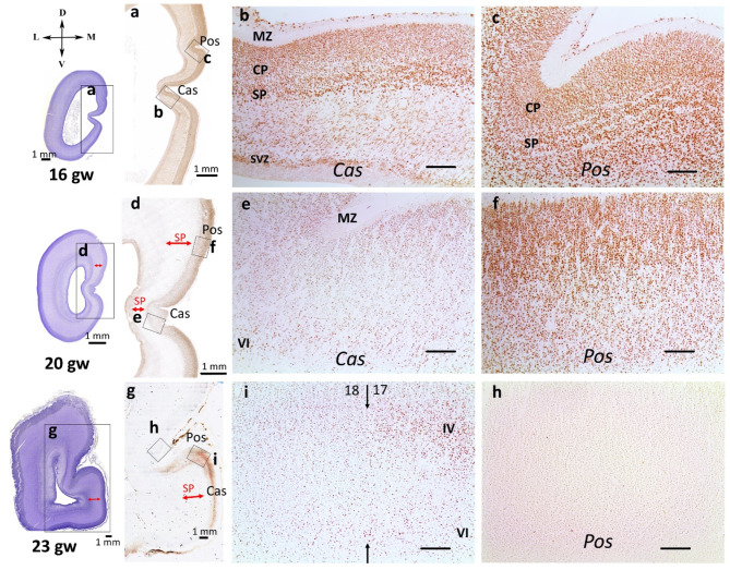
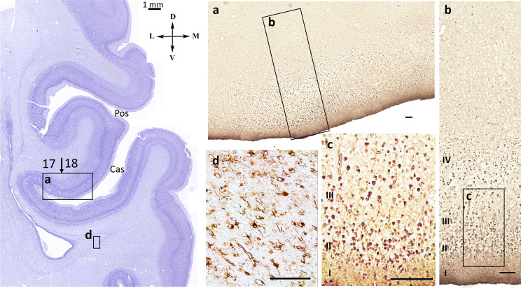
The human neocortex has a huge surface area with unique cytoarchitectonics, most of which is concealed in sulci. Some cytoarchitectonic fields are associated with macroscopic landmarks. In particular, the primary visual field 17 is associated with the calcarine sulcus. During the prenatal development of the human brain, neocortical gyri and sulci undergo changes and modifications after primary formation. To explore the morphogenetic processes in visual fields during the formation of the primary (provisional) and secondary (permanent) sulci, the occipital lobe of the human fetal brain was studied using immunohistochemical methods. The distribution of various glial and neuronal markers (S-100, β-III-tubulin, NeuN, reelin) in the calcarine sulcus and parietooccipital sulcus was compared. The heterogeneity in the formation of primary and secondary visual fields was demonstrated. The study revealed that the development of the primary visual field 17, linked with the calcarine sulcus, preceded the development of a shared anlage of fields 18 and 19 linked with the parietooccipital sulcus. The functional differentiation of the primary visual field begins during the period of thalamic afferent ingrowth. This process coincides with the temporal smoothing of the calcarine sulcus, indicating a simultaneous progression of functional specialization and structural modifications. At the late fetal period, cortical plate of gyri and sulci banks showed higher NeuN-labeling than inside the sulcus in the same cytoarchitectonic field.

Keywords: Calcarine sulcus; Gyrification; Human fetal development; Human fetuses; Occipital lobe; Parietooccipital sulcus; Visual fields.


Conflict of interest statement

Declarations. Ethics approval and consent to participate: The study was conducted in accordance with the Declaration of Helsinki. All protocols were approved by the local Ethics Committee of the Research Institute of Human Morphology(No. 3, January 17, 2006; No. 33(9), February 7, 2022). Informed consent: Not applicable. Conflict of interest: The authors declare no conflict of interest. The funders had no role in the design of the study; in the collection, analyses, or interpretation of data; in the writing of the manuscript; or in the decision to publish the results.

Figures

Fig. 1
Fig. 1
Stages of the development of the calcarine (Cas) and parietooccipital sulci (Pos) during the human prenatal ontogenesis. Medial surface of the hemispheres of samples 13, 20, 23 and 26 gw, on the right are Nissl-stained histological coronal sections, right hemispheres
Fig. 2
Fig. 2
Parietooccipital sulcus (Pos) at 12th gw. The forming cortical plate was clearly visible on Nissl staining (a) and in NeuN-immunohistochemical labeling (b). NeuN-immunopositive neuroblasts were already seen in the subventricular (SVZ), intermediate zone (IZ), cortical plate (CP) and marginal zone (MZ). Right hemisphere. Coronal section. Scale bar: 100 µm
Fig. 3
Fig. 3
NeuN-immunohistochemical staining, coronal sections, occipital lobe at 16 gw, right hemisphere (a, b, c), 20 gw, right hemisphere (d, e, f), 23 gw, right hemisphere (g, I, h). The red arrow indicates the subplate zone. SVZ – subventricular zone, IZ – intermediate zone, SP – subplate zone, CP – cortical plate, MZ – marginal zone. Scale bar: 100 μm
Fig. 4
Fig. 4
(a) – Percentage ratio of NeuN-immunoreactivity coefficient in the cortical plate of calcarine sulcus and parietooccipital sulcus. (b) – Percentage ratio of NeuN-immunoreactivity coefficient in the subplate under calcarine sulcus and parietooccipital sulcus. (с) – NeuN-immunoreactivity coefficient in early fetal (ef), middle fetal (mf) and late fetal (lf) periods. Significant differences between sulci are marked by square brackets and asterisks (**p = 0.005036, ***p = 0.000001). (d) – Boxplots and multiple comparison for ratio of the NeuN-immunoreactivity coefficient in the cortical plate of calcarine sulcus and parietooccipital sulcus. (e) – Boxplots and multiple comparison for ratio of the NeuN-immunoreactivity coefficient in the subplate of calcarine sulcus and parietooccipital sulcus. Comparable stages: provisional sulci; the parietooccipital sulcus smoothing stage (Pos-); two sulci, when both sulci exist; the calcarine sulcus smoothing stage (Cas-). Significant differences between stages are marked by square brackets and asterisks or presented in the table to the right of the bosxplots. (f) –Scheme of the distribution of NeuN-immunoreactive cells in the cortical plate at different stages of fetal development, Cas - calcarine sulcus, Pos - parietooccipital sulcus
Fig. 5
Fig. 5
Nissl staining section, the medial part of the occipital lobe, preterm 32 gw + 5 weeks. (a) – NeuN-immunohistochemical staining, coronal sections, (b) – cortical plate in the bank of the calcarine sulcus (Cas), (c) – cortical plate in the fundus of the calcarine sulcus (Cas), (d) – cortical plate in the fundus parietooccipital sulcus (Pos). Right hemisphere. Coronal section. Scale bar: 100 μm
Fig. 6
Fig. 6
Reelin-positive layer I of the fetal brain: (a) – in the calcarine sulcus at 16 gw (Cas); (b) – in the parietooccipital sulcus at 16 gw (Pos), right hemisphere, coronal section; (c) – in the calcarine sulcus at 26 gw (Cas); (d) – in the parietooccipital sulcus at 26 gw (Pos), right hemisphere, coronal section. Dark-stained Cajal–Retzius cells were revealed. Scale bar: 50 μm. (e) – Reelin-immunoreactivity coefficient in early fetal (ef), middle fetal (mf) and late fetal (lf) periods. (f) – Reelin-immunoreactivity coefficient at different stages: provisional sulci; the parietooccipital sulcus smoothing stage (Pos-); two sulci, when both sulci exist; the calcarine sulcus smoothing stage (Cas-); permanent sulci. (g) – Reelin-immunoreactivity coefficient of the layer I at various gestational ages for calcarine sulcus and parietooccipital sulcus. Significant differences between sulci are marked by square brackets and asterisks (***p < 0.0001). (h) – Reelin-immunopositive cells in the deep cortical layers, 20 gw. Scale bar: 10 μm
Fig. 7
Fig. 7
S-100-immunoreactivity in cells in the cortical plate at 26 gw, right hemisphere, coronal section: (a) –cells in the cortical plate of the calcarine sulcus (Cas) bank, scale bar: 50 μm; (b) – S-100-immunoreactivity coefficient of the layer I at various gestational ages for calcarine sulcus and parietooccipital sulcus. (c) - S-100-immunoreactivity in neuroblasts in the fundus of the Cas in the deep cortical layers IVc, V, and VI, scale bar: 100 μm. (d) – Scheme of the distribution of S-100-immunoreactive cells in the cortical plate at different stages of fetal development. At 26 gw, S-100-immunoreactive cells are located in deep layers of neuroblasts of the cortical plate in the center of the primary visual field in the calcarine fundus. At 28, 34, and 40 gw, S-100-immunoreactive cells are detected in the gyri, but not in sulci or the border of cytoarchitectonic fields. (e) – S-100-immunoreactivity coefficient at different stages: provisional sulci; the parietooccipital sulcus smothing stage (Pos-); two sulci, when both sulci exist; the calcarine sulcus smoothing stage (Cas-); permanent sulci. (f) – S-100-immunoreactivity coefficient in early fetal (ef), middle fetal (mf) and late fetal (lf) periods. Significant differences between sulci are marked by square brackets and asterisks (*** p < 0.001)
Fig. 8
Fig. 8
Nissl staining section, the medial part of the occipital lobe, right hemisphere, coronal section, preterm 32 gw + 5 weeks. (a, b, c) – S-100-immunohistochemical staining. Cortical area at the cytoarchitectonic boundary, S-100-immunoreactive neuroblasts, (d) – intermediate zone, astrocytes. Scale bar: 100 μm
Fig. 9
Fig. 9
β-III-tubulin-immunoreactive fibers in the intermediate zone (a) – dorsomedial cortex, (b) – radial β-III-tubulin-immunoreactive bundles of fibers are clearly visible under the smooth cortex of calcarine sulcus at 23 gw. Right hemisphere, coronal section. Scale bar: 100 μm

Similar articles

References

    1. Smart IH, McSherry GM. Growth patterns in the lateral wall of the mouse telencephalon. II. Histological changes during and subsequent to the period of isocortical neuron production. J Anat. 1982;134:415–52. - PMC - PubMed
    1. Takahashi T, Nowakowski R, Caviness V. Early ontogeny of the secondary proliferative population of the embryonic murine cerebral wall. J Neurosci. 1995;15:6058–68. - PMC - PubMed
    1. Letinic K, Zoncu R, Rakic P. Origin of GABAergic neurons in the human neocortex. 2002;645–9. - PubMed
    1. Rakic P. Mode of cell migration to the superficial layers of fetal monkey neocortex. J Comp Neurol. 1972;145:61–83. - PubMed
    1. Schmechel DE, Rakic P. Arrested proliferation of radial glial cells during midgestation in rhesus monkey. Nature. 1979;277:303–5. - PubMed

LinkOut - more resources