. 2024 Nov 28;19(1):91.
doi: 10.1186/s13024-024-00780-2.

NOTCH2NLC GGC intermediate repeat with serine induces hypermyelination and early Parkinson's disease-like phenotypes in mice

Affiliations

NOTCH2NLC GGC intermediate repeat with serine induces hypermyelination and early Parkinson's disease-like phenotypes in mice

Haitao Tu et al. Mol Neurodegener. .

Abstract

Background: The expansion of GGC repeats (typically exceeding 60 repeats) in the 5' untranslated region (UTR) of the NOTCH2NLC gene (N2C) is linked to N2C-related repeat expansion disorders (NREDs), such as neuronal intranuclear inclusion disease (NIID), frontotemporal dementia (FTD), essential tremor (ET), and Parkinson's disease (PD). These disorders share common clinical manifestations, including parkinsonism, dementia, seizures, and muscle weakness. Intermediate repeat sizes ranging from 40 to 60 GGC repeats, particularly those with AGC-encoded serine insertions, have been reported to be associated with PD; however, the functional implications of these intermediate repeats with serine insertion remain unexplored.

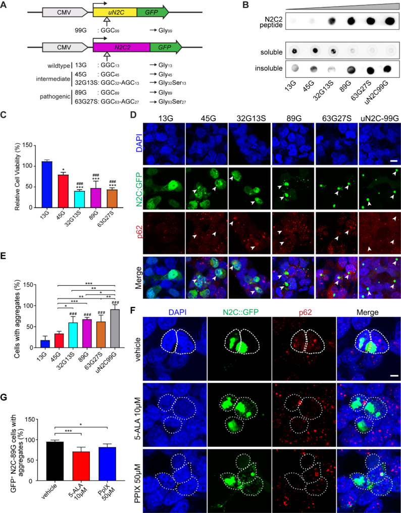
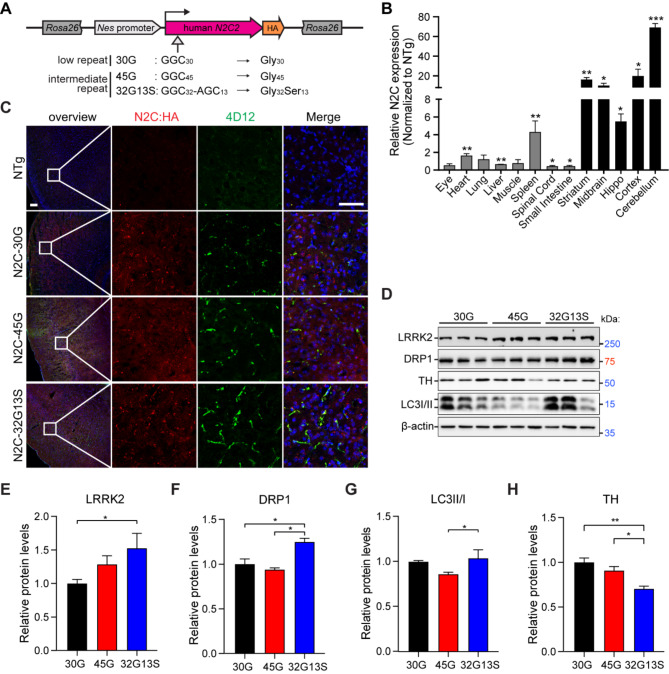
Methods: Here, we utilized cellular models harbouring different sizes of N2C variant 2 (N2C2) GGC repeat expansion and CRISPR-Cas9 engineered transgenic mouse models carrying N2C2 GGC intermediate repeats with and without serine insertion to elucidate the underlying pathophysiology associated with N2C intermediate repeat with serine insertion in NREDs.

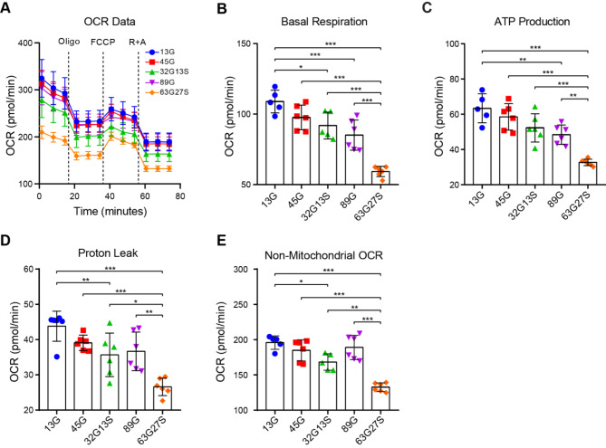
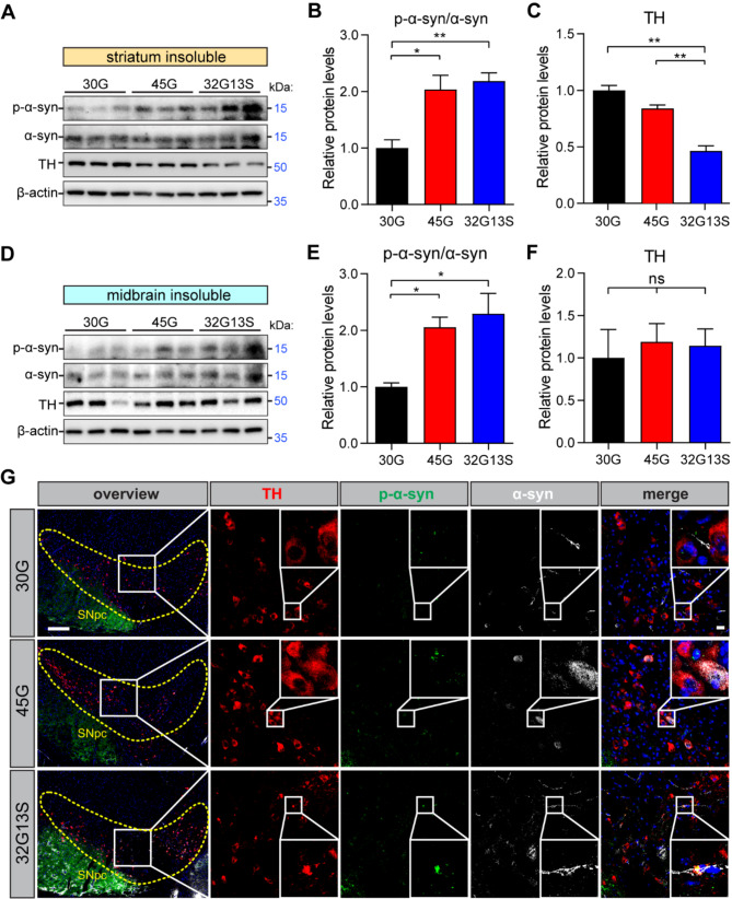
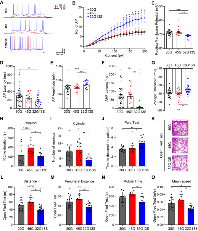
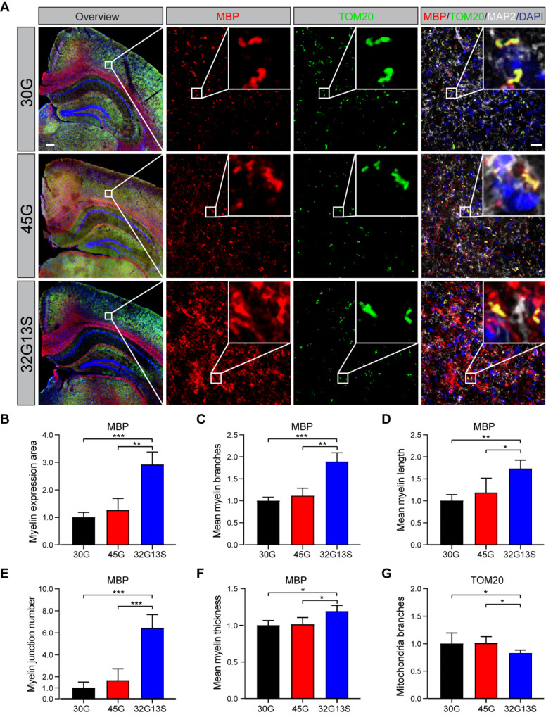
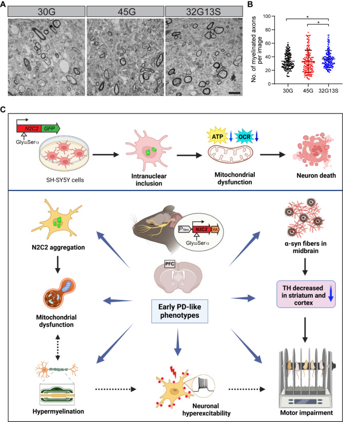
Results: Our findings revealed that the N2C2 GGC intermediate repeat with serine insertion (32G13S) led to mitochondrial dysfunction and cell death in vitro. The neurotoxicity was influenced by the length of the repeat and was exacerbated by the presence of the serine insertion. In 12-month-old transgenic mice, 32G13S intensified intranuclear aggregation and exhibited early PD-like characteristics, including the formation of α-synuclein fibers in the midbrain and the loss of tyrosine hydroxylase (TH)-positive neurons in both the cortex and striatum. Additionally, 32G13S induced neuronal hyperexcitability and caused locomotor behavioural impairments. Transcriptomic analysis of the mouse cortex indicated dysregulation in calcium signaling and MAPK signaling pathways, both of which are critical for mitochondrial function. Notably, genes associated with myelin sheath components, including MBP and MOG, were dysregulated in the 32G13S mouse. Further investigations using immunostaining and transmission electron microscopy revealed that the N2C intermediate repeat with serine induced mitochondrial dysfunction-related hypermyelination in the cortex.

Conclusions: Our in vitro and in vivo investigations provide the first evidence that the N2C-GGC intermediate repeat with serine promotes intranuclear aggregation of N2C, leading to mitochondrial dysfunction-associated hypermyelination and neuronal hyperexcitability. These changes contribute to motor deficits in early PD-like neurodegeneration in NREDs.

Keywords: NOTCH2NLC; AGC interruption; Early Parkinson’s disease; GGC repeat expansion; Hyperexcitability; Hypermyelination; Intermediate repeat; Mitochondrial dysfunction.


Conflict of interest statement

Declarations. Ethics approval and consent to participate: Informed consents were obtained from patients. The use of human samples in this study was properly assessed and approved by SingHealth Centralised Institutional Review Board (2017/2602). Animals were maintained under institutional guidelines, and all protocols were approved by the Institutional Animal Care and Use Committee (IACUC) of the National Neuroscience Institute (NNI) and Tan Tock Seng Hospital. Consent for publication: Not applicable. Competing interests: The authors report no competing interests.

Figures

Fig. 1
Fig. 1
N2C-polyG intermediate repeat with serine insertion promotes protein aggregation and increases neuronal cell death in vitro. (A) Illustration of the N2C expression constructs used in vitro. (B) Dot-blot image of pure N2C peptide (GRCWRSGCAARPP) (0, 6.125, 12.5, 25, 50, and 100 ng) and urea-soluble and insoluble fractions of the lysates of Lipofectamine LTX transfected SH-SY5Y carrying N2C-13G, -45G, -32G13S, -89G, -63G27S, and uN2C-99G. A customized N2C antibody targeting both N2C1 and N2C2 proteins was used for the detection of the N2C-polyG proteins. (C) WST1 cell viability assay was performed after transfecting SH-SY5Y cells with N2C-polyG plasmids. * represents comparisons between each of the other groups and 13G. # represents the comparisons between other groups and 45G. (D) SH-SY5Y cells were transfected with N2C vectors carrying different expansion repeats. Immunocytochemical staining was performed with a p62 antibody. White arrowheads represent cells colocalized with N2C: GFP and p62. Scale bar = 20 μm. (E) Quantification of N2C-polyG protein aggregation shown in D. N2C-polyG protein aggregations were defined as green fluorescence dots between 1 and 5 μm. Cell numbers counted: 13G = 118, 45G = 147, 32G13S = 57, 89G = 101, 63G27S = 90, and uN2C-99G = 105. # represents comparisons between other groups and 13G. (F) Treatment of N2C-89G transfected SH-SY5Y cells for 30 h with 50 µM PPIX and 10 µM 5-ALA. Scale bar = 10 μm. (G) Quantification of protein aggregations shown in F. Cell numbers counted: vehicle = 268, 10 µM 5-ALA = 259, 50 µM PPIX = 128. Data are presented as the mean ± SD, with n = 3 per group. *P < 0.05, **P < 0.01, and ***P < 0.001 by one-way ANOVA with Tukey’s post hoc test
Fig. 2
Fig. 2
N2C-polyG intermediate repeat with serine insertion impairs mitochondrial respiration in vitro. SH-SY5Y cells transfected with N2C-13G, -45G, -32G13S, -89G, and − 63G27S were seeded in a 96-well Seahorse XF cell culture microplate and cultured for 24 h. The OCR assay was performed using the Agilent Seahorse XF Cell Mito Stress Test Kit. The overall OCR data (A), basal respiration (B), ATP production (C), proton leak (D), and non-mitochondrial OCR (E) are shown. Data are presented as the mean ± SD, with n = 6 per group. *P < 0.05, **P < 0.01, and ***P < 0.001 by one-way ANOVA with Tukey’s post hoc test
Fig. 3
Fig. 3
N2C-32 S triggers N2C-polyG protein aggregation and resembles PD pathogenesis in transgenic mice. (A) Illustration of the N2C-polyG transgenic mice. The human N2C2 coding sequence, carrying 30G, 45G, or 32G13S, is driven by the human Nes promoter and fused with an HA-tag. This sequence is inserted into the Rosa26 locus of the C57BL/6 N mouse genome by CRISPR/Cas9-mediated genome editing. Heterozygous mice of both genders were used for experiments. (B) Tissues were collected from adult N2C-32G13S and non-transgenic (NTg) mice for qPCR analysis. Relative N2C expression levels in N2C-32G13S were normalized to NTg. Data are presented as the mean ± SD, with n = 3 per group. *P < 0.05, **P < 0.01, and ***P < 0.001 by two-tailed Student’s t test. (C) Cortical tissues from 8-month-old NTg, N2C-30G, -45G, and − 32G13S transgenic mice were used for immunostaining. Both HA (red) and 4D12 (green) antibodies were used to detect N2C-polyG protein. Scale bar = 100 μm. (D) Cortical tissues were used for immunoblotting. (E-H) Quantification of LRRK2 (E), DRP1 (F), LC3II to LC3I ratio (G), and TH (H) from the analysis in D. Data are presented as the mean ± SD; n = 3 per group. *P < 0.05 and **P < 0.01 by one-way ANOVA with Tukey’s post hoc test
Fig. 4
Fig. 4
N2C-polyG intermediate repeat with serine displays early PD-like phenotypes. (A) Western blot analysis of insoluble proteins from the striatum of 12-month-old N2C-30G, -45G, -32G13S mice. (B-C) Quantification of p-α-synuclein (S129) to α-synuclein (B) and TH (C) in A. (D) Western blot analysis of insoluble proteins from the midbrain of 12-month-old N2C-30G, -45G, -32G13S mice. (E-F) Quantification of p-α-synuclein (S129) to α-synuclein (E) and TH (F) in D. (G) IHC staining of 12-month-old N2C-30G, -45G, -32G13S mouse midbrain region by TH (red), p-α-synuclein (S129) (green), and α-synuclein (grey). The yellow dotted outline represents the substantia nigra pars compacta (SNpc) region. The scale bar in the left overview panel is 200 μm. The scale bar in the zoomed image is 20 μm. Data are presented as the mean ± SD; n = 3 per group. Ns = no significance. *P < 0.05 and **P < 0.01 by one-way ANOVA with Tukey’s post hoc test
Fig. 5
Fig. 5
N2C-polyG intermediate with serine insertion induces neuronal hyperexcitability and motor deficits in mice. (A-G) Prefrontal cortex cells from 16-month-old N2C-30G, -45G, or -32G13S transgenic mice were used for electrophysiological tests. (A) Representative voltage response obtained from the medial prefrontal cortex (mPFC) of mouse brains. (B) Number of action potentials (AP) at different voltages. * represents comparisons between 32G13S and 30G. # represents comparisons between 32G13S and 45G. (C-G) Basic intrinsic neuronal parameters: resting membrane potential (mV) (C), AP latency (D), AP amplitude (E), after hyperpolarisation (AHP) latency (F), and AP voltage threshold (G) are significantly different in 32G13S PFC compared to N2C-30G and N2C-45G neurons. No differences were observed in input resistance (Rin), AP duration, or AHP amplitude among N2C-30G, N2C-45G, or N2C-32G13S (Supplementary Fig. 5A-C). N2C-30G, n = 38; N2C-45G, n = 23; N2C-32G13S, n = 25. Three independent mice per genotype were used for data collection. One-way ANOVA with Tukey’s post hoc test was used for statistical analysis. Data are presented as the mean ± SEM. * P < 0.05, ** P < 0.01, and *** P < 0.001. (H-O) Behavioural tests were performed in 12-month-old N2C-30G, -45G, and − 32G13S mice in Rotarod (H), Cylinder (I), Pole Test (J), and Open Field Test (K-O). (K) Representative walking trails of mice in the Open Field Test. Total distance (L), peripheral distance (M), Mobile time (N), and Mean speed (O) travelled during the Open Field Test are shown. N2C-30G, n = 9; N2C-45G, n = 6; N2C-32G13S, n = 13. Data are presented as the mean ± SD. *P < 0.05 and **P < 0.01 by one-way ANOVA with Tukey’s post hoc test
Fig. 6
Fig. 6
Mitochondrial-related pathways and myelin sheath component genes are dysregulated in N2C-32G13S mice. Cortical tissues from 12-month-old N2C transgenic mice were used for bulk RNA sequencing. n = 3 mice per group. (A) Volcano plot showing differentially expressed genes between N2C-32G13S and N2C-45G mice at padj < 0.05 and fold change < 0.67 or > 1.5. Top dysregulated genes are highlighted, with those related to mitochondrial function shown in blue. (B) For differentially expressed genes between N2C-32G13S and N2C-45G mice, 54 top dysregulated genes are shown in biological process (BP), cellular component (CC), and molecular function (MF) by gene ontology (GO) term analysis. (C) Top dysregulated KEGG pathways between N2C-32G13S and N2C-45G mice. (D) Connections between top differentially expressed genes and cellular components. Genes highlighted in red are myelin sheath components. (E-H) Quantitative PCR was performed for myelin sheath component genes MBP (E), MOG (F), GJC2 (G), and ERMN (H) using cortical tissues from 12-month-old N2C mice. (I) Cortical tissues were used for immunoblotting. (J-K) Quantification of MBP (J) and MOG (K) from western blot in I. Data are presented as the mean ± SD, n = 3. *P < 0.05 and **P < 0.01 by one-way ANOVA with Tukey’s post hoc test
Fig. 7
Fig. 7
N2C-polyG intermediate repeat with serine induces mitochondrial dysfunction-associated hypermyelination in mice. (A) IHC staining of 12-month-old mouse brains. Immunofluorescence was detected using MBP (red), Tom20 (green), and Map2 (grey) antibodies. Scale bar = 200 μm in the overview image. Scale bar = 20 μm in the zoomed image. Quantification of myelin and mitochondria was conducted using an ImageJ plugin macro “Mitochondria Analyzer”. Cells quantified: 30G n = 4078, 45G n = 3949, 32G13S n = 4848. MBP and Tom20 signals represent myelin and mitochondria, respectively. (B-G) MBP expression area (B), myelin branches (C), myelin length (D), myelin junction number (E), mean myelin thickness (F), and mitochondrial branches (G) were measured and quantified by the Mitochondria Analyzer. Data are presented as the mean ± SD, *P < 0.05, **P < 0.01, and ***P < 0.001 by one-way ANOVA with Tukey’s post hoc test
Fig. 8
Fig. 8
N2C-polyG intermediate repeat with serine induces hypermyelination in mice. Mouse cortical tissues near the corpus callosum were used for transmission electron microscopy (TEM). (A) Representative images of 30G, 45G, and 32G13S mouse cortex are shown at a magnification of 4300×. The scale bar is 2 μm. (B) Quantification of the number of myelinated axons under TEM. Myelinated axons are identified as the axons with dark circles in the images shown in A. Three mice were used for each group. Each dot represents one TEM image, as shown in A. Total image numbers: 30G = 187, 45G = 181, 32G13S = 199. Total myelinated axons counted: 30G = 5894, 45G = 5572, 32G13S = 6272. Data are presented as the mean ± SD, *P < 0.05 by one-way ANOVA with Tukey’s post hoc test. (C) A graphical summary illustrates that N2C-polyG intermediate repeat with serine induces neurotoxicity and early PD-like neurodegeneration in vitro and in vivo. Overexpression of the N2C-polyG intermediate repeat with serine promotes the formation of intranuclear inclusions, leading to mitochondrial dysfunction and subsequent neuronal cell death in vitro. In N2C-32G13S transgenic mice, this intermediate N2C-polyG with serine triggers N2C protein aggregation and early PD-like pathophysiological changes. These include α-synuclein fiber-like aggregates in the midbrain, loss of TH neurons in the striatum and cortex, and neuronal hyperexcitability with mitochondrial dysfunction-related hypermyelination in the cortex, all contributing to motor impairment. Our findings offer novel pathophysiological insights into the clinical relevance of N2C-polyG intermediate repeats observed in early PD patients

References

    1. Okubo M, Doi H, Fukai R, Fujita A, Mitsuhashi S, Hashiguchi S, Kishida H, Ueda N, Morihara K, Ogasawara A, et al. GGC repeat expansion of NOTCH2NLC in adult patients with Leukoencephalopathy. Ann Neurol. 2019;86:962–8. - PubMed
    1. Sone J, Mitsuhashi S, Fujita A, Mizuguchi T, Hamanaka K, Mori K, Koike H, Hashiguchi A, Takashima H, Sugiyama H, et al. Long-read sequencing identifies GGC repeat expansions in NOTCH2NLC associated with neuronal intranuclear inclusion disease. Nat Genet. 2019;51:1215–21. - PubMed
    1. Tian Y, Wang JL, Huang W, Zeng S, Jiao B, Liu Z, Chen Z, Li Y, Wang Y, Min HX, et al. Expansion of human-specific GGC repeat in neuronal intranuclear inclusion Disease-Related disorders. Am J Hum Genet. 2019;105:166–76. - PMC - PubMed
    1. Jiao B, Zhou L, Zhou Y, Weng L, Liao X, Tian Y, Guo L, Liu X, Yuan Z, Xiao X, et al. Identification of expanded repeats in NOTCH2NLC in neurodegenerative dementias. Neurobiol Aging. 2020;89:142. e141-142 e147. - PubMed
    1. Ma D, Tan YJ, Ng ASL, Ong HL, Sim W, Lim WK, Teo JX, Ng EYL, Lim EC, Lim EW, et al. Association of NOTCH2NLC repeat expansions with Parkinson Disease. JAMA Neurol. 2020;77:1559–63. - PMC - PubMed

Substances

LinkOut - more resources