. 2024 Nov 28;10(1):57.
doi: 10.1186/s40813-024-00404-7.

TbpB-based oral mucosal vaccine provides heterologous protection against Glässer's disease caused by different serovars of Spanish field isolates of Glaesserella parasuis

Affiliations

TbpB-based oral mucosal vaccine provides heterologous protection against Glässer's disease caused by different serovars of Spanish field isolates of Glaesserella parasuis

Alba González-Fernández et al. Porcine Health Manag. .

Abstract

Background: Glaesserella parasuis (G. parasuis) is the primary agent of Glässer's disease, significantly affecting nursery and early fattening piglets. Current prophylactic measures, mainly serovar-specific bacterins administered to sows, are limited by maternal immunity, which can interfere with active immunization in piglets. Subunit vaccines containing G. parasuis-specific antigenic molecules show promise but are not yet commercially available. Transferrin-binding proteins (Tbp), which enable G. parasuis to acquire iron in low-iron environments like mucosal surfaces, have been proposed as potential vaccine antigens. The mucosal administration of a TbpB-based subunit vaccine could provide a promising solution to overcome the limitations posed by maternal immunity, offering an effective approach to control the disease in weaning piglets. This study, conducted in two phases, primarily evaluates (days 0-45) the immunogenicity of a two-dose oral mucosal TbpB-based subunit vaccine (TbpBY167A) administered to colostrum-deprived piglets, and subsequently (days 45-52), its heterologous protection by challenging these piglets with four G. parasuis clinical isolates from different TbpB clusters (I, III) and serovars (SV1, SV4, SV5, SV7) recovered from Spanish pig farms.

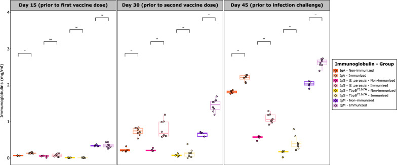
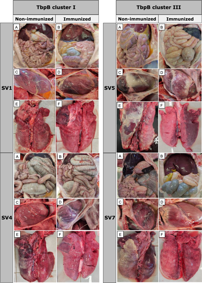
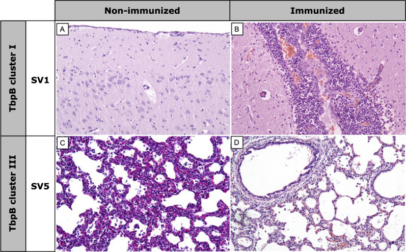
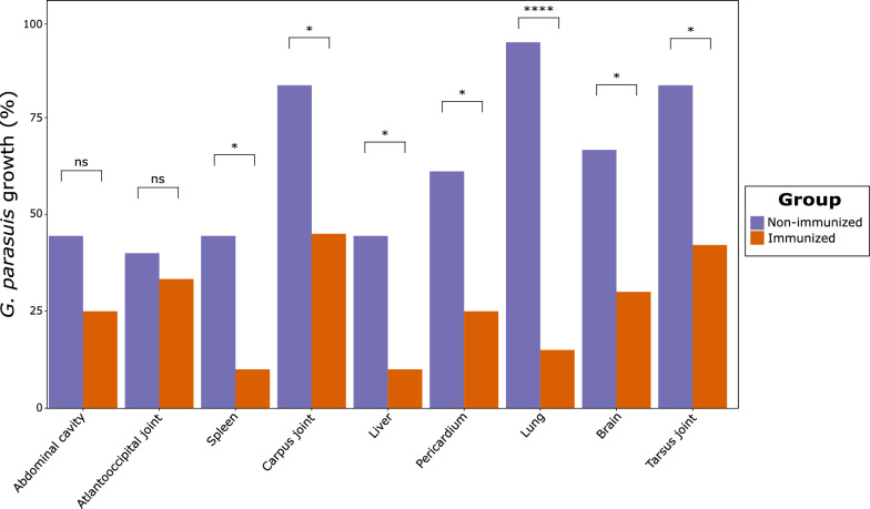
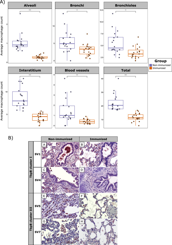
Results: The oral mucosal administration of the two-dose TbpB-based vaccine induced a robust humoral immune response in immunized colostrum-deprived piglets, significantly increasing IgA and IgM concentration 15 days after the second dose (p < 0.01). Upon challenge with four G. parasuis clinical isolates, the vaccine demonstrated heterologous protection, markedly improving survival rates (OR: 8.45; CI 95%: 4.97-14.36) and significantly reducing clinical signs and lesions, regardless of the TbpB cluster and serovar. The vaccine reduced G. parasuis colonization in the respiratory tract (p < 0.0001) and G. parasuis systemic target tissues, like tarsus and carpus joints, liver, and brain (p < 0.05). Immunohistochemical analysis showed a lower macrophage count in different lung locations of immunized piglets (p < 0.0001).

Conclusions: This study demonstrates that oral mucosal administration of the TbpBY167A subunit vaccine in piglets provides effective heterologous protection against diverse virulent European G. parasuis field isolates, significantly reducing bacterial colonization and dissemination. This vaccine offers a promising alternative to traditional bacterins, overcoming limitations due to maternal immunity, and represents a strong candidate for universal vaccination against Glässer's disease.

Keywords: Glaesserella parasuis; Cross-protection; Glässer’s disease; Humoral immune response; Immunization; Needle-free vaccination; Porcine respiratory disease complex; Swine; TbpBY167A subunit vaccine.


Conflict of interest statement

Declarations. Ethics approval and consent to participate: All procedures involving animals were approved by the institutional bioethical committee (Reference Number OEBA-ULE-003–2022) and performed according to European regulations regarding animal welfare and protection of animals used for experimental and other scientific purposes. Consent for publication: Not applicable. Competing interests: The authors declare no competing interests.

Figures

Fig. 1
Fig. 1
Two-phase experimental design for the evaluation of the immunogenicity and efficacy of the TbpB-based oral mucosal vaccine against Glässer’s disease. The first phase (marked in blue) consisted of the immunization of colostrum deprived piglets. Immunized piglets (n = 20) received two oral mucosal administrations of the TbpBY167A subunit vaccine, while non-immunized piglets (n = 18) were inoculated with PBS. The second phase (marked in red) began 15 days after the second dose of the vaccine and piglets were divided into four groups and intranasally challenged with four different G. parasuis field isolates. This challenge lasted for seven days, after which all remaining piglets were euthanized. Animals were daily monitored for clinical signs and temperature and those exhibiting severe distress were humanely euthanized before the end of the experiment. After euthanasia, a necropsy was performed to evaluate macroscopic lesions and collect samples. Serum samples were collected on days 15, 30 and 45. SV: serovar
Fig. 2
Fig. 2
Evolution of immunoglobulin concentration (mg/ml) in immunized and non-immunized colostrum-deprived piglets after oral mucosal administration of the two-dose TbpB-based vaccine. Each piglet is represented by a dot with horizontal jitter in the boxplot for visibility. The horizontal box lines represent the first quartile, the median, and the third quartile. Whiskers include the range of points within the 1.5 interquartile range. Differences per group were evaluated with the Wilcoxon rank-sum test
Fig. 3
Fig. 3
Evaluation of the efficacy of the oral mucosal TbpB-based vaccine in piglets immunized against Glässer’s disease throughout a seven-day experiment. A Survival rate, expressed as percentage, of immunized and non-immunized piglets challenged with four different serovars (SVs) of G. parasuis. B Clinical score average (0–3) and standard deviation of immunized and non-immunized piglets, itemized into six different clinical signs. No non-immunized piglet survived after the third day of challenge. Differences per group were evaluated with the Wilcoxon rank-sum test
Fig. 4
Fig. 4
Comparison of macroscopic lesions after G. parasuis infection challenge between immunized and non-immunized piglets with the oral mucosal TbpB-based vaccine. Abdominal and thoracic cavities are shown and itemized by G. parasuis TbpB cluster and serovar (SV). A and B, peritoneal serosae; C and D, pericardium; E and F, pleura
Fig. 5
Fig. 5
Histopathological differences after G. parasuis infection challenge between immunized and non-immunized piglets with the oral mucosal TbpB-based vaccine. Brain (A and B) and lung (C and D) are itemized by G. parasuis TbpB cluster and serovar (SV). Non-immunized piglets infected with TbpB cluster I (SV1) showed no brain lesions (A), while immunized ones revealed intense meningitis (B). Non-immunized animals challenged with TbpB cluster III (SV5) showed interstitial pneumonia (C), while absence or slight pneumonia was observed in immunized ones (D). X100
Fig. 6
Fig. 6
Glaesserella parasuis growth, expressed as percentage, in different anatomical regions after infection challenge in immunized and non-immunized piglets with the oral mucosal TbpB-based vaccine. Differences per group were evaluated with the Wilcoxon rank-sum test
Fig. 7
Fig. 7
Macrophage abundance in pulmonary tissue after infection challenge in immunized and non-immunized piglets with the oral mucosal TbpB-based vaccine. A Boxplots representing average macrophage count across various lung locations. Each piglet is represented by a dot with horizontal jitter in the boxplot for visibility. The horizontal box lines represent the first quartile, the median, and the third quartile. Whiskers include the range of points within the 1.5 interquartile range. Differences per group were evaluated with the Wilcoxon rank-sum test. B Immunohistochemical (IHC) observation of brown labeled CD163 positive macrophages across various lung locations, itemized by G. parasuis TbpB cluster and serovar (SV). A and B, bronchioles; C and D, bronchi; E and F, blood vessels and connective tissue; G and H, alveoli. Contrast of nuclei with Harris hematoxylin. Avidin–biotin-peroxidase complex. X100

Similar articles

Cited by

References

    1. Moller K, Kilian M. V factor-dependent members of the family Pasteurellaceae in the porcine upper respiratory tract. J Clin Microbiol. 1990;28(12):2711–6. - PMC - PubMed
    1. Peet RL, Fry J, Lloyd J, Henderson J, Curran J, Moir D. Haemophilus parasuis septicaemia in pigs. Aust Vet J. 1983;60(6):187–187. - PubMed
    1. White M, BVSc, LLB, DPM, MRCVS. Glässers Disease. Available online: https://www.nadis.org.uk/disease-a-z/pigs/glaessers-disease/. Accessed 17 Mar 2024.
    1. Cerdà-Cuéllar M, Naranjo JF, Verge A, Nofrarías M, Cortey M, Olvera A, et al. Sow vaccination modulates the colonization of piglets by Haemophilus parasuis. Vet Microbiol. 2010;145(3–4):315–20. - PubMed
    1. Harris DL, Ross RF, Switzer WP. Incidence of certain microorganisms in nasal cavities of swine in Iowa. Am J Vet Res. 1969;30(9):1621–4. - PubMed

LinkOut - more resources