. 2025 Mar;24(3):e14418.
doi: 10.1111/acel.14418. Epub 2024 Nov 28.

Knockdown of RUVBL2 improves hnRNPA2/B1-stress granules dynamics to inhibit perioperative neurocognitive disorders in aged mild cognitive impairment rats

Affiliations

Knockdown of RUVBL2 improves hnRNPA2/B1-stress granules dynamics to inhibit perioperative neurocognitive disorders in aged mild cognitive impairment rats

Zixuan Wang et al. Aging Cell. 2025 Mar.

Abstract

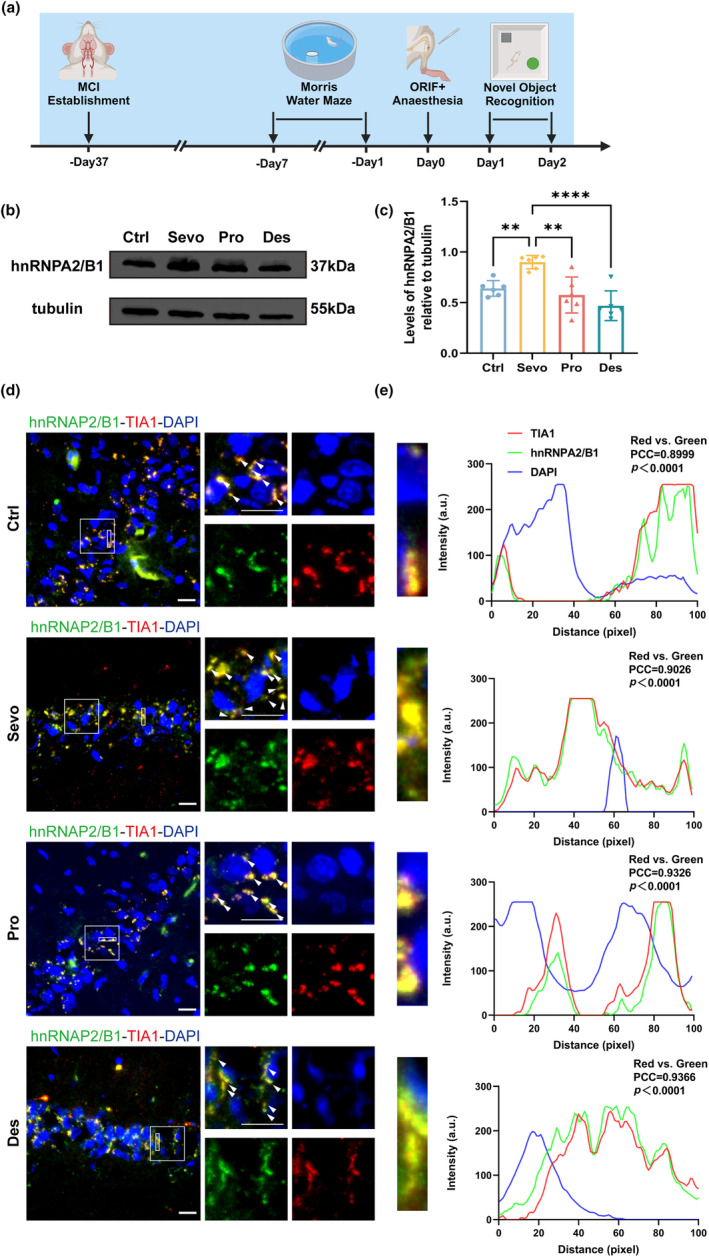
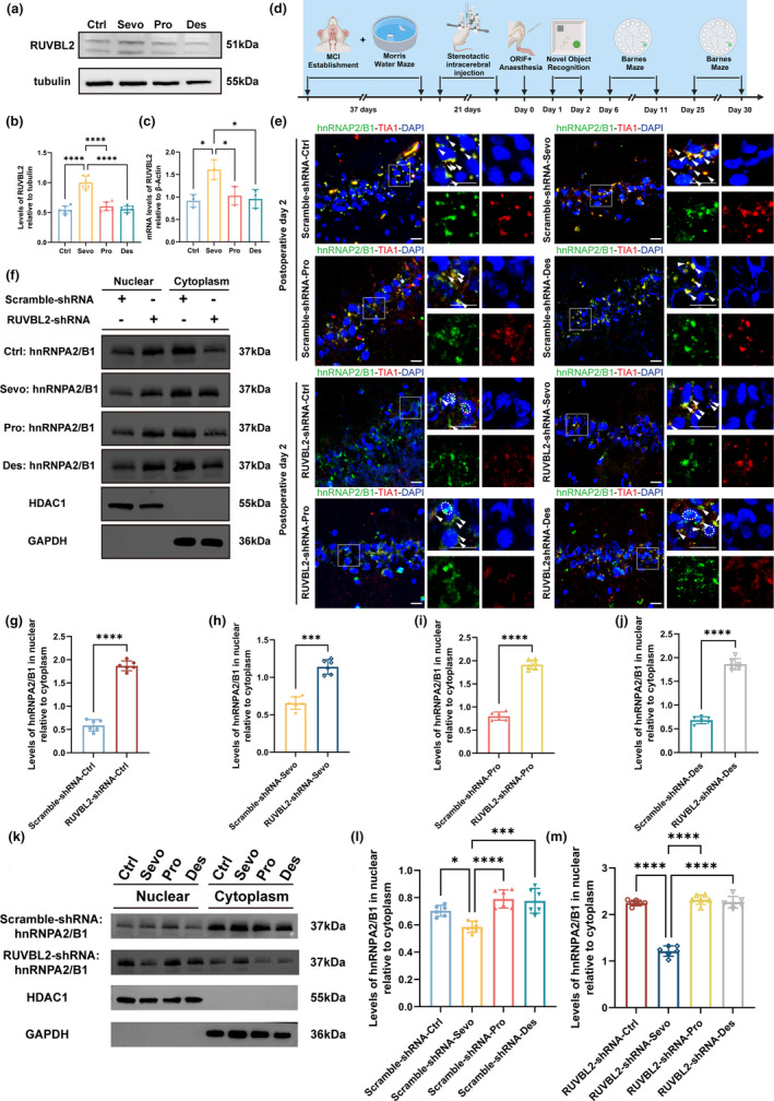
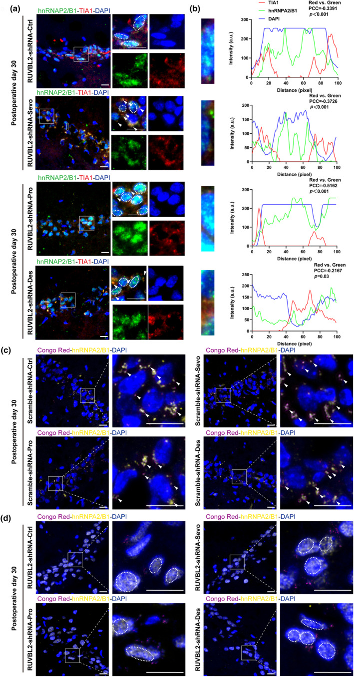
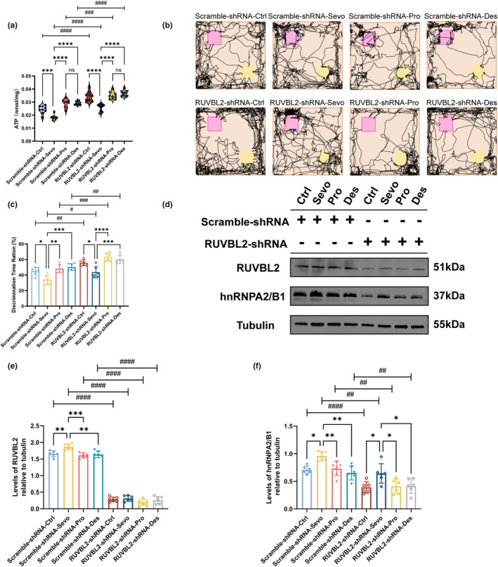
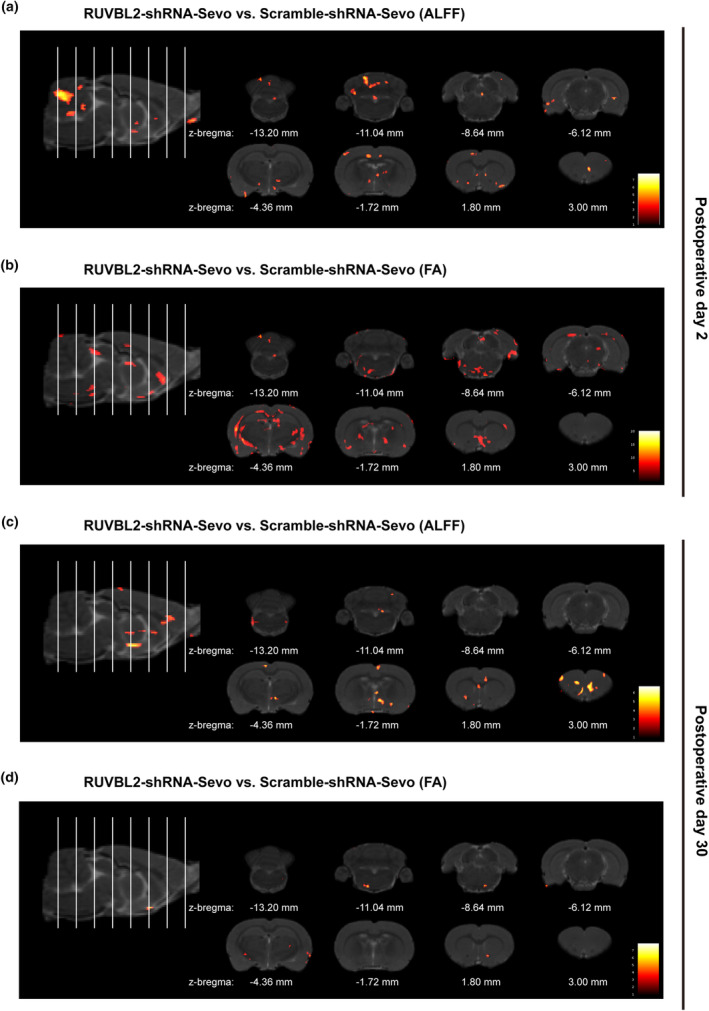
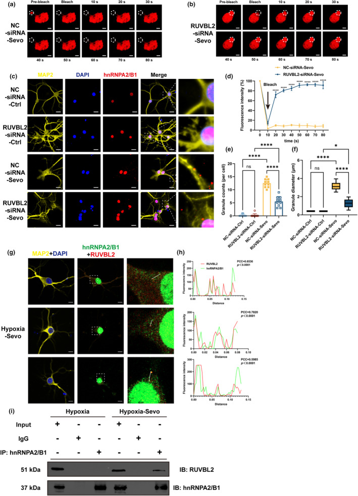
Perioperative neurocognitive disorders (PND) is common in aged mild cognitive impairment (MCI) patients and can accelerate the progression to dementia. This process involves heterogeneous nuclear ribonucleoprotein A2/B1 (hnRNPA2/B1)-mediated aggregates of stress granules (SGs), while RUVBL2 influences the dynamics of these SGs. Our research explored a new target for modulating hnRNAPA2/B1-SGs dynamics to accelerate their disassembly and potentially delay MCI progression due to PND. We assessed the effect of hippocampal RUVBL2 knockdown on hnRNPA2/B1-SGs in aged MCI rats through behavioral studies, biochemical experiments and MRI. We also examined hnRNPA2/B1-SGs dynamics using immunofluorescence staining and fluorescence recovery after photobleaching (FRAP) in rat primary hippocampal neurons. Our results revealed that hnRNPA2/B1 in the hippocampus of aged MCI rats translocates to the cytoplasm to form SGs following anesthesia. RUVBL2 knockdown promotes the disappearance of hnRNPA2/B1-SGs, allowing hnRNPA2/B1 to return to the nucleus and enhancing functional activity in the brain regions of aged MCI rats. In primary hippocampal neurons, RUVBL2 deletion facilitated hnRNPA2/B1-SGs transition from hydrogel to liquid, promoting disassembly. We compared three commonly used general anesthetics-3% sevoflurane, 40 mg·kg-1·h-1 propofol, and 9% desflurane. Sevoflurane upregulated RUVBL2, which decreased the intraneuronal pH and disrupted energy metabolism. These changes resulted in greater stabilization of hnRNPA2/B1- SGs. In conclusion, our findings indicated that the knockdown of RUVBL2 expression contributes to the transition of hnRNPA2/B1-SGs from the hydrogel phase to the liquid phase. Targeted interference with RUVBL2 may represent a novel approach to delay the progression to dementia due to PND in aged MCI patients.

Keywords: RUVBL2; aged mild cognitive impairment; dynamics; hnRNPA2/B1; perioperative neurocognitive disorders; stress granules.


Conflict of interest statement

The authors declare that they have no competing interests.

Figures

FIGURE 1
FIGURE 1
hnRNPA2/B1 localizes to SGs in MCI rats after surgery with diverse general anesthetics. (a) Timeline of the experiment. (b, c) Representative Western blot and statistical histogram of hippocampal lysates from sevoflurane‐, propofol‐, and desflurane‐anaesthetized MCI rats and postoperative aged MCI rats showing hnRNPA2/B1 expression. Western blots were labelled with hnRNPA2/B1 and β‐tubulin antibodies (n = 6 independent experiments, one‐way ANOVA followed by the post hoc Bonferroni multiple comparisons test). (d) Representative confocal microscopy images depicting the expression of hnRNPA2/B1 and TIA1 in the hippocampal CA1 region of aged MCI rats after sevoflurane, propofol and desflurane anesthesia and surgery. (e) Fluorescence co‐localization analysis of hnRNPA2/B1, TIA1 and DAPI within the rectangular region of interest in Figure 1c (PCC: Pearson's correlation coefficient). **p < 0.01, ****p < 0.0001. Values are presented as the means ± SEMs. Scale bar = 20 μm.
FIGURE 2
FIGURE 2
RUVBL2 knockdown promotes the return of hnRNPA2/B1 to the nuclei and dissipation of hnRNPA2/B1‐SGs. (a, b) Representative Western blot and statistical histogram of hippocampal lysates from sevoflurane‐, propofol‐, and desflurane‐anesthetized MCI rats and postoperative aged MCI rats showing RUVBL2 expression. Western blots were labelled with RUVBL2 and β‐tubulin antibodies (n = 6 independent experiments, one‐way ANOVA followed by the post hoc Bonferroni multiple comparisons test). (c) RUVBL2 mRNA levels after sevoflurane, propofol, and desflurane anesthesia and surgery (n = 3 independent experiments, one‐way ANOVA followed by the post hoc Bonferroni multiple comparisons test). (d) Timeline of the experiment. (e) Representative confocal microscopy images of hnRNPA2/B1 and TIA1 expression in the hippocampal CA1 region of aged MCI rats in the Scramble‐shRNA group on Day 2 after anesthesia and surgery. (f) Representative Western blot of hippocampal nuclei and cytoplasmic lysates showing hnRNPA2/B1 expression in aged MCI rats on Day 2 after sevoflurane, propofol, or desflurane‐anesthetized and surgery. Western blots were labelled with hnRNPA2/B1, HDAC1 and GAPDH antibodies. HDAC1 was used as a nuclei internal reference protein, and GAPDH was used as a cytoplasmic internal reference protein. (g–j) hnRNPA2/B1 nucleoplasmic ratio on Day 2 after the post‐anesthesia and surgery Day 2 (n = 6 independent experiments, unpaired t test). (k–m) Representative Western blot and statistical histogram of hippocampal nuclei and cytoplasmic lysates showing hnRNPA2/B1 expression in aged MCI rats on Day 2 after sevoflurane, propofol, or desflurane‐anesthetized and surgery. Western blots were labelled with hnRNPA2/B1, HDAC1, and GAPDH antibodies. HDAC1 was used as a nuclei internal reference protein, and GAPDH was used as a cytoplasmic internal reference protein. (n = 6 independent experiments; one‐way ANOVA followed by the post hoc Bonferroni multiple comparisons test). *p < 0.05, ***p < 0.001 and ****p < 0.0001; values are presented as the means ± SEMs.
FIGURE 3
FIGURE 3
RUVBL2 knockdown reduces hnRNPA2/B1 and TIA1 co‐localization, promotes hnRNPA2/B1 nucleation, and improves hnRNPA2/B1 fibril formation (a) Representative confocal microscopy images of hnRNPA2/B1 and TIA1 expression in the hippocampal CA1 region of the RUVBL2‐shRNA group of aged MCI rats on Day 30 after anesthesia and surgery. (b) Fluorescence co‐localization analysis of hnRNPA2/B1, TIA1 and DAPI within the rectangular region of interest in (a) (PCC: Pearson's correlation coefficient). Scale bar = 20 μm. (c) Representative confocal microscopy images of Congo Red staining and hnRNPA2/B1 expression in the hippocampal CA1 region of the Scramble‐shRNA group of aged MCI rats on Day 30 after anesthesia and surgery. (d) Representative confocal microscopy images of Congo Red staining and hnRNPA2/B1 expression in the hippocampal CA1 region of the RUVBL2‐shRNA group of aged MCI rats on Day 30 after anesthesia and surgery. Scale bar = 20 μm.
FIGURE 4
FIGURE 4
RUVBL2 knockdown increases ATP levels and improves learning and memory in MCI rats after anesthesia and surgery. (a) ATP levels in the hippocampus of aged MCI rats on Day 2 after post‐anesthesia and surgery (n = 9 independent experiments, unpaired t test and one‐way ANOVA followed by the post hoc Bonferroni multiple comparisons test). (b) NOR test trajectory map of aged MCI rats on Day 2 after anesthesia and surgery. (c) Ratio of the discrimination time for novel objects in the NOR test on Day 2 after anesthesia and surgery (n = 6 independent experiments, unpaired t test and one‐way ANOVA followed by the post hoc Bonferroni multiple comparisons test). (d–f) Representative Western blots and statistical histogram of hippocampal lysates showing the expression of hnRNPA2/B1 and RUVBL2 from aged MCI rats expressing hnRNPA2/B1 on Day 2 after anesthesia and surgery. Western blots were labelled with hnRNPA2/B1, RUVBL2 and β‐tubulin antibodies (n = 6 independent experiments, unpaired t test and one‐way ANOVA followed by the post hoc Bonferroni multiple comparisons test). *p < 0.05, **p < 0.01, ***p < 0.001, ****p < 0.0001, #p < 0.05, ##p < 0.01, ###p < 0.001 and ####p < 0.0001; values are presented as the means ± SEMs.
FIGURE 5
FIGURE 5
Functional magnetic resonance imaging (ALFF) results and diffusion tensor imaging (FA) of aged MCI rats. (a) Representative maps showing significant differences in ALFF values between the RUVBL2‐shRNA‐Sevo group and Scramble‐shRNA‐Sevo group on postoperative Day 2 (n = 3, two‐sample t test); (b) Representative maps showing significant differences in FA values between the RUVBL2‐shRNA‐Sevo group and Scramble‐shRNA‐Sevo group on postoperative Day 2 (n = 3, two‐sample t test); (c) Representative maps showing significant differences in ALFF values between the RUVBL2‐shRNA‐Sevo group and Scramble‐shRNA‐Sevo group on postoperative Day 30 (n = 3, two‐sample t test); (d) Representative maps showing significant differences in FA values between the RUVBL2‐shRNA‐Sevo group and Scramble‐shRNA‐Sevo group on postoperative Day 2 (n = 3, two‐sample t test). The white line on the sagittal image indicates the section of the corresponding representative coronal image. p < 0.01, cluster size >50. Warm colors indicate brain regions with enhanced functional activity.
FIGURE 6
FIGURE 6
Knockdown of RUVBL2 mediates the transition of hnRNPA2/B1‐SGs from the hydrogel phase to the liquid phase and disassembly of hnRNPA2/B1‐SGs, and RUVBL2 co‐localizes and interacts with hnRNPA2/B1. (a, b) FRAP analysis of hnRNPA2/B1‐SGs formed in response to sevoflurane exposure in NC‐siRNA and RUVBL2‐siRNA rat primary hippocampal neurons. Scale bar = 3 μm. (d) Change in the fluorescence intensity in the ROI over time (n = 3 independent experiments, two‐way ANOVA followed by post hoc Bonferroni multiple comparisons test). (c) Disassembly dynamics of hnRNPA2/B1‐SGs in primary hippocampal neurons cultured in fresh medium for 1 h. Scale bar = 20 μm. (e, f) Statistical histogram of the counts and diameter of hnRNPA2/B1‐SGs (n = 10 independent experiments, one‐way ANOVA followed by post hoc Bonferroni multiple comparisons test). (g) Representative confocal images of hnRNPA2/B1 and RUVBL2 co‐localization. White lines indicate the ROI where fluorescence intensity profiles of hnRNPA2/B1 and RUVBL2 (Scale bar = 10 μm). (h) The fluorescence intensity distribution of hnRNPA2/B1 and RUVBL2 co‐localization within the ROI in (g) (PCC: Pearson's correlation coefficient). (i) Co‐immunoprecipitation of hnRNPA2/B1 and RUVBL2 after exposure to Hypoxia and Hypoxia‐Sevo. *p < 0.05, ****p < 0.0001; values are presented as the means ± SEMs.

Similar articles

References

    1. Aulas, A. , Stabile, S. , & Vande, V. C. (2012). Endogenous TDP‐43, but not FUS, contributes to stress granule assembly via G3BP. Molecular Neurodegeneration, 7, 54. 10.1186/1750-1326-7-54 - DOI - PMC - PubMed
    1. Blamire, A. M. (2018). MR approaches in neurodegenerative disorders. Progress in Nuclear Magnetic Resonance Spectroscopy, 108, 1–16. 10.1016/j.pnmrs.2018.11.001 - DOI - PubMed
    1. Buchan, J. R. , Kolaitis, R. M. , Taylor, J. P. , & Parker, R. (2013). Eukaryotic stress granules are cleared by autophagy and Cdc48/VCP function. Cell, 153(7), 1461–1474. 10.1016/j.cell.2013.05.037 - DOI - PMC - PubMed
    1. Cao, S. J. , Zhang, Y. , Zhang, Y. X. , Zhao, W. , Pan, L. H. , Sun, X. D. , Jia, Z. , Ouyang, W. , Ye, Q. S. , Zhang, F. X. , Guo, Y. Q. , Ai, Y. Q. , Zhao, B. J. , Yu, J. B. , Liu, Z. H. , Yin, N. , Li, X. Y. , Ma, J. H. , Li, H. J. , … Wang, D. X. (2023). Delirium in older patients given propofol or sevoflurane anaesthesia for major cancer surgery: A multicentre randomised trial. British Journal of Anaesthesia, 131(2), 253–265. 10.1016/j.bja.2023.04.024 - DOI - PubMed
    1. Chen, B. , Qin, G. , Xiao, J. , Deng, X. , Lin, A. , & Liu, H. (2022). Transient neuroinflammation following surgery contributes to long‐lasting cognitive decline in elderly rats via dysfunction of synaptic NMDA receptor. Journal of Neuroinflammation, 19(1), 181. 10.1186/s12974-022-02528-5 - DOI - PMC - PubMed

LinkOut - more resources