. 2024 Dec 16;4(12):100909.
doi: 10.1016/j.crmeth.2024.100909. Epub 2024 Nov 27.

Patient-derived tumor organoid and fibroblast assembloid models for interrogation of the tumor microenvironment in esophageal adenocarcinoma

Affiliations

Patient-derived tumor organoid and fibroblast assembloid models for interrogation of the tumor microenvironment in esophageal adenocarcinoma

Benjamin P Sharpe et al. Cell Rep Methods. .

Abstract

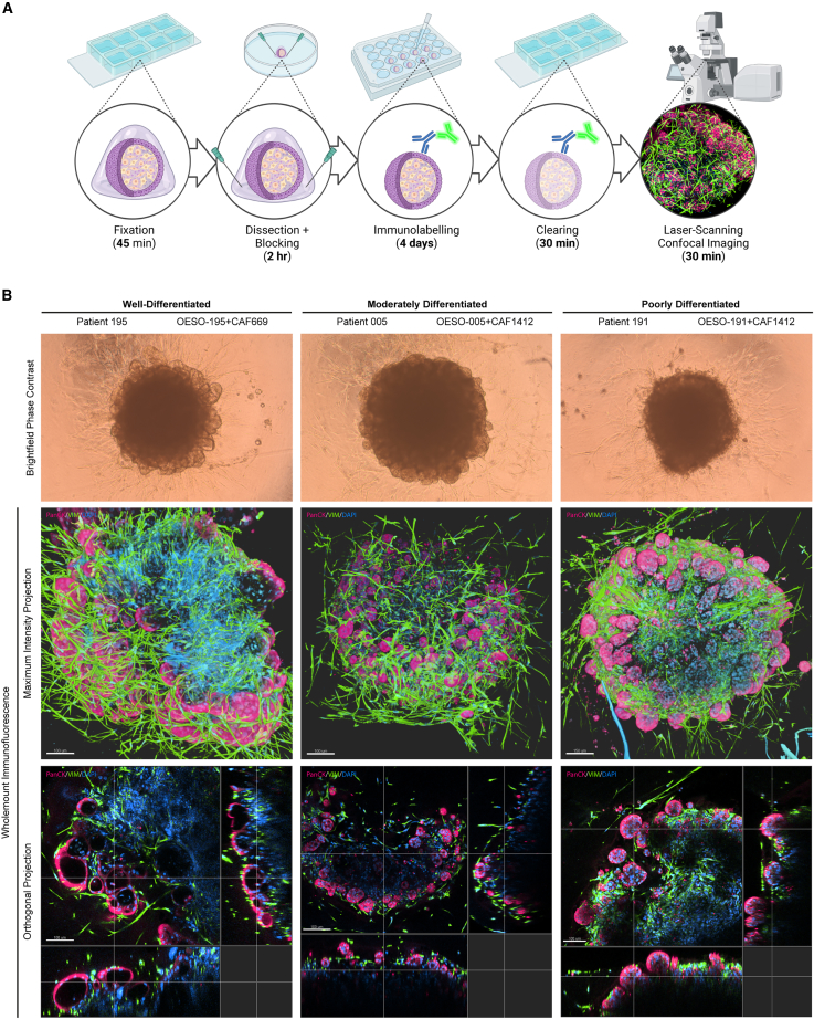
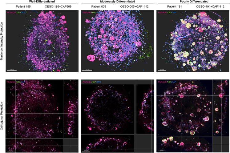
The tumor microenvironment (TME) comprises all non-tumor elements of cancer and strongly influences disease progression and phenotype. To understand tumor biology and accurately test new therapeutic strategies, representative models should contain both tumor cells and normal cells of the TME. Here, we describe and characterize co-culture tumor-derived organoids and cancer-associated fibroblasts (CAFs), a major component of the TME, in matrix-embedded assembloid models of esophageal adenocarcinoma (EAC). We demonstrate that the assembloid models faithfully recapitulate the differentiation status of EAC and different CAF phenotypes found in the EAC patient TME. We evaluate cell phenotypes by combining tissue-clearing techniques with whole-mount immunofluorescence and histology, providing a practical framework for the characterization of cancer assembloids.

Keywords: CP: Cancer biology; CP: Stem cell; assembloids; cancer-associated fibroblasts; esophageal adenocarcinoma; organoids; tumor microenvironment.


Conflict of interest statement

Declaration of interests The authors declare no competing interests.

Figures

None
Graphical abstract
Figure 1
Figure 1
Derivation of primary models and the workflow for creation of EAC assembloids (A) Two example EAC organoids derived from different patients with EAC observed with a 4× objective under phase contrast microscopy. Scale bars: 250 μm. (B) Two example CAFs derived from different patients with EAC observed with a 20× objective under phase contrast microscopy. Scale bars: 50 μm. (C) Workflow for production of EAC assembloids. EAC organoids and primary CAFs are expanded prior to model creation, dissociated to single cells, counted, and mixed together at a 2:1 ratio of CAFs to organoid cells. The cell suspension is then plated out at 75,000 cells per well in an ultra-low attachment 96-well U-bottom plate. The next day, assembloids are plated in a 3:1 mixture of collagen I:BME2, and complete DMEM media are overlayed once set. Assembloids are cultured for a further 7 days prior to harvest. (D) Structure formation of EAC assembloids imaged with a 4× objective under phase contrast microscopy every day for 7 days after matrix embedding. Scale bars: 500 μm.
Figure 2
Figure 2
Histological and immunohistochemical characterization of EAC assembloid phenotypes compared to the corresponding patient tumors (A) Immunohistochemistry of 5 μm organoid sections grown in monoculture and stained for pan-cytokeratin (red) and vimentin (green). Nuclei were counterstained with DAPI (blue). (B) Immunocytochemistry of primary fibroblasts grown in monoculture and stained for pan-cytokeratin (red) and vimentin (green). Actin cytoskeleton is counterstained with phalloidin (grayscale), and nuclei are counterstained with DAPI (blue). Scale bars: 100 μm. (C) 5 μm sections of EAC assembloids and resected patient tumors stained with tinctorial stains for overall histology (H&E), mucin secretion (Alcian blue-PAS, mucins in blue/purple), and collagen fibers (picrosirius red, collagen in orange) viewed under polarized light. Signet ring-like cells are indicated in the inset of OESO-191+CAF1412 with arrowheads. (D) Immunofluorescence of EAC assembloids and resected patient tumor sections stained for markers of epithelial and fibroblast cell identity—pan-cytokeratin and vimentin, keratin 7 (glandular epithelium) and keratin 20 (colonic epithelium), and α-SMA and POSTN (myofibroblast differentiation). Nuclei are counterstained with DAPI. Insets show regions of interest at higher resolution in (A), (C), and (D). Scale bars: 150 μm.
Figure 3
Figure 3
Whole-mount immunofluorescent protocol for 3D visualization of EAC assembloid organization (A) Workflow of the 5-day whole-mount staining protocol. EAC assembloids are fixed in 4% PFA, excess collagen is removed to improve antibody penetration, and then assembloids are stained with primary and fluorescent-conjugated secondary antibodies (with DAPI) for 2 days each. Assembloids are cleared in fructose-glycerol clearing buffer and then imaged with a fluorescent laser scanning confocal microscope. (B) Example assembloids from each organoid/CAF combination at day 8 of culture imaged in bright-field phase contrast (scale bars: 500 μm). Shown below are corresponding Z-projections of whole-mount-stained and cleared assembloids for pan-cytokeratin (organoid cells, red) and vimentin (fibroblasts, green) in top-down maximum intensity projections and orthogonal projections cutting through outer epithelial buds (scale bars: 100 μm unless indicated otherwise). Rendering was performed in Imaris under blend mode. See also Videos S1, S2, and S3.
Figure 4
Figure 4
Visualization of cell proliferation in 3D using whole-mount immunofluorescence Z-projections of whole-mount-stained and cleared 8-day-old assembloids for Ki-67 (proliferating cells, green) counterstained with Phalloidin-iFluor594 (cell bodies, red) in top-down maximum intensity projections and orthogonal projections cutting through outer epithelial buds (scale bars: 200 μm). Rendering was performed in Imaris under blend mode.

References

    1. Sung H., Ferlay J., Siegel R.L., Laversanne M., Soerjomataram I., Jemal A., Bray F. Global Cancer Statistics 2020: GLOBOCAN Estimates of Incidence and Mortality Worldwide for 36 Cancers in 185 Countries. CA A Cancer J. Clin. 2021;71:209–249. doi: 10.3322/caac.21660. - DOI - PubMed
    1. Uhlenhopp D.J., Then E.O., Sunkara T., Gaduputi V. Epidemiology of esophageal cancer: update in global trends, etiology and risk factors. Clin. J. Gastroenterol. 2020;13:1010–1021. doi: 10.1007/s12328-020-01237-x. - DOI - PubMed
    1. Al-Batran S.E., Homann N., Pauligk C., Goetze T.O., Meiler J., Kasper S., Kopp H.G., Mayer F., Haag G.M., Luley K., et al. Perioperative chemotherapy with fluorouracil plus leucovorin, oxaliplatin, and docetaxel versus fluorouracil or capecitabine plus cisplatin and epirubicin for locally advanced, resectable gastric or gastro-oesophageal junction adenocarcinoma (FLOT4): a randomised, phase 2/3 trial. Lancet. 2019;393:1948–1957. doi: 10.1016/s0140-6736(18)32557-1. - DOI - PubMed
    1. Eyck B.M., van Lanschot J.J.B., Hulshof M.C.C.M., van der Wilk B.J., Shapiro J., van Hagen P., van Berge Henegouwen M.I., Wijnhoven B.P.L., van Laarhoven H.W.M., Nieuwenhuijzen G.A.P., et al. Ten-Year Outcome of Neoadjuvant Chemoradiotherapy Plus Surgery for Esophageal Cancer: The Randomized Controlled CROSS Trial. J. Clin. Oncol. 2021;39:1995–2004. doi: 10.1200/jco.20.03614. - DOI - PubMed
    1. Bang Y.J., Van Cutsem E., Feyereislova A., Chung H.C., Shen L., Sawaki A., Lordick F., Ohtsu A., Omuro Y., Satoh T., et al. Trastuzumab in combination with chemotherapy versus chemotherapy alone for treatment of HER2-positive advanced gastric or gastro-oesophageal junction cancer (ToGA): a phase 3, open-label, randomised controlled trial. Lancet. 2010;376:687–697. doi: 10.1016/s0140-6736(10)61121-x. - DOI - PubMed

MeSH terms

Supplementary concepts

LinkOut - more resources