. 2024 Dec;102(3):763-777.
doi: 10.1177/13872877241291175. Epub 2024 Nov 29.

DJ-1-mediated repression of the RNA-binding protein FMRP is predicted to impact known Alzheimer's disease-related protein networks

Affiliations

DJ-1-mediated repression of the RNA-binding protein FMRP is predicted to impact known Alzheimer's disease-related protein networks

Ayse Uneri et al. J Alzheimers Dis. 2024 Dec.

Abstract

Background: RNA-binding proteins (RBPs) modulate the synaptic proteome and are instrumental in maintaining synaptic homeostasis. Moreover, aberrant expression of an RBP in a disease state would have deleterious downstream effects on synaptic function. While many underlying mechanisms of synaptic dysfunction in Alzheimer's disease (AD) have been proposed, the contribution of RBPs has been relatively unexplored.

Objective: To investigate alterations in RBP-messenger RNA (mRNA) interactions in AD, and its overall impact on the disease-related proteome.

Methods: We first utilized RNA-immunoprecipitation to investigate interactions between RBP, DJ-1 (Parkinson's Disease protein 7) and target mRNAs in controls and AD. Surface Sensing of Translation - Proximity Ligation Assay (SUnSET-PLA) and western blotting additionally quantified alterations in mRNA translation and protein expression of DJ-1 targets. Finally, we utilized an unbiased bioinformatic approach that connects AD-related pathways to two RBPs, DJ-1 and FMRP (Fragile X messenger ribonucleoprotein 1).

Results: We find that oligomeric DJ-1 in AD donor synapses were less dynamic in their ability to bind and unbind mRNA compared to synapses from cognitively unimpaired, neuropathologically-verified controls. Furthermore, we find that DJ-1 associates with the mRNA coding for FMRP, Fmr1, leading to its reduced synaptic expression in AD. Through the construction of protein-protein interaction networks, aberrant expression of DJ-1 and FMRP are predicted to lead to the upregulation of key AD-related pathways, such as thyroid hormone stimulating pathway, autophagy, and ubiquitin mediated proteolysis.

Conclusions: DJ-1 and FMRP are novel targets that may restore established neurobiological mechanisms underlying AD.

Keywords: Alzheimer's disease; DJ-1; KEGG pathways; RNA networks; RNA-binding proteins; autophagy; fragile x messenger ribonucleoprotein (FMRP); synapse; thyroid hormone stimulating pathway; ubiquitin mediated proteolysis.


Conflict of interest statement

Declaration of conflicting interestsThe authors declared no potential conflicts of interest with respect to the research, authorship, and/or publication of this article.

Figures

Figure 1.
Figure 1.. DJ-1 oligomerization is increased and is stable in AD cortical synapses.
A. (Left) Representative Western blots from control (CTL) and Alzheimer’s Disease (AD) donor dorsolateral prefrontal cortex synaptoneurosomes, showing the DJ-1 monomer at ∼23 kDa, and higher molecular weight (HMW) species at ∼34 kDa, and ∼60 kDa. (Right) Line graph showing fold change of DJ-1 and its HMW species in synaptoneurosomes from control (CTL) and Alzheimer’s Disease (AD) donors. Two-way ANOVA (F2,22 = 3.996; Interaction p = 0.03). Fisher LSD Control vs. AD: 23 kDa p = 0.65; 34 kDa p = 0.11; 60kDa = 0.0003. (b) (Top) Quantification of Western Blot showing increased 60 kDa DJ-1 expression in synaptoneurosomes prepared from APP/PS1 mice, compared to wildtype (WT) mice. (WT = 1.000 ± 0.1668, n = 4; APP/PS1 = 1.690 ± 0.06621, n = 3; p = 0.0100; student’s t-test). (Below) Representative western blot. (c) Quantification (fold change) of the 34 kDa DJ-1 band comparing RNase A treatment of CTL (black; CTL/A) or AD (purple; AD/A) by a single t-test to the untreated band (CTL, p = 0.026, AD, p = 0.52) (d) Quantification of the 60 kDa DJ-1 band comparing RNase A treatment of CTL (black; CTL/A) or AD (purple; AD/A) by a single t-test to the untreated band (dotted line, normalized to 1 within sample; CTL, p = 0.0044; AD, p = 45). (e) Ratio of the monomer (∼23 kDa) to HMW species (∼60 kDa) of DJ-1 with (CTL+, dotted black; AD+, dotted purple) and without (CTL−, solid black; AD−, solid purple) RNAse A treatment (CTL− compared to CTL+; p = 0.0010; CTL + compared to AD−; p = 0.0005, CTL + compared to AD+; p = 0.0007; and AD− compared to AD+; p = 0.9833). One-way ANOVA (F(3, 8) = 22.43, p = 0.0003).
Figure 2.
Figure 2.. Predicted targets of DJ-1 contain a cluster enriched in Alzheimer’s disease KEGG genes.
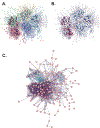
(a) DJ-1 predicted targets seeded into STRING presented 5 clusters. Yellow cluster with 175 proteins out of 912; green cluster, 180 proteins; orange cluster 193 proteins; blue cluster 210 proteins; purple cluster 154 proteins. (b) AD KEGG genes highlighted within the predicted DJ-1 targets (orange). (c) Orange cluster from A with labeled gene names, with ∼23% known AD-related genes, p = 9.54×10⁻³². Inclusion criteria was experiments, databases, and co-expression only.
Figure 3.
Figure 3.. DJ-1 binds to Fmr1 mRNA.
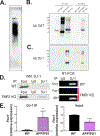
(a). Immunoblotting for biotin-labeled RNA of Fmr1 5’UTR synthesized by T7 RNA polymerase. The in vitro transcription product was resolved by 6% PAGE (0.5x TBE) gel and then transferred onto nitrocellulose membrane. A HRP-conjugated anti-biotin antibody (Cell Signaling) was used for detecting the RNA. (b,-c). RNA pull-down assay for assessing the in vitro binding of recombinant GST-tagged DJ-1 with biotinylated Fmr1 5’UTR RNA. GST-UBC9 and GST-SUMO1 were used as negative controls. The immunoblotting was performed with anti-GST (b) and anti-DJ1 (c) antibodies, respectively. (d) (Left) Representative Western blot of DJ-1 RIP; note DJ-1 protein specifically precipitates with DJ-1 antibody but not IgG. (Right) RT-PCR of RIP from WT and Fmr1KO cortical lysates; note specific amplification of Fmr1 from WT but not FMR1KO, with Akt1 as a positive control. (e) (Left) qPCR of Fmr1 from DJ-1 RIP using WT and APP/PS1 cortical lysates; students t-test, one-tailed (WT = 0.1914 ± 0.1229, APP/PS1 = 3.286 ± 1.586, p = 0.04, n = 5). (Right) Comparison of input Fmr1 mRNA in cortices from WT and APP/PS1 (WT = 1.001 ± 0.0005947; APP/PS1 = 0.5581 ± 0.2048, p = 0.09, n = 5).
Figure 4.
Figure 4.. DJ-1 represses de novo synthesis of FMRP.
(a) (Left, top panel) SuNSET-PLA of rat cortical neurons transfected with RFP as control, showing newly synthesized FMRP (puromycin-PLA, white), map2 staining (purple), and the merged image of both, yellow arrows pointing to sites of new FMRP. (Left, bottom) SuNSET-PLA of neurons overexpressing DJ-1 showing decreased newly synthesized FMRP. (Right) Quantification of the puromycin-FMRP puncta normalized to ROI, p = 0.0016, students t-test. CTL, number of neurons = 27; OE, number of neurons = 38. (b) (Top) Quantification of Western Blot showing decreased FMRP expression in synaptoneurosomes prepared from APP/PS1 mice, compared to wildtype (WT) mice. (WT = 1.000 ± 0.03548, n = 8; APP/PS1 = 0.8418 ± 0.05106, n = 8; p = 0.0117; student’s t-test) (Below) Representative western blot. (c) (Top) Quantification of Western Blot showing decreased FMRP expression in synaptoneurosomes prepared from AD donors (AD), compared to neurocognitively normal control (CTL). N = 5, p = 0.04, student’s t-test. (Below) Representative western blot.
Figure 5.
Figure 5.. Dysregulation of DJ-1 and thus FMRP can lead to network-wide disruption in protein expression in ad, affecting multiple pathways.
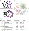
(a) Network model. DJ-1 and FMRP are both RBP that repress the translation of their target mRNAs. (Above) In healthy synapses, DJ-1 and FMRP should be translated at an equilibrium, where their target mRNA levels are then balanced. (Below) In AD synapses, this equilibrium is tilted due to increased expression of DJ-1 at synapses, resulting in over-repression of its target mRNAs, one of which is Fmr1. Therefore, it would be expected that the mRNAs that would normally be repressed by FMRP are now free to be translated, which could mean an overexpression of those proteins. (b) Protein-Protein Interaction (PPI) network of unique FMRP target list predicted to be dysregulated in AD. FMRP target mRNAs that do not bind DJ-1 were seeded into STRING to create a PPI network, linking FMRP to several AD-related pathways. Minimum interaction score of 0.04 was applied, inclusion criteria consisted of co-expression, experiments, and datasets. (c) KEGG pathways that are highlighted through FMRP and published works connecting them to AD. All the KEGG pathways identified through the three clusters determined by k-means clustering algorithm in STRING, and the citations for papers that indicate their disruption in AD.

References

    1. Kishore S, Luber S and Zavolan M. Deciphering the role of RNA-binding proteins in the post-transcriptional control of gene expression. Briefings in functional genomics 2010; 9: 391–404. - PMC - PubMed
    1. Licatalosi DD and Darnell RB. RNA processing and its regulation: global insights into biological networks. Nature Reviews Genetics 2010; 11: 75–87. - PMC - PubMed
    1. Morris AR, Mukherjee N and Keene JD. Systematic analysis of posttranscriptional gene expression. Wiley Interdisciplinary Reviews: Systems Biology and Medicine 2010; 2: 162–180. - PubMed
    1. Keene JD and Lager PJ. Post-transcriptional operons and regulons co-ordinating gene expression. Chromosome research 2005; 13: 327–337. - PubMed
    1. Heaney CF, Namjoshi SV, Uneri A, et al. Role of FMRP in rapid antidepressant effects and synapse regulation. Molecular Psychiatry 2021; 26: 2350–2362. DOI: 10.1038/s41380-020-00977-z. - DOI - PMC - PubMed

LinkOut - more resources