Review
. 2024 Nov 6;6(6):fcae395.
doi: 10.1093/braincomms/fcae395. eCollection 2024.

Spinal cord evaluation in multiple sclerosis: clinical and radiological associations, present and future

Affiliations
Review

Spinal cord evaluation in multiple sclerosis: clinical and radiological associations, present and future

B Mark Keegan et al. Brain Commun. .

Abstract

Spinal cord disease is important in most people with multiple sclerosis, but assessment remains less emphasized in patient care, basic and clinical research and therapeutic trials. The North American Imaging in Multiple Sclerosis Spinal Cord Interest Group was formed to determine and present the contemporary landscape of multiple sclerosis spinal cord evaluation, further existing and advanced spinal cord imaging techniques, and foster collaborative work. Important themes arose: (i) multiple sclerosis spinal cord lesions (differential diagnosis, association with clinical course); (ii) spinal cord radiological-pathological associations; (iii) 'critical' spinal cord lesions; (iv) multiple sclerosis topographical model; (v) spinal cord atrophy; and (vi) automated and special imaging techniques. Distinguishing multiple sclerosis from other myelopathic aetiology is increasingly refined by imaging and serological studies. Post-mortem spinal cord findings and MRI pathological correlative studies demonstrate MRI's high sensitivity in detecting microstructural demyelination and axonal loss. Spinal leptomeninges include immune inflammatory infiltrates, some in B-cell lymphoid-like structures. 'Critical' demyelinating lesions along spinal cord corticospinal tracts are anatomically consistent with and may be disproportionately associated with motor progression. Multiple sclerosis topographical model implicates the spinal cord as an area where threshold impairment associates with multiple sclerosis disability. Progressive spinal cord atrophy and 'silent' multiple sclerosis progression may be emerging as an important multiple sclerosis prognostic biomarker. Manual atrophy assessment is complicated by rater bias, while automation (e.g. Spinal Cord Toolbox), and artificial intelligence may reduce this. Collaborative research by the North American Imaging in Multiple Sclerosis and similar groups with experts combining distinct strengths is key to advancing assessment and treatment of people with multiple sclerosis spinal cord disease.

Keywords: atrophy; magnetic resonance imaging; multiple sclerosis; pathology; spinal cord.


Conflict of interest statement

B.M.K.: consulting EMD Serono, royalties Oxford University Press Mayo Clinic Cases in Neuroimmunology. M.A.: consulting Biogen, Abata Therapeutics, Sanofi-Genzyme and GSK. E.P.F.: grants or contracts UCB, NIH and Medimmune/Viela Bio/Horizon Therapeutics; royalties UpToDate; honoraria Pharmacy Times; Patents DACH1-IgG as a biomarker of paraneoplastic autoimmunity; participation on a Data Safety Monitoring Board Alexion, Genentech, Horizon Therapeutics and UCB, MOG project; and leadership or fiduciary roles Journal of the Neurological Sciences and Neuroimmunology Reports. R.G.H.: grants or contracts Roche/Genentech and Atara; consulting Roche/Genentech Boston Pharma QIA and LLC; honoraria Sanofi; and participation on a Data Safety Monitoring Board Roche and Novartis. E.C.K: grants or contracts AbbVie, Biogen, Genentech; consulting Banner Life Sciences, EMD Serono, Galen/Atlantica Genentech, Greenwich Biosciences, INmune Bio, Myrobalan Therapeutics, OM1 and TG Therapeutics, honoraria BioPharm Communications, MJH Lifesciences; and participation on a Data Safety Monitoring Board Tiziana Life Sciences. S.K: grants Roche and Biogen. S.K.: grants or contracts Biogen BMS Novartis Sanofi; consulting Baim Institute Biogen Cycle EMD Serono Genentech Novartis Ootave Genzyme/Sanofi TG Therapeutics; and honoraria Biogen EMD Serono Genentech TG Therapeutics. N.P.: research support Race to Erase MS. A.T.: grants or contracts Hilton Foundation, Roche, AbbVie, honoraria Biogen and Roche, support for attending meetings EMD Serono; and participation on a Data Safety Monitoring Board Sanofi Genzyme, Roche. J.C.-A., C.L., J.A.L., S.M., J.O. and S.A.S.: no competing interests.

Figures

Graphical Abstract
Graphical Abstract
Figure 1
Figure 1
Pathology and MRI of secondary progressive multiple sclerosis. Histological staining and post-mortem 7 T MRI of a secondary progressive multiple sclerosis patient (female, 78 years old, 25-year disease duration). Focal lesions (arrows) are visible on cervical, thoracic and lumbar sections, highlighted by myelin (Luxol fast blue) and axon (Bielschowsky) staining, as well as on ex vivo 7 T T2-weighted and myelin water fraction MRI scans. Modified from Laule et al. with permission. MWF, myelin water fraction.
Figure 2
Figure 2
MRI differential diagnosis of myelitis in multiple sclerosis. (A) Myelin oligodendrocyte glycoprotein antibody-associated disease. (B) Aquaporin 4-IgG positive neuromyelitis optica spectrum disorder (AQP4+ neuromyelitis optica spectrum disorder). (C) Spinal cord sarcoidosis. (D) Cervical spondylotic myelopathy. Thoracic spine MRI reveals a sagittal T2 lesion in the lower thoracic spine involving the conus and extending <3 vertebral segments (Ai) with axial T2 images revealing a central T2 lesion (Aii) in a patient with myelin oligodendrocyte glycoprotein antibody-associated disease. Cervical spine sagittal MRI reveals a longitudinally extensive T2 lesion extending >3 vertebral segments (Aiii) with T2 hyperintensity restricted to the central GM on axial T2 images forming a H sign (Aiv, arrow) in a patient with myelin oligodendrocyte glycoprotein antibody-associated disease. Cervical spine sagittal MRI reveals a longitudinally extensive T2 lesion extending >3 vertebral segments (Bi) with central T2 lesion on axial images (Bii) with an elongated ring of enhancement on T1-weighted images post-gadolinium (Biii) with an open ring on axial T1-weighted images post-gadolinium (Biv) in a patient with AQP4+ neuromyelitis optica spectrum disorder. Cervical spine sagittal MRI reveals a longitudinally extensive T2 lesion extending >3 vertebral segments (Ci) with central T2 lesion on axial images (Cii) accompanied by linear dorsal subpial enhancement on sagittal and axial T1-weighted images post-gadolinium (Ciii, Civ) in spinal cord sarcoidosis. Cervical spine sagittal MRI reveals a T2 lesion extending <3 vertebral segments (Di) with axial T2 images showing some T2 hyperintensity (Dii) and evidence of a transverse band or ‘pancake-like’ enhancement with the width greater than the height (Diii) circumferentially involving the WM (Div).
Figure 3
Figure 3
Critical demyelinating lesions of the spinal cord. ‘Critical’ demyelinating lesions of the spinal cord are anatomically associated with progressive motor impairment in people with multiple sclerosis; they are typically laterally based in the corticospinal tract and have focal spinal cord atrophy. MRI cervical spine sagittal (A) and axial (B) images showing ‘critical’ demyelinating lesion with T2 signal abnormality in the right lateral-dorsal cord with focal atrophy (black arrows and arrowhead) in a 53-year-old man with a 5-year history of progressive right upper motor neuron face-sparing hemiparesis. CSF showed four unique oligoclonal bands. MRI cervical spine sagittal (C) and axial (D) images showing ‘critical’ demyelinating lesion with T2 signal abnormality in the left lateral cord (arrows) with focal atrophy (open arrowhead) in a 59-year-old woman with a prior history of relapsing-remitting multiple sclerosis and an 8-year history of progressive left face-sparing upper motor neuron hemiparesis. Additional demyelinating lesion was seen in the lower thoracic spine with no volume loss (not shown). MRI thoracic spine sagittal (E) and axial (F) images showing ‘critical’ demyelinating lesion with T2 signal abnormality right hemispinal cord (arrows and open arrowhead) in a 58-year-old man with multiple sclerosis and progressive right lower extremity monoparesis.
Figure 4
Figure 4
Advanced imaging techniques in the spinal cord. (A) MT imaging can involve a scan with (MTon) and without (MToff) an off-resonance pulse that is applied to partially saturate the macromolecular pool prior to imaging; the normalized ratio (MToff–MTon/MToff) provides the MTR. A representative cervical cord axial MTR map in an RIS participant shows a decrease in MTR within a wedge-shaped lesion in the right ventral spinal cord relative to a matched control. A T1-weighted (T1w) image can be used to calculate MT saturation, which reduces T1 dependence to increase inter-scanner agreement. Quantitative MT aims to measure properties of the bound pool. (B) Myelin water fraction imaging isolates signal from water between the myelin bilayers based on T2 relaxation times. (C) PSIR helps highlight lesions; it should be noted that this sagittal protocol is based on a sequence different from the axial technique that has been shown to be important for area quantification and is based on a real reconstruction of a spin-echo inversion recovery sequence. (D) Chemical exchange saturation transfer indirectly detects amide protons associated with proteins and peptides quantified by amide proton transfer maps. Many different mathematical and biophysical models exist to extract diffusion-related metrics and microstructural features including (E) diffusion tensor imaging, spherical mean technique and neurite orientation dispersion and density imaging to produce metrics such as axial diffusivity, FA, mean diffusivity, radial diffusivity, diffusion coefficient of the isotropic compartment, extra-neurite microscopic mean diffusivity, extra-neurite transverse microscopic diffusivity, intra-cellular volume fraction, isotropic compartment fraction, intracellular volume fraction and orientation dispersion index. (F) Resting-state functional MRI demonstrates local alterations in connectivity, which sometimes correlates with diffusion microstructural metrics, suggesting increased connectivity may represent compensatory mechanisms in response to structural damage. Quantitative MT in A, F in Smith et al., used with permission. Diffusion imaging in E modified from fig. 6 in Schilling et al., used with permission. AD, axial diffusivity; DIFF, diffusion coefficient of the isotropic compartment; DTA, diffusion tensor imaging; EXTRA-MD, extra-neurite microscopic mean diffusivity; EXTRA-TRANS, extra-neurite transverse microscopic diffusivity; FA, fractional anisotropy; FICVF, intracellular volume fraction; FISO, isotropic compartment fraction; INTRA, intra-cellular volume fraction; MD, mean diffusivity; ODI, orientation dispersion index; qMT, quantitative MT; RD, radial diffusivity.

References

    1. Kuhlmann T, Moccia M, Coetzee T, et al. Multiple sclerosis progression: Time for a new mechanism-driven framework. Lancet Neurol. 2023;22(1):78–88. - PMC - PubMed
    1. Thompson AJ, Banwell BL, Barkhof F, et al. Diagnosis of multiple sclerosis: 2017 revisions of the McDonald criteria. Lancet Neurol. 2018;17(2):162–173. - PubMed
    1. Kerbrat A, Gros C, Badji A, et al. Multiple sclerosis lesions in motor tracts from brain to cervical cord: Spatial distribution and correlation with disability. Brain. 2020;143(7):2089–2105. - PMC - PubMed
    1. Petrova N, Carassiti D, Altmann DR, Baker D, Schmierer K. Axonal loss in the multiple sclerosis spinal cord revisited. Brain Pathol. 2018;28(3):334–348. - PMC - PubMed
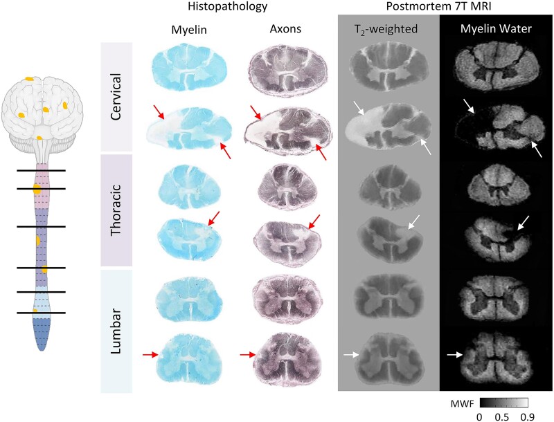
    1. Laule C, Yung A, Pavolva V, et al. High-resolution myelin water imaging in post-mortem multiple sclerosis spinal cord: A case report. Mult Scler. 2016;22(11):1485–1489. - PubMed

LinkOut - more resources