. 2025 Feb;25(2):e2400411.
doi: 10.1002/mabi.202400411. Epub 2024 Nov 29.

Dexamethasone Acetate-Loaded PLGA Nanospheres Targeting Liver Macrophages

Affiliations

Dexamethasone Acetate-Loaded PLGA Nanospheres Targeting Liver Macrophages

Barbora Boltnarova et al. Macromol Biosci. 2025 Feb.

Abstract

Glucocorticoids are potent anti-inflammatory drugs, although their use is associated with severe side effects. Loading glucocorticoids into suitable nanocarriers can significantly reduce these undesirable effects. Macrophages play a crucial role in inflammation, making them strategic targets for glucocorticoid-loaded nanocarriers. The main objective of this study is to develop a glucocorticoid-loaded PLGA nanocarrier specifically targeting liver macrophages, thereby enabling the localized release of glucocorticoids at the site of inflammation. Dexamethasone acetate (DA)-loaded PLGA nanospheres designed for passive macrophage targeting are synthesized using the nanoprecipitation method. Two types of PLGA NSs in the size range of 100-300 nm are prepared, achieving a DA-loading efficiency of 19 %. Sustained DA release from nanospheres over 3 days is demonstrated. Flow cytometry analysis using murine bone marrow-derived macrophages demonstrates the efficient internalization of fluorescent dye-labeled PLGA nanospheres, particularly into pro-inflammatory macrophages. Significant down-regulation in pro-inflammatory cytokine genes mRNA is observed without apparent cytotoxicity after treatment with DA-loaded PLGA nanospheres. Subsequent experiments in mice confirm liver macrophage-specific nanospheres accumulation following intravenous administration using in vivo imaging, flow cytometry, and fluorescence microscopy. Taken together, the data show that the DA-loaded PLGA nanospheres are a promising drug-delivery system for the treatment of inflammatory liver diseases.

Keywords: PLGA nanospheres; biodegradable nanoparticles; glucocorticoids; liver inflammation; macrophages.


Conflict of interest statement

The authors declare that no conflict of interest.

Figures
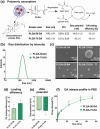
Figure 1
Figure 1
Characterization of dexamethasone acetate (DA)‐loaded PLGA nanospheres (NSs) a) Schematic diagram of prepared biodegradable PLGA NSs and overview of NSs characterization; b) Size distribution by the intensity of DA‐loaded NSs as determined using Zeta Sizer; c) SEM pictures of DA‐loaded NSs, scale bar 300 nm d) Drug loading efficiency of NSs measured by HPLC; e) Zeta potential measured by dynamic light scattering method; f) DA release profile of DA‐loaded NSs in phosphate buffered saline (PBS) pH 7.4 over 72 h. n = 3.
Figure 2
Figure 2
In vitro experiments with bone marrow‐derived macrophages (BMMs) treated with DA‐loaded NSs a) Viability of BMMs in the presence of DA‐loaded NSs after 24 h b) Effect of DA‐loaded NSs on mRNA expression of cytokines genes Il‐1β and Tnf‐α after 4 h c) Effect of PLGA‐50‐DA in 100 nm concentration of DA on the mRNA expression of cytokines genes Il‐1β and Tnf‐α at 1, 2, 4, 6 and 8 h compared to the effect of DA (100 nm) solution d) Effect of PLGA‐75‐DA in 100 nm concentration of DA on mRNA expression of cytokines genes Il‐1β and Tnf‐α at 1, 2, 4, 6 and 8 h compared to the effect of DA (100 nm) solution. LPS (100 ng mL−1) was used in the experiments to stimulate pro‐inflammatory M1 polarization (b–d). Il‐1β and Tnf‐α mRNA expression have been analyzed using RT‐qPCR with a reference gene and data are presented as fold change to untreated cells (control). * p < 0.05, ** p < 0.01, *** p < 0.001, statistically significant difference between samples and 100 ng mL−1 LPS‐treated control (b–d); # p < 0.05, ## p < 0.01, ### p < 0.001 statistically significant difference between empty NSs and DA‐loaded NSs (a,b) or significant difference between DA solution (100 nm) and DA‐loaded NSs using the DA concentration 100 nm (c,d). n = 3.
Figure 3
Figure 3
Uptake of DiL‐labeled and DA‐loaded NSs in bone marrow‐derived macrophages. Images from confocal/fluorescence microscopy after 10, 60, and 90 min; nuclei stained by Hoechst 33342 (blue color); PLGA‐75‐DA‐DiL (red color).
Figure 4
Figure 4
Flow cytometry analysis of single‐cell suspensions of mouse liver and bone marrow‐derived macrophages (BMMs) after treatment with DiR dye‐labeled (PLGA‐75‐DA‐DiR) NSs. a) The analysis of the single‐cell suspensions of mouse liver by flow cytometric scatter plots indicates the cell size, granularity, and viability of DiR‐positive cells. Representative flow cytometry histograms show the DiR‐labeled nanoparticles’ accumulation in the mouse liver's single‐cell suspension 1.5 h after PLGA‐75‐DA‐DiR application (0.3 mg mL−1 PLGA). b) Representative flow cytometry histograms showing the DiR‐labeled nanoparticles accumulation in BMMs after in vitro treatment with PLGA‐75‐DA‐DiR application (1.5 h, 0.3 mg mL−1 PLGA). The shift in peak area indicates increased cellular uptake after LPS treatment (100 ng mL−1). The cell size, granularity, and viability of BMMs correspond with the scatter plots of DiR‐positive cells of mouse liver. * p < 0.05 statistically significant difference between DiR positive BMMs and LPS treated DiR positive BMMs.
Figure 5
Figure 5
Accumulation of fluorescent DiR/DiL dye‐labeled/DA‐loaded PLGA‐75 NSs in mice after intravenous administration a) Pictures of mice obtained from IVIS Spectrum In Vivo Imaging System after i.v. application of PLGA‐75‐DA‐DiR in different time points; PLGA‐75‐DA‐DiR (red color) b) Fluorescence microscopy of tissue slides prepared 1.5 h after i.v. application of PLGA‐75‐DA‐DiL; nuclei stained by Hoechst 33342 (blue color); PLGA‐75‐DA‐DiL (orange color); scale bar 25 µm.

References

    1. Gasmi B., Kleiner D. E., Clin. Liver Dis. 2020, 24, 61. - PMC - PubMed
    1. Gassler N., Press A., Rauchfuß F., Theis B., Kaemmerer E., AME. Med. J. 2022, 7.
    1. Wen Y., Lambrecht J., Ju C., Tacke F., Cell. Mol. Immunol. 2021, 18, 45. - PMC - PubMed
    1. Diaz‐Jimenez D., Kolb J. P., Cidlowski J. A., Front. Immunol. 2021, 12. - PMC - PubMed
    1. Patel U., Rajasingh S., Samanta S., Cao T., Dawn B., Rajasingh J., Drug Discov. Today 2017, 22, 186. - PMC - PubMed

LinkOut - more resources