. 2024 Nov;13(11):e70014.
doi: 10.1002/jev2.70014.

Monocyte-derived extracellular vesicles, stimulated by Trypanosoma cruzi, enhance cellular invasion in vitro via activated TGF-β1

Affiliations

Monocyte-derived extracellular vesicles, stimulated by Trypanosoma cruzi, enhance cellular invasion in vitro via activated TGF-β1

Ephraim A Ansa-Addo et al. J Extracell Vesicles. 2024 Nov.

Abstract

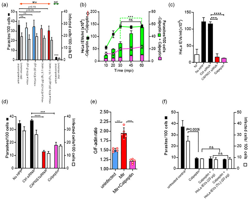
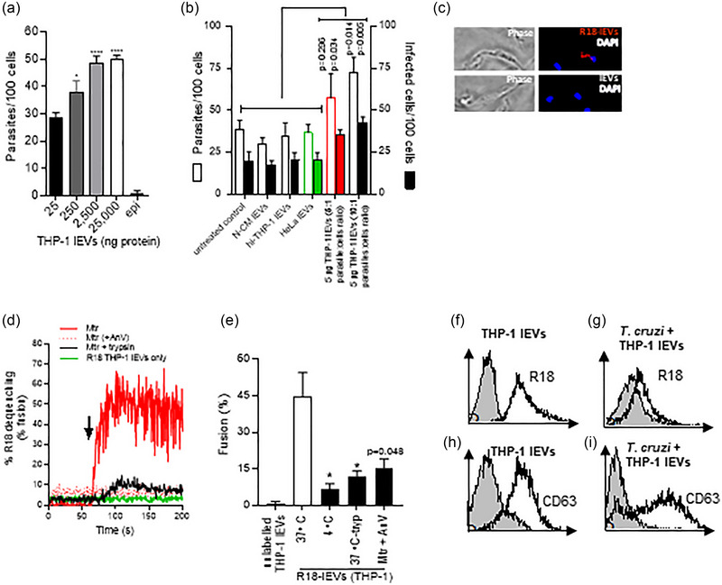
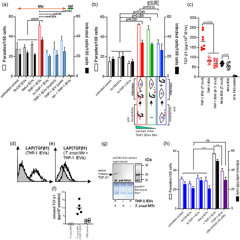
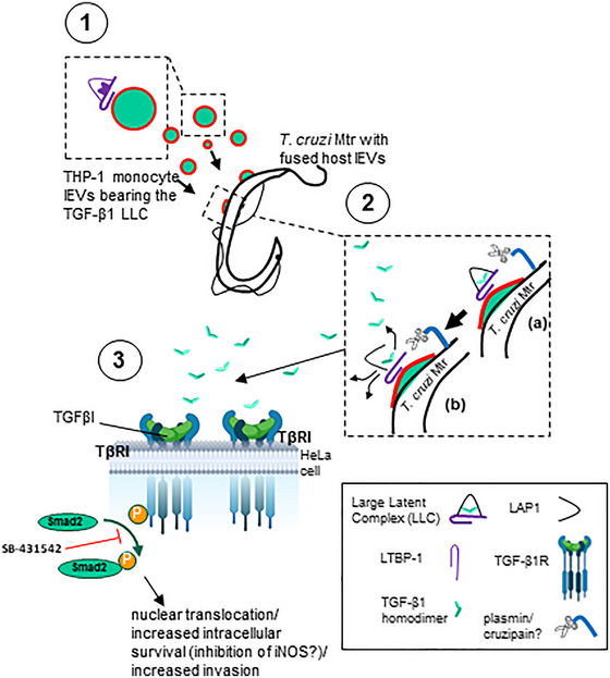
During cell invasion, large Extracellular Vesicle (lEV) release from host cells was dose-dependently triggered by Trypanosoma cruzi metacyclic trypomastigotes (Mtr). This lEV release was inhibited when IP3-mediated Ca2+ exit from the ER and further Ca2+ entry from plasma membrane channels was blocked, but whilst any store-independent Ca2+ entry (SICE) could continue unabated. That lEV release was equally inhibited if all entry from external sources was blocked by chelation of external Ca2+ points to the major contributor to Mtr-triggered host cell lEV release being IP3/store-mediated Ca2+ release, SICE playing a minor role. Host cell lEVs were released through Mtr interaction with host cell lipid raft domains, integrins, and mechanosensitive ion channels, whereupon [Ca2+]cyt increased (50 to 750 nM) within 15 s. lEV release and cell entry of T. cruzi, which increased up to 30 and 60 mpi, respectively, as well as raised actin depolymerization at 60 mpi, were all reduced by TRPC inhibitor, GsMTx-4. Vesicle release and infection was also reduced with RGD peptide, methyl-β-cyclodextrin, knockdown of calpain and with the calpain inhibitor, calpeptin. Restoration of lEV levels, whether with lEVs from infected or uninfected epithelial cells, did not restore invasion, but supplementation with lEVs from infected monocytes, did. We provide evidence of THP-1 monocyte-derived lEV interaction with Mtr (lipid mixing by R18-dequenching; flow cytometry showing transfer to Mtr of R18 from R18-lEVs and of LAP(TGF-β1). Active, mature TGF-β1 (at 175 pg/×105 in THP-1 lEVs) was detected in concentrated lEV-/cell-free supernatant by western blotting, only after THP-1 lEVs had interacted with Mtr. The TGF-β1 receptor (TβRI) inhibitor, SB-431542, reduced the enhanced cellular invasion due to monocyte-lEVs.

Keywords: Trypanosoma cruzi; cell uptake; endocytosis; extracellular vesicles.


Conflict of interest statement

The authors report no conflict of interest.

Figures

FIGURE 1
FIGURE 1
Trypanosoma cruzi induces a calcium‐dependent lEV release from epithelial HeLa cells which can be reduced by blocking mechanosensitive surface channels. (a), HeLa cells (1.0 × 106) were loaded with Fura 2‐AM at 37°C in nominal Ca2+‐free buffer and for all experiments left to equilibrate by preincubating at 37°C for 5 min, from which point t = 0 s was taken. After 60 s, metacyclic trypomastigotes (Mtr) (5.0 × 106) were added (red arrow) and [Ca2+]cyt was monitored using a spectrophotometer over the ensuing 90 s. Mtr infected cells were also treated with EGTA (5 mM) and BAPTA‐AM (10 µM) and epimastigote and non‐infected (NI) controls were included. (b), HeLa cells harvested by trypsinization (1 × 106/well in triplicate) were stimulated at 37°C for 30 min with T. cruzi Mtr (5:1, parasites‐to‐cell ratio) also in the presence of calpeptin, CPT (10 µg/mL). The dose‐response curves were fitted with a 4‐parameter logistic equation. Additional analysis used 2‐way ANOVA with Sidak's post‐test. ***p < 0.001, **p < 0.01 (= 6). lEVs released were isolated and analysed as described in Materials and Methods. (c), Transmission electron microscopy of HeLa cell‐derived lEVs being released after stimulation with T. cruzi Mtr 5:1 (parasites‐to‐cell) ratio at 37°C for 30 min or of resting, uninfected cells, (d). HeLa cells showing increased injury (%PI+), in representative histogram following 30 min exposure of HeLa to metacyclic trypomastigotes (Mtr) (red curves), compared to uninfected cells (green curves), in the presence of propidium iodide, (e). In (f), HeLa cells were injured by a 5:1 ratio of T. cruzi metacyclic forms (30 min/37°C) in the presence or absence of Ca2+. (g), HeLa cells (1.0 × 106) were loaded with Fura 2‐AM at 37°C in nominal Ca2+‐free buffer and left to equilibrate for 5 min, from which point t = 0 s was taken. After 60 s, in calcium‐free buffer, thapsigargin (TG) was added and [Ca2+]cyt monitored using a spectrophotometer over the ensuing 600 s, whereupon cells were incubated in 1.8 mM Ca2+ to stimulate SOCE (green line). HeLa were also infected with Mtr, (with no TG) but in the presence of extracellular Ca2+ (1.8 mM) (blue line). Also in the absence of TG, cells were also infected and then 240 s later, in the presence of Ca2+, treated with 2‐APB (5 µM) (red line). In (h), the effect on lEV release from infected HeLa cells of removing extracellular Ca2+ with EGTA was monitored. Other treatments included 2‐APB (SOCE modulator), YM‐58483 (CRAC channel inhibitor), U‐73122 (PLC inhibitor) and BAPTA‐AM (cell‐permeant Ca2+ chelator). HeLa cells were also stimulated with sublytic MAC (C5b‐9) to release lEVs. HeLa cells (1 × 105/well) when pretreated with the Stretch‐Activated Calcium TRPC blocker, GsMTx‐4, showed marked reduction in infection with Mtr. HeLa cells (1 × 10⁶/well were infected with Mtr and showed reduced invasion with GsMTx‐4 (i)). To measure the G‐actin to F‐actin ratios in uninfected versus T. cruzi‐infected HeLa, G‐actin was measured by flow cytometry with DNase 1‐Alexa Fluor 488 conjugate (j) and F‐actin by Phalloidin Alex Fluor 660 (k) as described in Materials and Methods. Results in (l) show mean ± S.D from a representative experiment performed in triplicate, for the G/F‐actin ratios of HeLa cells infected (60 mpi), with or without GsMTx‐4. Upon infection of HeLa cells (1 × 106/well) with Mtr (m), the greatest reduction in host cell mEV release was for GdCl3 and GsMTx‐4. EGTA (5 mM) was used as a negative control for parasite‐stimulated Ca2+‐mediated mEV release. The inhibitors used to limit T. cruzi‐mediated lEV release did not work when the stimulus for lEV release was sublytic complement (n). Data represents the mean ± SD of three independent experiments performed in triplicate. *p < 0.05, **p < 0.01, and ***p < 0.001 were considered statistically significant.
FIGURE 2
FIGURE 2
Inhibition of lEV release from host HeLa cells using the actin remodelling drug calpeptin, is not restored by supplementation of host lEVs, whether from infected cells or not. (a), There is no increase of cellular invasion of HeLa cells with Mtr, whether HeLa lEVs are added from infected or uninfected cells. (b), Kinetic analysis of lEV release (lines) and T. cruzi metacyclic invasion (bars) before and after pretreatment (37°C for 30 min) with calpeptin (20 µM). Without treatment (green line), T. cruzi‐elicited release of lEVs increased over the first 30 min, but parasite invasion (green bars) continued beyond this point. However, calpeptin inhibited lEV release (pink line) and abrogated parasite entry (pink bars). Calpeptin and CAPNS1 siRNA block lEV release (c) and inhibit invasion (d). The G/F‐actin ratios of calpeptin‐treated, Mtr‐infected HeLa cells, 60 mpi, are significantly reduced, (e). Addition of lEVs (107/mL) from infected or uninfected HeLa did not restore invasion levels reduced by calpeptin, (f).
FIGURE 3
FIGURE 3
lEVs from THP‐1 monocytes carrying latent TGF‐β1 fuse with Mtr, activated TGF‐β1 being released to enhance cellular invasion. (a), Mtr infection of HeLa increases dose dependently with increasing added THP‐1 lEVs. (b), invasion assays in which HeLa were treated with 5 µg THP‐1 lEVs (5:1 and 10:1 parasite:cell ratio). N‐CM (non‐conditioned medium); hi‐THP‐1 lEVs (heat‐inactivated lEVs). (c), T. cruzi Mtr were incubated with R18‐labelled and unlabelled THP‐1 lEVs as described in Materials and Methods and observed by fluorescence microscopy after fixation and mounting with DAPI‐Vectashield. (d), Using the time scan R18 dequenching assay, unlabelled T. cruzi Mtr or trypsinized Mtr (106) (0.25% trypsin/5 min/37°C) were incubated after 60 s (indicated by arrow) with R18‐labelled THP‐1 lEVs (2.5 µg). As control, R18‐lEVs were incubated without Mtr (green line). Fluorescence readings were obtained (excitation/emission 560 nm/590 nm) for the period prior to and then for the 100 s after addition of R‐18 lEVs to give a % dequenching over time. Maximum fluorescence was obtained by adding Triton X‐100 (1% v/v). (e), Percentage fusion was calculated using the formula in Materials and Methods and showed that fusion was temperature and lEV surface protein‐dependent (lEVs treated with 0.25% trypsin (10 min/37°C)) as well as dependent on Mtr‐PtdSer. (f and g), shows acquisition of R18‐ and in (h) and (i) of anti‐CD63‐labelled lEVs to the surface of Mtr by flow cytometry. Unlabelled lEVs are represented by filled histograms in (f) and (h) and T. cruzi with added unlabelled lEVs shown in (g) and (i).
FIGURE 4
FIGURE 4
THP‐1 monocyte lEVs carrying latent TGF‐β1 release activated TGF‐β1 upon fusion with Mtr to enhance cellular invasion. In (a), cellular infection levels are shown with added intact THP‐1 monocyte lEVs, and as control: lEVs from non‐conditioned medium (N‐CM), heat inactivated lEVs, trypsin‐treated lEVS, and a lEV lysate (0.1% Triton X‐100 + protease inhibitor cocktail); infection with epimastigotes was also used as control. (b), Infections were carried out in which either Mtr were preincubated with THP‐1 lEVs (red bars), or in which there was no preincubation (green bars), and in which HeLa cells to be infected were incubated with THP‐1 lEVs, which were then removed at the point of infection (blue bars). (c), ELISA measurements of activated TGF‐β1 (10 min with 1N HCl, as per Materials and Methods) for lysed lEVs from HeLa stimulated with T. cruzi or T. cruzi (heat inactivated) and from lEVs released from untreated HeLa and THP‐1 cells. (d), shows detectable LAP‐TGF‐β1 on the surface of THP‐1 lEVs, also observed on Mtr upon incubation with THP‐1 lEVs, (e), as well as released TGF‐β1 detected by ELISA, (f). (g), western analysis of TGF‐β1 released into the serum‐free cell culture supernatant (rendered cell‐ and lEV‐free by differential centrifugation) and concentrated using a Microcon‐10 kDa Centrifugal Filter Unit with Ultracel‐10 membrane; samples of equal protein concentration were loaded for western analysis using anti‐TGF‐β1. (h), Reduced invasion levels are demonstrated upon addition of lEVs from T. cruzi‐stimulated THP‐1 monocytes, when TβRI signalling is blocked with SB‐431542.
FIGURE 5
FIGURE 5
Model for possible activation of host TGF‐β1 from monocyte cells by T. cruzi metacyclic trypomastigotes. (1) Monocytes carrying TGF‐β1, within the large latent complex (LLC), a tripartite complex of TGF‐β, LAP, and LTBP‐1 attach to the metacyclic trypomastigotes surface where (2) they are activated by surface proteases such as cruzipain or plasmin (from uPA‐activated plasminogen), releasing the TGF‐β1 homodimer to (3) interact with its cognate TGF‐β1 receptor, TβRI, whereupon Smad2 is activated and phosphorylated and then translocating into the nucleus, resulting in increased survival/invasion.

References

    1. Ahmed, N. , Riley, C. , Rice, G. E. , Quinn, M. A. , & Baker, M. S. (2002). Alpha(v)beta(6) integrin‐A marker for the malignant potential of epithelial ovarian cancer. The Journal of Histochemistry and Cytochemistry: Official Journal of The Histochemistry Society, 50, 1371–1380. - PubMed
    1. Al‐Nedawi, K. , Meehan, B. , Micallef, J. , Lhotak, V. , May, L. , Guha, A. , & Rak, J. (2008). Intercellular transfer of the oncogenic receptor EGFRvIII by microvesicles derived from tumour cells. Nature Cell Biology, 10, 619–624. - PubMed
    1. Ambudkar, I. S. , de Souza, L. B. , & Ong, H. L. (2017). TRPC1, Orai1, and STIM1 in SOCE: Friends in tight spaces. Cell Calcium, 63, 33–39. - PMC - PubMed
    1. Ansa‐Addo, E. , Lange, S. , Stratton, D. , Antwi‐Baffour, S. , Cestari, I. , Ramirez, M. , McCrossan, M. , & Inal, J. (2010). Human plasma membrane‐derived vesicles halt proliferation and induce differentiation of THP‐1 acute monocytic leukemia cells. Journal of Immunology, 185, 5236–5246. - PubMed
    1. Antwi‐Baffour, S. , Malibha‐Pinchbeck, M. , Stratton, D. , Jorfi, S. , Lange, S. , & Inal, J. (2020). Plasma mEV levels in Ghanain malaria patients with low parasitaemia are higher than those of healthy controls, raising the potential for parasite markers in mEVs as diagnostic targets. Journal of Extracellular Vesicles, 9, 1697124. - PMC - PubMed

MeSH terms

LinkOut - more resources