Editorial
. 2024 Dec;6(6):e240415.
doi: 10.1148/ryct.240415.

Top 2024 Images in Cardiothoracic Imaging

Affiliations
Editorial

Top 2024 Images in Cardiothoracic Imaging

Domenico Mastrodicasa et al. Radiol Cardiothorac Imaging. 2024 Dec.
No abstract available


Conflict of interest statement
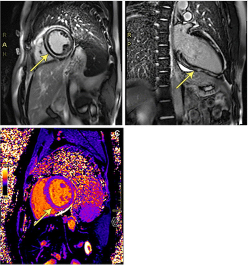
Disclosures of conflicts of interest: D.M. Research grant from the National Institute of Biomedical Imaging and Bioengineering (no. 5T32EB009035); consulting fees from Segmed; trainee deputy editor member of Radiology: Cardiothoracic Imaging trainee editorial board; stock or stock options in Segmed. S.G. NIH K99; trainee deputy editor member of Radiology: Cardiothoracic Imaging trainee editorial board. S.A. Funding from the NIHR Biomedical Research Centre Sheffield; grants from the NIHR, Academy of Clinical Science, South Yorkshire Digital Hub, and NHS England; co-chair of the British Society of Cardiovascular Imaging Trainee Committee Member of the Academic Committee of the Royal College of Radiologists; trainee deputy editor member of Radiology: Cardiothoracic Imaging trainee editorial board. K.H. Honoraria from Bayer; associate editor for Radiology and Radiology: Cardiothoracic Imaging; chair of the trainee editorial board for Radiology: Cardiothoracic Imaging. G.S.G. Trainee deputy editor member of Radiology: Cardiothoracic Imaging trainee editorial board.

Figures

Volume-rendered CT images in the patient show (A) an ascending aortic aneurysm with (B) severe tortuosity of the descending aorta. (C) Coronal maximum intensity projection image shows severe tortuosity and corkscrew appearance of the branches of the superior mesenteric artery (yellow arrow). Source.—Reference 4.
Figure 1:
Volume-rendered CT images in the patient show (A) an ascending aortic aneurysm with (B) severe tortuosity of the descending aorta. (C) Coronal maximum intensity projection image shows severe tortuosity and corkscrew appearance of the branches of the superior mesenteric artery (yellow arrow). Source.—Reference .
Cardiac MR images in a 21-year-old male patient with filamin C variant–associated cardiomyopathy. Postcontrast T1-weighted images acquired with a 1.5-T MRI scanner in (A) short-axis view and (B) vertical long-axis view demonstrate marked concentric (or “ringlike”) midmyocardial late gadolinium enhancement of the left ventricle (yellow arrows). (C) Native T1 mapping image in short-axis view demonstrates an elevated T1 value (1120 msec). Source.—Reference 5.
Figure 2:
Cardiac MR images in a 21-year-old male patient with filamin C variant–associated cardiomyopathy. Postcontrast T1-weighted images acquired with a 1.5-T MRI scanner in (A) short-axis view and (B) vertical long-axis view demonstrate marked concentric (or “ringlike”) midmyocardial late gadolinium enhancement of the left ventricle (yellow arrows). (C) Native T1 mapping image in short-axis view demonstrates an elevated T1 value (1120 msec). Source.—Reference .
Images in a 41-year-old male patient with tracheal Ewing sarcoma with characteristic EWSR-FLI1 fusion protein. (A–C) Contrast-enhanced CT pulmonary angiography images obtained after intravenous injection of 100 mL of iopamidol (Isovue-370; Bracco) in (A) axial soft-tissue window and (B) axial and (C) sagittal lung windows demonstrate polypoidal intraluminal mass arising from posterior right lateral trachea at the level of thyroid gland (white arrow in A and black arrow in B and C). (D) Cinematic volume-rendered CT angiography image in Pulmonary3D object setting (Siemens Healthineers) demonstrates an intraluminal filling defect (white arrow). (E) Photomicrograph of a tracheal biopsy specimen shows cellular tumor with diffuse, sheetlike distribution of small- to medium-sized round, blue primitive neoplastic cells with scant vacuolated cytoplasm (black arrow), consistent with Ewing sarcoma. (Hematoxylin-eosin stain; original magnification, ×40.) (F–H) Initial staging fluorine 18 (18F) fluorodeoxyglucose (FDG) PET/CT images. (F) Maximum intensity projection, (G) transaxial fused PET/CT, and (H) unenhanced axial CT images with abnormal focus of increased FDG uptake in the neck (black arrow in F) corresponding to the intraluminal soft-tissue tracheal lesion (white arrow in G and H). (I) Restaging 18F-FDG PET/CT maximum intensity projection image shows postsurgical changes following resection of the upper tracheal soft-tissue mass lesion with no residual FDG uptake at the resection site to suggest metabolically active residual or recurrent tumor. Persistent focal uptake at the right alveolar margin is related to dental disease and faint uptake in surgical bed is related to the thyroid tracer uptake. (J) Bronchoscopic image shows pedunculated mass in the upper trachea (white arrow). Source.—Reference 6.
Figure 3:
Images in a 41-year-old male patient with tracheal Ewing sarcoma with characteristic EWSR-FLI1 fusion protein. (A–C) Contrast-enhanced CT pulmonary angiography images obtained after intravenous injection of 100 mL of iopamidol (Isovue-370; Bracco) in (A) axial soft-tissue window and (B) axial and (C) sagittal lung windows demonstrate polypoidal intraluminal mass arising from posterior right lateral trachea at the level of thyroid gland (white arrow in A and black arrow in B and C). (D) Cinematic volume-rendered CT angiography image in Pulmonary3D object setting (Siemens Healthineers) demonstrates an intraluminal filling defect (white arrow). (E) Photomicrograph of a tracheal biopsy specimen shows cellular tumor with diffuse, sheetlike distribution of small- to medium-sized round, blue primitive neoplastic cells with scant vacuolated cytoplasm (black arrow), consistent with Ewing sarcoma. (Hematoxylin-eosin stain; original magnification, ×40.) (F–H) Initial staging fluorine 18 (18F) fluorodeoxyglucose (FDG) PET/CT images. (F) Maximum intensity projection, (G) transaxial fused PET/CT, and (H) unenhanced axial CT images with abnormal focus of increased FDG uptake in the neck (black arrow in F) corresponding to the intraluminal soft-tissue tracheal lesion (white arrow in G and H). (I) Restaging 18F-FDG PET/CT maximum intensity projection image shows postsurgical changes following resection of the upper tracheal soft-tissue mass lesion with no residual FDG uptake at the resection site to suggest metabolically active residual or recurrent tumor. Persistent focal uptake at the right alveolar margin is related to dental disease and faint uptake in surgical bed is related to the thyroid tracer uptake. (J) Bronchoscopic image shows pedunculated mass in the upper trachea (white arrow). Source.—Reference .

References

    1. Mastrodicasa D , Gunasekaran S , Alabed S , Gulsin GS , Hanneman K . Top 2023 Images in Cardiothoracic Imaging . Radiol Cardiothorac Imaging 2023. ; 5 ( 6 ): e230259 . - PMC - PubMed
    1. Consul N , Li P , Lennartz S , Chernyak V . 2023 Top Images in Radiology: Radiology In Training Editors’ Choices . Radiology 2024. ; 310 ( 1 ): e233112 . - PubMed
    1. Lennartz S , Li P , Consul N , Lee SI . 2022 Top Images in Radiology: Radiology In Training Editors’ Choices . Radiology 2023. ; 306 ( 2 ): e229031 . - PubMed
    1. Radhakrishnan J , Valakkada J , Ayyappan A , Bellala PK . Corkscrew Mesenteric Arteries and Tortuous Descending Aorta in Autosomal Recessive Cutis Laxa . Radiol Cardiothorac Imaging 2023. ; 5 ( 6 ): e230138 . - PMC - PubMed
    1. Weller S , Bartz-Overman C , Fuss C , Wu EH . Filamin C-associated Cardiomyopathy on Cardiac MR Images . Radiol Cardiothorac Imaging 2023. ; 5 ( 6 ): e230165 . - PMC - PubMed

Publication types

LinkOut - more resources