. 2024 Nov 26;16(22):13505-13525.
doi: 10.18632/aging.206159. Epub 2024 Nov 26.

When do the pathological signs become evident? Study of human mesenchymal stem cells in MDPL syndrome

Affiliations

When do the pathological signs become evident? Study of human mesenchymal stem cells in MDPL syndrome

Spitalieri Paola et al. Aging (Albany NY). .

Abstract

Aging syndromes are rare genetic disorders sharing the features of accelerated senescence. Among these, Mandibular hypoplasia, Deafness and Progeroid features with concomitant Lipodystrophy (MDPL; OMIM #615381) is a rare autosomal dominant disease due to a de novo in-frame deletion in POLD1 gene, encoding the catalytic subunit of DNA polymerase delta. Here, we investigated how MSCs may contribute to the phenotypes and progression of premature aging syndromes such as MDPL. In human induced pluripotent stem cells (hiPSCs)-derived MSCs of three MDPL patients we detected several hallmarks of senescence, including (i) abnormal nuclear morphology, (ii) micronuclei presence, (iii) slow cell proliferation and cell cycle progression, (iv) reduced telomere length, and (v) increased levels of mitochondrial reactive oxygen species (ROS). We newly demonstrated that the pathological hallmarks of senescence manifest at an early stage of human development and represent a warning sign for the progression of the disease. Dissecting the mechanisms underlying stem cell dysfunction during aging can thereby contribute to the development of timely pharmacological therapies for ameliorating the pathological phenotype.

Keywords: MDPL syndrome; MSCs; POLD1 gene; aging; hiPSCs.


Conflict of interest statement

CONFLICTS OF INTEREST: The authors declare no conflicts of interest related to this study.

Figures

Figure 1
Figure 1
Schematic overview of the findings from the characterization of mesenchymal stem cells (MSCs) derived from hiPSCs in MDPL patients and healthy donors.
Figure 2
Figure 2
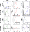
Expression of MSC markers in hiPSC-derived MSCs. Representative histograms of the flow cytometric analysis of the markers CD44, CD90 and CD73 in WT and MDPL MSCs. Grey solid histograms represent the fluorescence of isotype controls, while the empty black histograms represent the specific fluorescence of the indicated MSC markers. The MSC WT histogram is representative of one of two healthy controls because values were comparable. The analyses were performed at passage 3.
Figure 3
Figure 3
Analysis of POLD1 protein expression levels in MDPL and WT-MSCs. (A) Protein extracts of MDPL-MSCs, compared with healthy controls. (B) Densitometric analysis of POLD1 protein normalized on GAPDH levels; *p < 0.05, **p < 0.01, ***p < 0.001, by one-way ANOVA test.
Figure 4
Figure 4
Cell viability of MSCs. (A) Population doubling levels of WT (blue) and MDPL MSCs (orange, green and light blue) from 0 to 96 hours. (B) Cell death count in MSCs of WT and MDPL MSCs from 0 to 96 h. (C) MTS assay, histogram representing the percentage of cell viability at 48 h of WT and MDPL MSCs at 5 × 10³ and 10 × 103 cell seeded. The WT data are derived from the mean of the values obtained from the two healthy control samples. Error bars indicate the standard deviation ± standard error of the mean (SEM). *p < 0.1, **p < 0.01, by one-way ANOVA test.
Figure 5
Figure 5
MSCs altered nuclear morphology analysis. (A) Histogram representing the percentage of aberrant nuclear morphology. (B) A representative image of MSCs WT and MDPL obtained by immunofluorescence for anti-Lamin A/C antibody (red). (C) Percentage of micronuclei (%MN) and percentage of Lamin B1 positive (% Lamin B1+) MN in MSCs MDPL and WT. (D) Representative immunofluorescence images showing the presence of micronuclei (MN) positive (arrow yellow) or negative (arrow white) for anti-Lamin B1 antibody (red) in MDPL cells. Hoechst nuclear staining (blue). Scale bar 100 µm. The WT data are derived from the mean of the values obtained from the two healthy control samples. Experiments were carried out in triplicates; 300 nuclei were counted per individual. Error bars indicate the standard deviation ± standard error of the mean (SEM). *p < 0.1, **p < 0.01 by one-way ANOVA test.
Figure 6
Figure 6
Cell cycle analysis. Histogram plots showing cells in G0/G1, S and G2/M phases of the cell cycle performed with the DNA intercalating dye propidium iodide.
Figure 7
Figure 7
MDPL MSCs senescence and telomere shortening. (A) The histogram shows the average percentage of β-galactosidase-positive cells in WT and MDPL mesenchymal stem cells. (B) Representative image of senescence-associated β-galactosidase assay. A greater amount of intensely positive blue cells is displayed in MDPL-MSCs than in WT controls. Arrows indicated altered cell shape. Scale bar = 200 µm. Experiments were carried in triplicates; 100 nuclei were counted per individual. Error bars indicate the standard deviation ± standard error of the mean (SEM), *p < 0.1, **p < 0.01. (C) Relative telomere length was evaluated by real-time q-PCR in MSCs MDPL and WT. Experiments were carried in triplicates. Error bars indicate the standard deviation ± standard error of the mean (SEM). ***p < 0.001, ****p < 0.0001 by one-way ANOVA test.
Figure 8
Figure 8
Mitochondrial dysfunction. Flow cytometry quantification of total reactive oxygen species using CM-H2DCFDA in MSC WT and MDPL ones. (A) Histogram representing the relative fluorescence intensity (FITCH-A), data are shown as mean ± SD of N = 3 biological replicates. Adjusted p-value was calculated using Student’s t-test. (B) Representative histogram plots in log scale (WT blue). Flow cytometry quantification of mitochondrial superoxide using MitoSOX Red in MSC WT and MDPL ones. (C) Histogram representing the relative fluorescence intensity (PE-A), data are shown as mean ± SD of N = 3 biological replicates. Adjusted p-value was calculated using Student’s t-test. (D) Representative histogram plots in log scale. (E) Histogram representing the quantification of red/green fluorescence intensity ratio, data are shown as mean ± SD of N = 3 biological replicates. Adjusted p-value was calculated using Student’s t-test. (F) Representative dot plots in log scale of JC1 stained MSC WT and MDPL cells. The WT data are derived from the mean of the values obtained from the two healthy control samples; *p < 0.1, **p < 0.01.

References

    1. Lessel D, Kubisch C. Hereditary Syndromes with Signs of Premature Aging. Dtsch Arztebl Int. 2019; 116:489–96. 10.3238/arztebl.2019.0489 - DOI - PMC - PubMed
    1. Coppedè F. Mutations Involved in Premature-Ageing Syndromes. Appl Clin Genet. 2021; 14:279–95. 10.2147/TACG.S273525 - DOI - PMC - PubMed
    1. Weedon MN, Ellard S, Prindle MJ, Caswell R, Lango Allen H, Oram R, Godbole K, Yajnik CS, Sbraccia P, Novelli G, Turnpenny P, McCann E, Goh KJ, et al.. An in-frame deletion at the polymerase active site of POLD1 causes a multisystem disorder with lipodystrophy. Nat Genet. 2013; 45:947–50. 10.1038/ng.2670 - DOI - PMC - PubMed
    1. Shastry S, Simha V, Godbole K, Sbraccia P, Melancon S, Yajnik CS, Novelli G, Kroiss M, Garg A. A novel syndrome of mandibular hypoplasia, deafness, and progeroid features associated with lipodystrophy, undescended testes, and male hypogonadism. J Clin Endocrinol Metab. 2010; 95:E192–7. 10.1210/jc.2010-0419 - DOI - PMC - PubMed
    1. Novelli G, Muchir A, Sangiuolo F, Helbling-Leclerc A, D'Apice MR, Massart C, Capon F, Sbraccia P, Federici M, Lauro R, Tudisco C, Pallotta R, Scarano G, et al.. Mandibuloacral dysplasia is caused by a mutation in LMNA-encoding lamin A/C. Am J Hum Genet. 2002; 71:426–31. 10.1086/341908 - DOI - PMC - PubMed

MeSH terms

LinkOut - more resources