. 2024 Nov 29;14(1):121.
doi: 10.1186/s13550-024-01157-8.

Imaging tumor and ascites-associated macrophages in a mouse model of metastatic ovarian cancer

Affiliations

Imaging tumor and ascites-associated macrophages in a mouse model of metastatic ovarian cancer

Catherine A Foss et al. EJNMMI Res. .

Abstract

Background: Tumor-Associated Macrophages (TAMs) play a critical role in the pathogenesis and progression of ovarian cancer, a lethal gynecologic malignancy. [124I]iodo-DPA-713 is a PET radiotracer that is selectively trapped within reactive macrophages. We have employed this radioligand here as well as a fluorescent analog to image TAMs associated with primary tumors, secondary pulmonary metastases and gastrointestinal tract-associated macrophages, associated with ascites accumulation in a syngeneic mouse model of metastatic ovarian cancer. Intact female C57BL/6 mice were engrafted with ID8-Defb29-VEGF tumor pieces. One month after engraftment, the mice were selected for positive bioluminescence to show primary and secondary tumor burden and were then scanned by PET/MRI with [124I]iodo-DPA-713, observing a 24 h uptake time. PET data were overlayed with T2-weighted MRI data to facilitate PET uptake tissue identity. Additionally, mice were imaged ex vivo using Near IR Fluorescence (NIRF), capturing the uptake and sequestration of DPA-713-IRDye800CW, a fluorescent analog of the radioligand used here. Additionally, cell culture uptake of DPA-713-IRDye680LT in ID8-DEFb29-VEGF, IOSE hTERT and RAW264.7 cells was conducted to measure tracer uptake in ovarian cancer cells, ovarian epithelial cells and macrophage.

Results: PET/MRI data show an intense ring of radiotracer uptake surrounding primary tumors. PET uptake is also associated with lung metastases, but not healthy lung. Mice displaying ascites also display PET uptake along the gastrointestinal tract while sham-operated mice show minimal gastrointestinal uptake. All mice show specific kidney uptake. Mice imaged by NIRF confirmed TAMs uptake mostly at the rim of primary tumors while 1 mm secondary tumors in the lungs displayed robust, homogeneous uptake of the radio- and fluorescent analog. Ex vivo biodistribution of [124I]iodo-DPA-713 showed that contralateral ovaries in middle-stage disease had the highest probe uptake with tissues sampled in mid- and late-stage disease showing increasing uptake.

Conclusion: [124I]iodo-DPA-713 and DPA-713-IRDye800CW sensitively identify and locate TAMs in a syngeneic mouse model of metastatic ovarian cancer.

Keywords: Ascites; ID8-Defb29-VEGF; Macrophages; Ovarian cancer; TAMs; iodo-DPA-713.


Conflict of interest statement

Declarations. Ethics approval and consent to participate: The study was performed in accordance with the ethical standards as laid down in the 1964 Declaration of Helsinki and its later amendments or comparable ethical standards. All methods were carried out in accordance with relevant guidelines and regulations. All rodent studies were approved by the Johns Hopkins IACUC. The study was carried out in compliance with the ARRIVE guidelines Consent for publication: Not applicable. Competing interests: C.A.F. holds a patent share in DPA-713-IRDye800CW, currently licensed to D&D Pharmatek. No other competing interests.

Figures
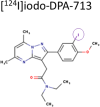
Fig. 1
Fig. 1
Chemical structures of [124I]iodoDPA-713 and DPA-713-IRDye800CW. I-124 radiolabel is circled [21]
Fig. 2
Fig. 2
In vivo PET/CT, PET/MRI, BLI and ex vivo imaging of reactive macrophages in orthotopic OvCa and sham operated mice. Mice on upper left in top (BLI and PET/CT) show a mouse with moderate disease and no metastatic spread. Tumor uptake or radiolabeled shown as a MIP reveals a torus-shaped uptake around where the orthotopic tumor resides. The top right two mice (PET/MRI) show radiotracer uptake in sham operated mouse (sham, O) and a mouse with a 1 cm orthotopic tumor (T), showing a ring-spaped uptake of radiotracer around the tumor. [124I]iodoDPA uptake in the top panel shows stomach (S, metabolic radioiodine uptake), gastrointestinal tract and uptake by TAMs, which encircle the tumor. The sham surgery shows radiotracer uptake similar to background. The lower panel shows mirror uptake of the fluorescent analog where the tumor (T) is the brightest tissue. The sham mouse again shows background uptake of the fluorescent analog along with gastrointestinal tract and liver uptake
Fig. 3
Fig. 3
In vivo macrophage imaging in primary and metastatic OvCa. A [124I]iodoDPA PET/MRI imaging of a mouse bearing an 8 mm primary orthotopic ID8-Defb29-VEGF tumor (T), peritoneal ascites (white arrows) and several 1 mm pulmonary metastases (red arrows). The primary tumor shows peritumoral uptake of radiotracer in TAMs (dotted lines), even 1 mm metastases with recruited macrophages are easily imaged (B). B Rightmost panel shows magnified views of pulmonary metastases with probe uptake (top) and T2 MRI (middle) and a magnified view of primary orthotopic probe uptake by PET with MRI (bottom). Peri-tumoral TAMs uptake in the primary tumor is shown in dotted lines. Scale bar indicates kBq/cc
Fig. 4
Fig. 4
Near IR fluorescence ex vivo imaging of orthotopic, metastatic and subcutaneous ID8-Defb29-VEGF tumors. DPA-713-IRDye800CW (green) exhibits very high whole-tumor uptake in the orthotopic model (1˚), much brighter than the < 1 mm pulmonary metastases (red arrows). The mouse bearing a subcutaneous tumor shows classic peri-tumoral uptake (subcutaneous ID8-Defb29-VEGF in green and in merged 700 + 800 nm channels)
Fig. 5
Fig. 5
Ex vivo Biodistribution of [124I]iodo-DPA-713 in C57Bl/6 mice ± ID8-Defb29-VEGF tumors. Three groups of three to five mice bearing early growth (red bars) orthotopically implanted ID8-Defb29-VEGF, mid growth (green bars) ID8-Defb29-VEGF, late growth (purple bars) ID8-Defb29-VEGF (with ascites) or sham operated were injected with 110 µCi of [124I]iodo-DPA-713 followed by a 24 h uptake period. The mice were then euthanized and the indicated tissues were collected. The mice with mid- and late-growth ID8-Defb29-VEGF tumors displayed the highest uptake but not just in the tumors. The mice appear to be systemically inflamed, with elevated uptake across tissues. Contralateral ovary took up a large amount of radiotracer while large intestine was also displaying high uptake, possibly due to inflamed peritoneal macrophages
Fig. 6
Fig. 6
Live cell uptake of DPA-713-IRDye680LT with subsequent anti-CD68 antibody staining. All three cell types took up DPA-713-IRDye680LT probe (red) as expected while ID8-Defb29-VEGF carcinoma and RAW264.7 macrophages displayed the highest uptake of DPA probe and CD68 antibody (green)

References

    1. Mathieu KB, et al. Screening for ovarian cancer: imaging challenges and opportunities for improvement. Ultrasound Obstet Gynecol. 2018;51(3):293–303. - PMC - PubMed
    1. Beiderwellen K, et al. 18)F]FDG PET/MRI vs. PET/CT for whole-body staging in patients with recurrent malignancies of the female pelvis: initial results. Eur J Nucl Med Mol Imaging. 2015;42(1):56–65. - PubMed
    1. Gupta V, Yull F, Khabele D. Bipolar Tumor-Associated macrophages in Ovarian Cancer as targets for Therapy. Cancers (Basel), 2018. 10(10). - PMC - PubMed
    1. Takaishi K, et al. Involvement of M2-polarized macrophages in the ascites from advanced epithelial ovarian carcinoma in tumor progression via Stat3 activation. Cancer Sci. 2010;101(10):2128–36. - PMC - PubMed
    1. Etzerodt A et al. Tissue-resident macrophages in omentum promote metastatic spread of ovarian cancer. J Exp Med, 2020. 217(4). - PMC - PubMed

LinkOut - more resources