. 2024 Nov 29;10(48):eadr4227.
doi: 10.1126/sciadv.adr4227. Epub 2024 Nov 29.

Molecular principles of the assembly and construction of a carboxysome shell

Affiliations

Molecular principles of the assembly and construction of a carboxysome shell

Peng Wang et al. Sci Adv. .

Abstract

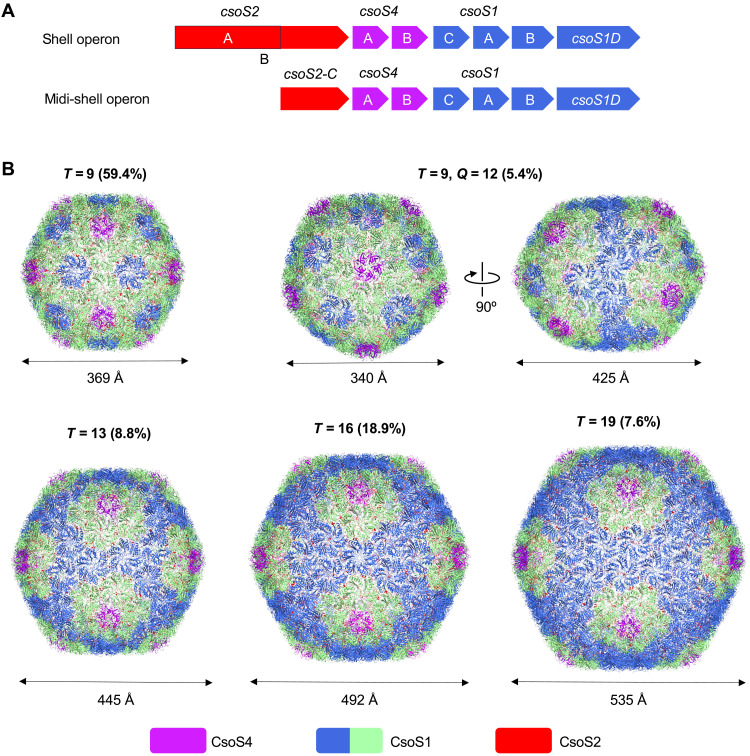
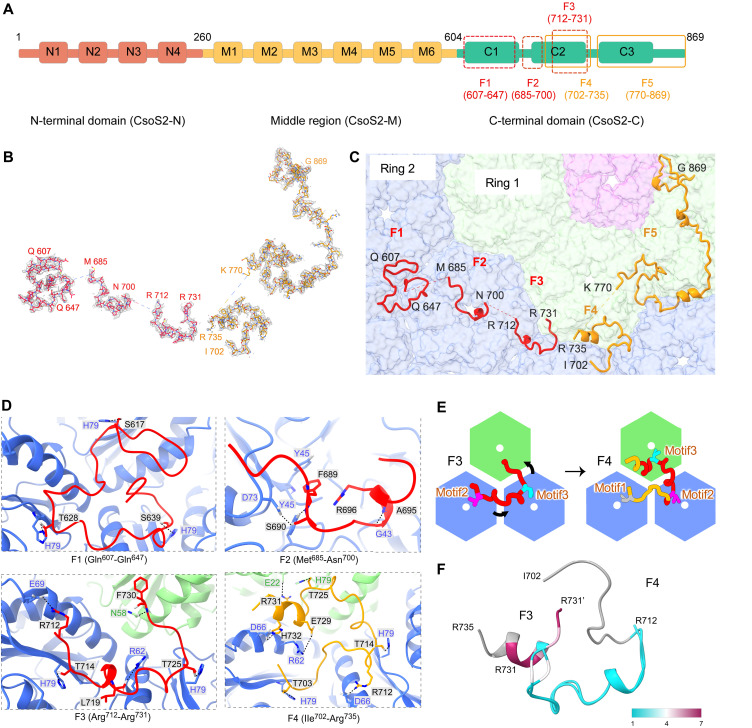
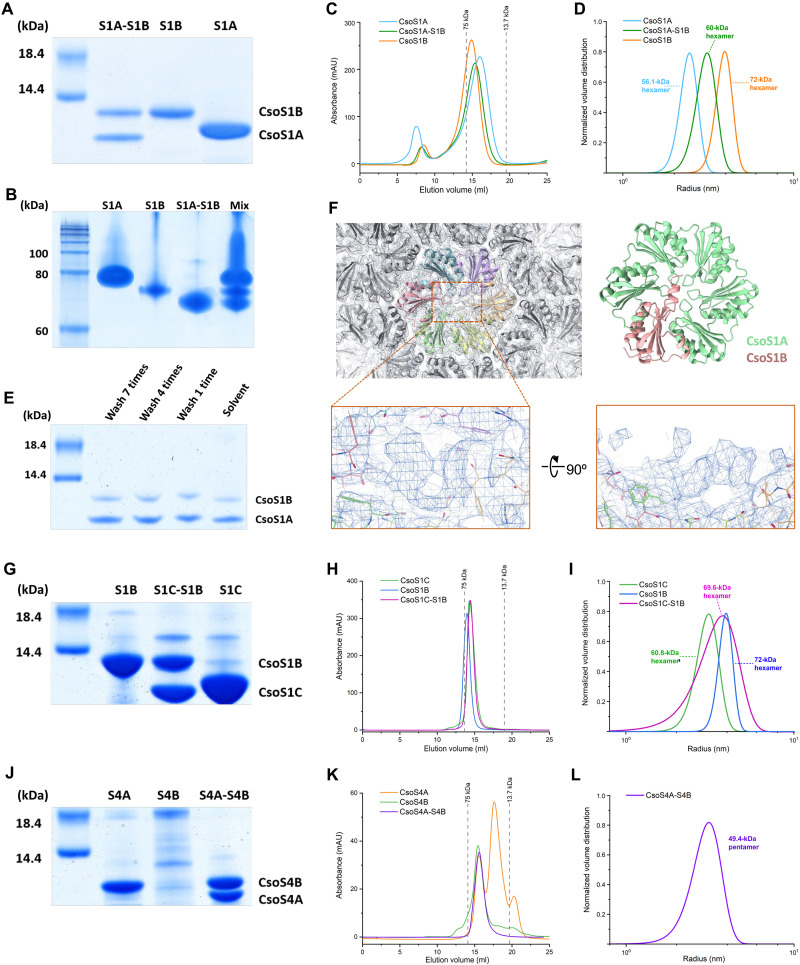
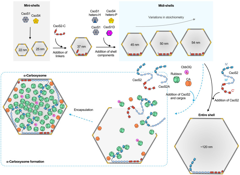
Intracellular compartmentalization enhances biological reactions, crucial for cellular function and survival. An example is the carboxysome, a bacterial microcompartment for CO2 fixation. The carboxysome uses a polyhedral protein shell made of hexamers, pentamers, and trimers to encapsulate Rubisco, increasing CO2 levels near Rubisco to enhance carboxylation. Despite their role in the global carbon cycle, the molecular mechanisms behind carboxysome shell assembly remain unclear. Here, we present a structural characterization of α-carboxysome shells generated from recombinant systems, which contain all shell proteins and the scaffolding protein CsoS2. Atomic-resolution cryo-electron microscopy of the shell assemblies, with a maximal size of 54 nm, unveil diverse assembly interfaces between shell proteins, detailed interactions of CsoS2 with shell proteins to drive shell assembly, and the formation of heterohexamers and heteropentamers by different shell protein paralogs, facilitating the assembly of larger empty shells. Our findings provide mechanistic insights into the construction principles of α-carboxysome shells and the role of CsoS2 in governing α-carboxysome assembly and functionality.


Figures

Fig. 1.
Fig. 1.. Design and overall cryo-EM structures of midi-shells.
(A) Genetic organizations of shell operon and midi-shell operon. (B) Cryo-EM structures of different midi-shell forms of T = 9, T = 9 Q = 12, T = 13, T = 16, and T = 19, at the resolution of 2.3, 2.99, 2.93, 2.75, and 3.04 Å, respectively. The relative ratios and the diameters of different types of shells are indicated. The diameters of the shells are indicated. Shell components are colored purple (CsoS4 pentamer), blue/green (CsoS1 hexamer), and red (CsoS2). Hexamers at different relative positions are represented by different colors: green, shell hexamers adjacent to pentamers; blue, hexamers that are not adjacent to pentamers. The T and Q numbers are parameters for describing the geometry of polyhedral structures. T represents the area of a triangular facet within an icosahedral shell, whereas Q denotes the area of an elongated face within a prolate shell. Their respective computational formulas are as follows: T = h2 + hk + k2, Q = hh′ + hk′ + kk′. Here, h and k indicate the counts of two-step connections linking adjacent pentamers along the principal directions of the hexagonal lattice, respectively; h′ and k′ represent the counts of two-step connections linking diagonal pentamers within the prolate shell, respectively.
Fig. 2.
Fig. 2.. The shell proteins and interfaces in midi-shells.
(A) Overall organization of T = 9, T = 13, T = 16, and T = 9 Q = 12 shells, with labeled assembly interfaces between capsomeres. (B) Details of interacting residues in the 10 different assembly interfaces of the T = 19 shell. (C) Overlay of interface 1 from T = 9 (yellow), T = 13 (green), T = 16 (blue), T = 19 (purple), and T = 9 Q = 12 (orange) shells. (D) Side view of the overlay interfaces 2 to 10 from the T = 19 shell. (E) Vertical and horizontal angle differences of hexamer-hexamer interfaces 2 to 10 from the T = 19 shell. The hexamers are simplified to averaged planes.
Fig. 3.
Fig. 3.. CsoS2 fragments and their interaction sites with shell proteins in midi-shells.
(A) Domain arrangement of CsoS2. The N-terminal, middle and C-terminal domains are colored green, yellow, and pink, respectively. The four dashed boxes and one solid box indicate the fragments resolved in the T = 19 midi-shell. (B) Cryo-EM densities of newly identified CsoS2 F1 to F5 with atom models. (C) CsoS2 interactions with shell components, viewed from inside. CsoS4 pentamers are colored purple. Fragments belonging to two different CsoS2 chains are colored red and yellow, respectively. The first (hexamers adjacent to the pentamer) and second rings of CsoS1 hexamers are colored green and blue, respectively. (D) Interaction interfaces between CsoS2 F1 to F4 fragments and CsoS1A hexamers. (E) A pattern to indicate the relative position change of F3 and F4 on the shell inner surface. Residues of 712-731 in F3 and F4 are colored in red, and the others are in yellow. The residues of Thr703, Thr714, and Thr725 are marked in gray, magenta, and cyan, respectively. (F) Cartoon view of the structural alignment of F3 and F4 colored by Cα RMSDs values.
Fig. 4.
Fig. 4.. The hetero-oligomer formation between CsoS1 and CsoS4 Paralogs.
(A and B) SDS-PAGE (A) and native-PAGE (B) analysis of individually expressed His-tagged CsoS1A (S1A), His-tagged CsoS1B (S1B), and coexpressed CsoS1A and His-tagged CsoS1B (S1A-S1B). The MIX lane sample in native-PAGE consists of a blend of individually purified proteins and coexpressed protein products. (C and D) Gel filtration chromatography (C) and DLS (D) results of His-tagged CsoS1A (blue), His-tagged CsoS1B (orange), and coexpressed CsoS1A and His-tagged CsoS1B (green). (E) SDS-PAGE analysis of crystals obtained from the co-expressed sample after various washing durations. (F) Extra density identified at the center and terminal regions of the CsoS1 hexamer in the C1 symmetry T = 16 shell map. (G to I) SDS-PAGE (G), gel filtration chromatographic (H), and DLS (I) analysis of CsoS1C (green), CsoS1B (blue), and coexpressed CsoS1C and CsoS1B (purple). (J and K) SDS-PAGE (J), gel filtration and chromatographic (K) analysis of CsoS4A (orange), CsoS4B (green), and coexpressed CsoS4A and CsoS4B (purple). (L) DLS analysis of coexpressed CsoS4A and CsoS4B (purple).
Fig. 5.
Fig. 5.. Proposed model of CsoS2-mediated assembly of α-carboxysome shells and intact α-carboxysomes.
The combination of minimal shell components, consisting of a single type of hexamer and pentamer, can generate mini-shells with a maximal size of 25 nm. The presence of CsoS2-C, which stabilizes the interfaces between shell proteins, enables the expansion of mini-shells to 37 nm. Further incorporation of a full set of shell components, including homomultimers and heteromultimers, leads to formation of midi-shells with a maximal size of 54 nm. Replacing CsoS2-C with full-length CsoS2 yields large synthetic shells with an average size of ~120 nm, resembling native carboxysome shells. Furthermore, in the presence of CsoS2 and cargos in vivo, the shorter CsoS2 isoform (CsoS2A) assists the full-length CsoS2 isoform (CsoS2B) in recruiting Rubisco, along with the encapsulation of CA and the Rubisco activases CbbOQ, ultimately leading to the formation of an intact α-carboxysome (blue dashed arrows). Note that the numbers of carboxysome components do not represent the actual stoichiometry of α-carboxysome proteins.

References

    1. Chen A. H., Silver P. A., Designing biological compartmentalization. Trends Cell Biol. 22, 662–670 (2012). - PubMed
    1. Greening C., Lithgow T., Formation and function of bacterial organelles. Nat. Rev. Microbiol. 18, 677–689 (2020). - PubMed
    1. Kerfeld C. A., Aussignargues C., Zarzycki J., Cai F., Sutter M., Bacterial microcompartments. Nat. Rev. Microbiol. 16, 277–290 (2018). - PMC - PubMed
    1. Vacca I., Insights into bacterial microcompartments. Nat. Rev. Microbiol. 15, 451–451 (2017). - PubMed
    1. Stewart K. L., Stewart A. M., Bobik T. A., Prokaryotic Organelles: Bacterial microcompartments in e. coli and salmonella. EcoSal Plus 9, (2020). - PMC - PubMed

MeSH terms

LinkOut - more resources